
Diplophos vicinia, Koeda & Ho, 2019
|
publication ID |
https://doi.org/ 10.11646/zootaxa.4702.1.15 |
|
publication LSID |
lsid:zoobank.org:pub:8A8FD64D-DE1B-4379-BC96-D71AD0C43B04 |
|
DOI |
https://doi.org/10.5281/zenodo.5631338 |
|
persistent identifier |
https://treatment.plazi.org/id/24C71ECF-60B3-49DD-8EC8-8922917785F8 |
|
taxon LSID |
lsid:zoobank.org:act:24C71ECF-60B3-49DD-8EC8-8922917785F8 |
|
treatment provided by |
Plazi |
|
scientific name |
Diplophos vicinia |
| status |
sp. nov. |
Diplophos vicinia sp. nov.
English name: Neighbor Portholefish
Figures 1–3 View FIGURE 1 View FIGURE 2 View FIGURE 3 ; Tables 1–2 View TABLE 1 View TABLE 2
Holotype. NMMB-P 29097, 127.6 mm SL, off Dong-gang , Pingtung , South China Sea, Taiwan, 21 Mar. 2018, obtained at fish-landing ground at Dong-gang; collected by commercial mid-water trawl.
Paratypes. Collection locality and method same as holotype: ASIZP 64190 View Materials , 2 specimens, 84.4–95.0 mm SL, 24 Mar. 2004 ; NMMB-P9246 , 99.7 mm SL, NMMB-P 30714, 101.9 mm SL, 18 Sept. 2008 ; NMMB-P 11930, 129.7 mm SL, 28 Feb. 2011 ; NMMB-P 27286, 100.5 mm SL, 14 Oct. 2017 . South China Sea (22°04ʹN, 120°27ʹE): ASIZP 64180 View Materials , 73.9 mm SL, 25 March 2004 GoogleMaps . Between Papua New Guinea and Micronesia (02°42ʹN, 150°03ʹE): ASIZP 73845, 143.5 mm SL, 27 August 2010 GoogleMaps .
Diagnosis. A species of Diplophos with the following combination of characters: anal-fin rays 57–59; branchiostegal rays 14; gill rakers 4 + 10–11=14–15; abdominal vertebrae 30–32; caudal vertebrae 44–45; total vertebrae 74–76. Photophores: IP 13–14, PV 18–19, IV 31–32, VAV 12–13, AC 37–39 + 2, IC 83–86; last two AC photophores grouped, separation less than half that of preceding photophores; dorsal-fin origin closer to snout than caudal-fin base, pre-dorsal-fin length 45.2–47.3% SL.
Description. Counts and measurements are given in Tables 1 View TABLE 1 and 2 View TABLE 2 . The following data are given for the holotype, with values of paratypes in parentheses, unless otherwise indicated.
Body strongly elongated, its greatest depth (usually at base of pelvic-fin) 10.3 (8.7–13.8) in SL; dorsal and ventral outlines almost straight along abdomen, gradually becoming shallower along tail. Body and head strongly compressed laterally, progressively more so toward the caudal peduncle; snout to anus less than half body length. Scales on body lost in all specimens.
Head small, 6.6 (6.0–6.8) in SL; snout short, moderately pointed, its length greater than eye diameter. Eye moderately large, circular, its diameter 4.1–5.0 in HL. Olfactory laminae inside nostril 21–23, visible externally. Mouth large, weakly oblique, ventral margin of maxilla strongly curved, posteriorly nearly reaching the margin of preoperculum. Single row of large caniniform teeth on all jaws, one to three small sharp teeth between each large caniniform tooth; teeth present from near symphysis of premaxilla to posterior end of maxilla, largest on premaxilla, slightly larger than those on maxilla and dentary; single middle-sized tooth row on anterolateral aspect of dentary; two small conical teeth on vomer; single row of three to seven small conical teeth on palatine. Two small teeth on anterior part of basihyal. Four gill arches present, a well-developed slit behind fourth; gill rakers on first to third gill arches comb-like, but on fourth gill arche knob-like; lowermost upper gill raker and uppermost lower gill raker closely attached to each other. Pseudobranch small with 7 (3–10) short filaments.
All fin rays short, transparent and fragile; dorsal-fin base short, originating anterior to origin of anal fin, ending slightly posterior to anal-fin origin; anal-fin base long, its length 7.4 (7.3–10.6) times dorsal-fin base length; pectoral fin small, inserted level with posteroventral portion of gill slit; pelvic fin small, closer to anal-fin origin than closest point of pectoral-fin base; caudal fin small, its maximum length less than caudal-peduncle length.
Head photophores ( Fig. 3 View FIGURE 3 ). Single supraorbital photophore (ORB) at anterodorsal corner of eye between eye and nostril, small, embedded under skin; 12 small photophores in horizontal series along ventral margin of eye; one large photophore below anteroventral portion of eye; 9 small photophores in horizontal linear series below middle of eye; one medium-sized photophore below posteroventral portion of eye; one large photophore (anteriormost one of OP) beneath middle of preopercle, followed by 6 small photophores in horizontal linear series along dorsal margin of premaxilla; one large photophore posterior to and just below dorsal margin of preopercle; 5 small photophores in vertical series beneath border of preopercle and opercle; 5 small photophores in longitudinal series beneath ventral margin of preopercle; 2 large elongated photophores (posterior two of OP) beneath border of subopercle and interopercle; two series of 5 or 6 small photophores on interopercle; single row of ca. 26 small photophores, embedded in black membrane (just below base of teeth) along lateral aspect of dentary, posteriormost curving upward.
Main body photophores. Photophores vertically elongated except two posteriormost AC. Latter smaller than preceding photophores, not attached to each other, separated by less than half and distance between preceding photophores. A single specimen (ASIZP 64190) with nine VALC photophores (others lost; all missing, presumably lost, in other specimens).
Accessory body photophores. Most specimens with skin abrasion during capture, making precise photophore counts difficult; 33 and 35 abdominal lateral-line photophores (LLA) apparent in ASIZP 64180 and 61490, respectively. Caudal lateral-line photophores completely missing, presumably lost, in all specimens. Eleven (maximum) principal rows of small photophores on body paralleling IC and OA: five on dorsolateral aspect of trunk, uppermost along mid-dorsal line (except on dorsal-fin base); one of small photophores on lateral line slightly larger than other accessory photophores; four on ventral half of trunk; one between IC and OA, from just posterior to gill opening to below end of VALB (anteriormost two photophores in front of pectoral-fin base as large as main photophores). Single medium-sized photophore on midventral line midway between anus and anal-fin origin.
Fresh coloration ( Fig. 1 View FIGURE 1 ). Body uniformly creamy-white due to almost complete skin and scale abrasion, faintly brownish dorsally. Main photophores silver with black anterior margin; accessory body photophores on lateral line silver with black margin. Head black with silver reflection on skin remaining on cheek and opercular; buccal cavity and inner surface of opercle black; iris silver; fins semi-translucent.
Preserved coloration. Head and body uniformly light brown, faintly dusky dorsally. Main photophores silver with black anterior margin; accessory body photophores on lateral line silver with black margin; other accessory body photophores black. Head blackish, opercular and isthmus solid black. Buccal cavity, inner surface of opercle, gill rakers dusky. Iris brown with silver reflection. Peritoneum black.
Etymology. The specific name, vicinia (meaning “neighbor” in Latin), is given in reference to the grouping of the last two AC photophores.
Distribution. Diplophos vicinia sp. nov. has been collected mainly off southwestern Taiwan, although the single specimens collected in the northern South China Sea and from north of Papua New Guinea.
Remarks. Of the six nominal species recognized in the genus Diplophos , the new species described herein is distinct in having greater numbers of total gill rakers (14–15 vs. 12–13, usually 12, in congeners), and VAV photophores (12 vs. 11 in D. pacificus , 14–17 in other species). Ozawa et al. (1990) divided the genus into two groups, based on grouping or separation of the last two AC photophores, with only D. rebainsi and Manducus greyae (Johnson 1970) (previously included in the genus Diplophos by Smith et al. 1991) included in the latter group. Diplophos vicinia is distinguished from the other five species that also have the last two AC photophores grouped as follows: abdominal vertebrae 30–32 in D. vicinia (vs. 33–41); caudal vertebrae 44–45 (vs. 48–60); total vertebrae 74–76 (vs. 83–100); IP 13–14 (vs. 15–19); PV 18–20 (vs. 23–29); IV 31–34 (vs. 38–48); AC 38–40 (vs. 43–53); IC 85 (92–115) (comparative data adopted from Ozawa et al. 1990). Compared with D. rebainsi (last two AC photophores well separated), D. vicinia has anal-fin rays 57–58 (vs. 47–53); branchiostegal rays 14 (vs. 10–11); VAV 12 (vs. 15–17); AC 38–40 (vs. 32–34); BR 11 (vs. 7–8); dorsal-fin origin closer to snout than caudal-fin base, pre-dorsal-fin length 45.2–47.3% SL (vs. closer to caudal-fin base than snout, and pre-dorsal-fin length 61.2–64.4% SL) (data adopted from Schaefer et al. 1986; Harold 1999; Kenaley & Stewart 2015).
Most of the Diplophos specimens examined here had been previously identified as D. taenia or D. orientalis . Except for a single specimen (ASIZP 75043: Fig. 4 View FIGURE 4 ) of D. taenia collected from open waters of the South China Sea, all of the Diplophos specimens collected in Taiwanese waters reported on herein are identified as D. vicinia .
Comparative material. Diplophos taenia : ASIZP 75043, 114.5 mm SL, South China Sea (22°03ʹN, 118°54ʹE) GoogleMaps .
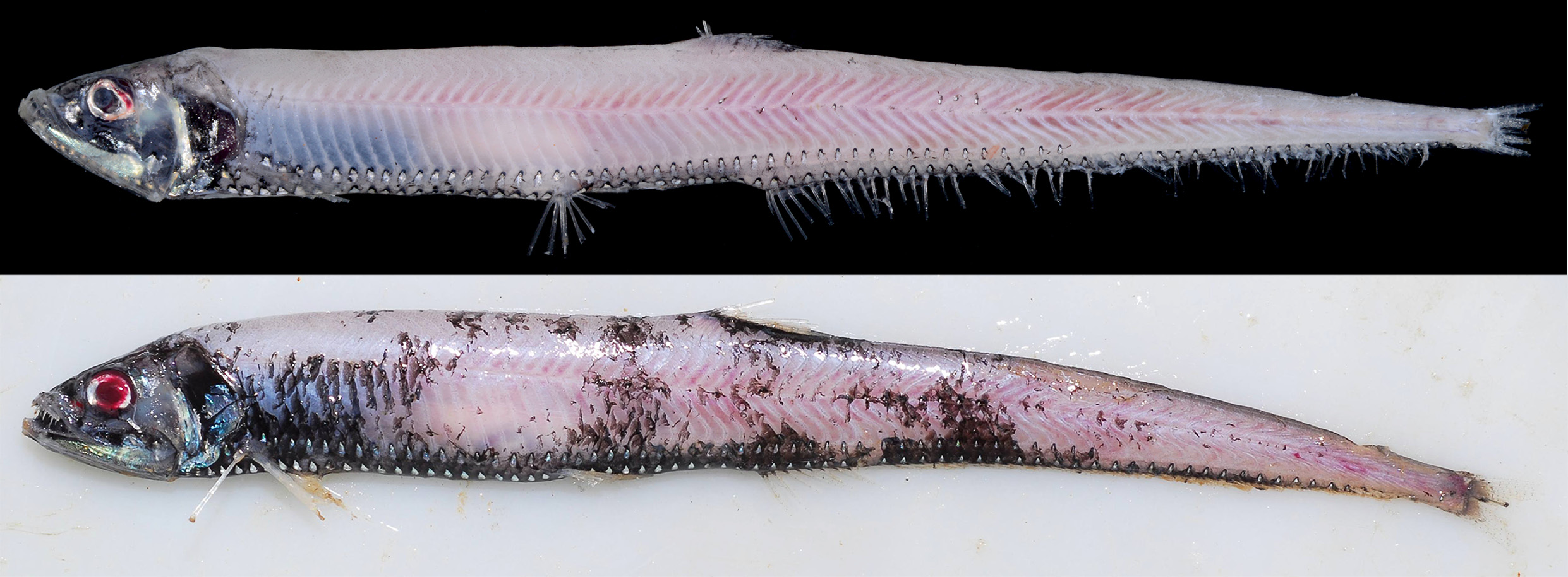
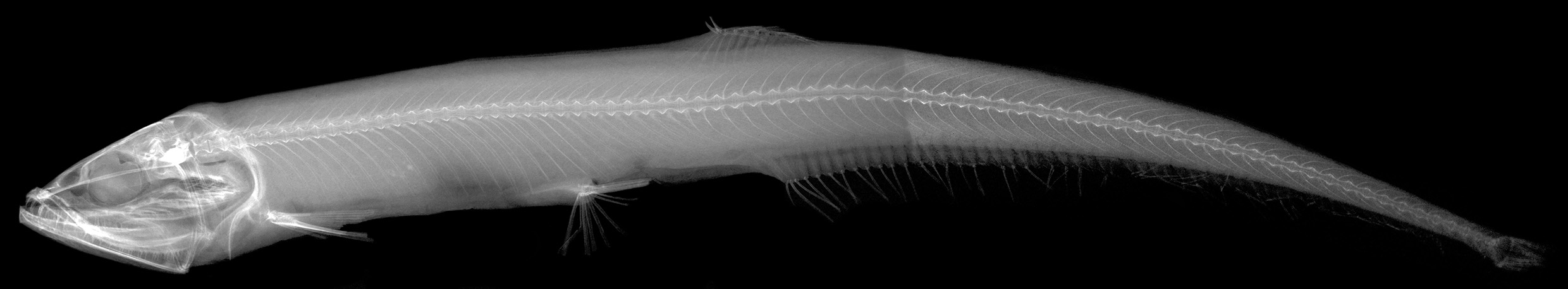

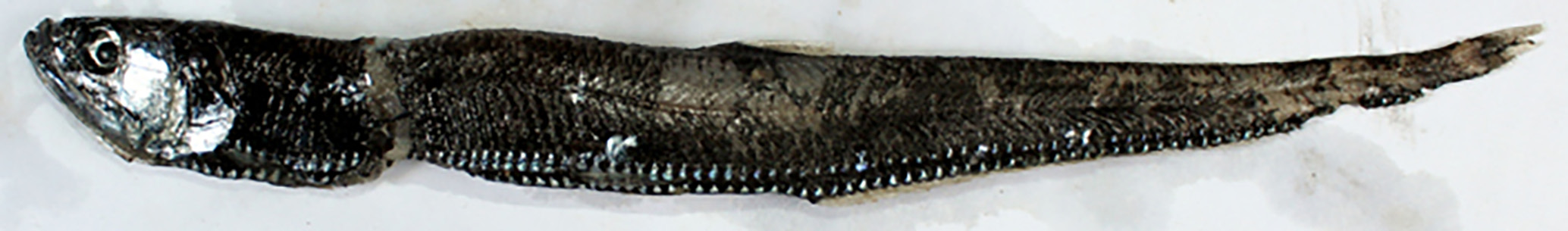
TABLE 1. Meristic data of the type series of Diplophos vicinia sp. nov. and D. taenia.
| D. vicinia | D. taenia | ||
|---|---|---|---|
| Holotype | Paratypes | Non-type | |
| NMMB-P29097 | n=8 | ASIZP 75043 | |
| Standard length | 127.6 | 73.9–143.5 | 114.5 |
| Counts | |||
| Dorsal-fin rays | 12 | 11–12 | 10 |
| Anal-fin rays | 57 | 57–59 | damaged |
| Pectoral-fin rays | 10 | 9–10 | 9 |
| Pelvic-fin rays | 8 | 8 | 8 |
| Caudal-fin rays | 10 + 9 | 10 + 9 | 10 + 9 |
| Branchiostegal rays | 14 | 14 | 14 |
| Gill rakers | 4 + 10=14 | 4 + 10–11=14–15 | 3 + 9=12 |
| Abdominal vertebrae | 30 | 30–32 | 38 |
| Caudal vertebrae | 44 | 44–45 | 55 |
| Total vertebrae | 74 | 74–76 | 93 |
| Photophores | |||
| IP | 13 | 13–14 | 19 |
| PV | 18 | 18–19 | 22+ |
| IV | 31 | 31–32 | 41+ |
| VAV | 13 | 12–13 | 17 |
| AC | 37 + 2=39 | 37–39 + 2=39–41 | damaged |
| IC | 83 | 83–86 | N/A |
| OV | 18 | 18–19 | 25+ |
| VALA | 13 | 12–13 | 16 |
| OAA | 31 | 31–32 | 41+ |
| VALB | 17 | 17–19 | 28 |
| OAB | 48 | 48–50 | 69+ |
| BR | 11 | 11–12 | 12 |
TABLE 2. Morphometric data of type series of Diplophos vicinia sp. nov. and D. taenia (Mean values in parentheses).
| D. vicinia | D. taenia | ||
|---|---|---|---|
| Holotype | Paratypes | Non-type | |
| NMMB-P29097 | n=8 | ASIZP 75043 | |
| Standard length (mm) | 127.6 | 73.9–143.5 | 114.5 |
| %S L | |||
| Body depth | 9.7 | 7.3–11.5 (9.3) | 9.1 |
| Body width | 3.6 | 2.6–4.1 (3.2) | 2.9 |
| Caudal-peduncle depth | 1.9 | 1.7–2.3 (2.0) | 2.0 |
| Caudal-peduncle length | 3.5 | 2.9–3.2 (2.8) | damaged |
| Head length | 15.1 | 14.8–16.8 (15.8) | 16.3 |
| Head depth | 10.1 | 10.0–11.1 (10.5) | 10.2 |
| Postorbital head depth | 8.4 | 8.4–9.5 (9.1) | 9.5 |
| Snout length | 4.0 | 3.9–4.6 (4.2) | 3.8 |
| Eye diameter | 3.5 | 3.0–4.1 (3.6) | 3.8 |
| Interorbital width | 2.8 | 2.4–3.1 (2.9) | 2.5 |
| Upper jaw length | 11.2 | 10.5–12.0 (11.3) | 11.1 |
| Premaxillary length | 4.4 | 3.7–5.3 (4.5) | 5.3 |
| Toothed maxillary length | 6.6 | 6.0–7.6 (6.9) | 6.2 |
| Lower jaw length | 11.5 | 11.0–12.7 (12.0) | 11.9 |
| Dorsal-fin base | 6.2 | 4.9–6.0 (5.4) | 4.0 |
| Anal-fin base | 45.5 | 45.4–51.7 (47.3) | damaged |
| Pre-dorsal-fin length | 45.2 | 45.7–47.3 (46.7) | 46.6 |
| Pre-anal-fin length | 49.5 | 49.5–51.2 (50.5) | 53.5 |
| Pre-pelvic-fin length | 35.4 | 36.0–37.4 (36.5) | 38.5 |
| Pectoral-fin base to pelvic-fin base | 20.0 | 19.8–22.1 (20.7) | 23.3 |
| Pelvic-fin base to anal-fin origin | 13.4 | 13.0–14.8 (13.8) | 15.3 |
| ASIZP |
Academia Sinica Institute of Zoology, Ichthyology Collection |
No known copyright restrictions apply. See Agosti, D., Egloff, W., 2009. Taxonomic information exchange and copyright: the Plazi approach. BMC Research Notes 2009, 2:53 for further explanation.
|
Kingdom |
|
|
Phylum |
|
|
Class |
|
|
Order |
|
|
Family |
|
|
Genus |