
Torvosaurus tanneri Galton & Jensen, 1979
|
publication ID |
https://doi.org/ 10.11646/zootaxa.3759.1.1 |
|
publication LSID |
lsid:zoobank.org:pub:E9F2EADE-3745-468B-9344-1ECCA4C2EA22 |
|
DOI |
https://doi.org/10.5281/zenodo.5038702 |
|
persistent identifier |
https://treatment.plazi.org/id/03C38794-FFE2-080A-53E4-FE8AB978FC7F |
|
treatment provided by |
Felipe |
|
scientific name |
Torvosaurus tanneri Galton & Jensen, 1979 |
| status |
|
Torvosaurus tanneri Galton & Jensen, 1979
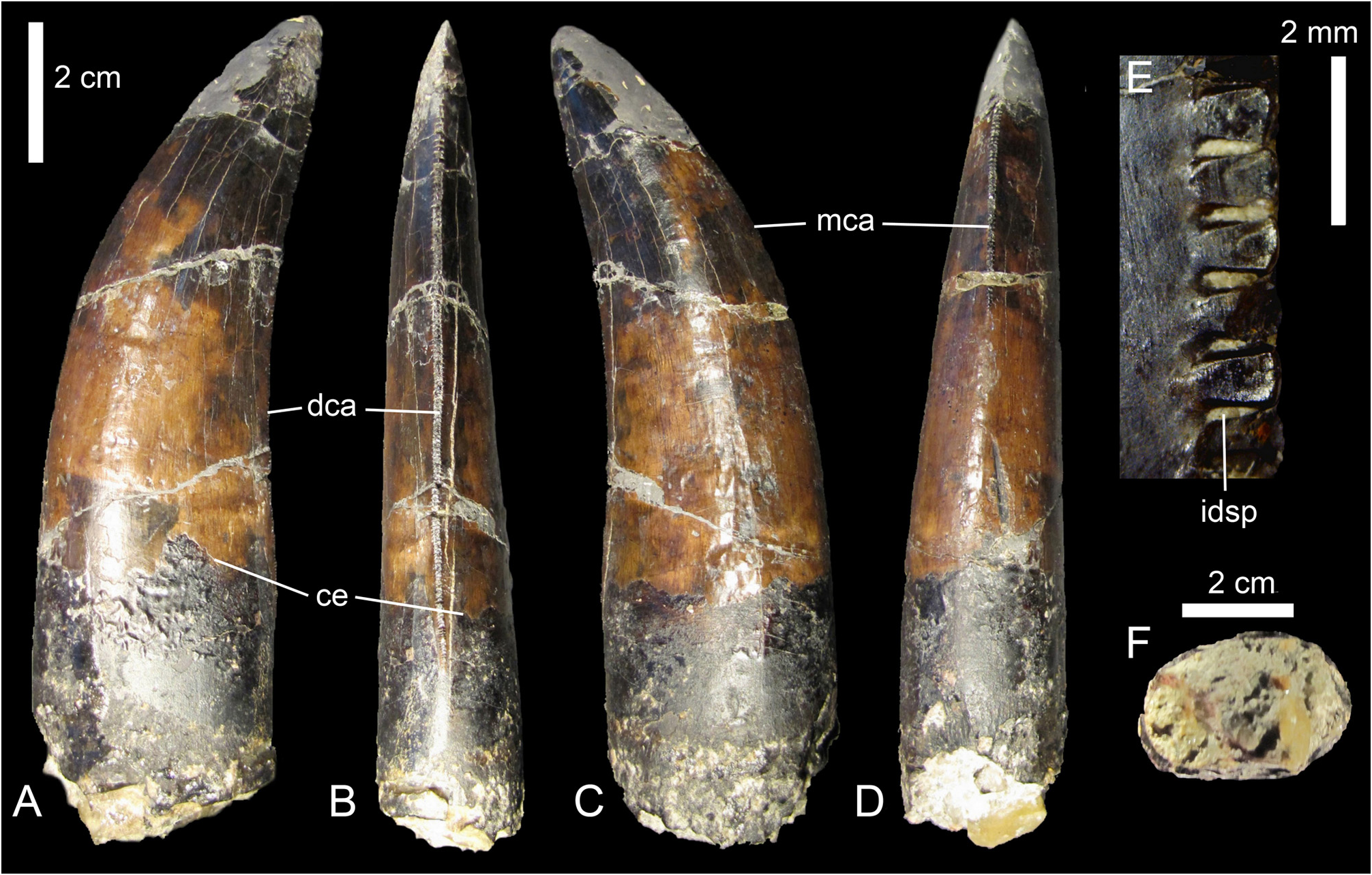
Referred material. ML 962 ( Fig. 9 View FIGURE 9 ).
Locality and horizon. Cliffs of Praia da Area Branca North, Praia da Area Branca, Lourinhã, Portugal. Bombaral Member, Lourinhã Formation, Tithonian, Upper Jurassic.
Description. ML 962 is an elongated tooth lacking the mesial part of the tip. Although most of the mesial and distal denticles are damaged and missing, their bases are still present so that it was possible to count the number of denticles basally, apically and at the mid-crown.
Crown. The tooth is particularly large, ( CH of 85 mm) and the general shape of the tooth resembles the ‘typical' blade-like theropod tooth by being labiolingually compressed, distally curved and having serrated carinae. However, the base is particularly narrow mesio-distally (CBL of 31.5 mm) and quite large labio-lingually (CBW of 20.2 mm) so that the crown-base has an ovoid cross-section (CBR of 0.64).
In lateral view, the mesial and distal margins of the root and basal half of the crown are roughly straight whereas the distal half of the crown is bent distally. The curvature of the crown is larger mesially than distally and the base of the crown is slightly larger than the mid-crown mesio-distally.
In distal view, the distal carina is medially positioned, slightly curved and bowed labially. The carina bears denticles all along the crown edge, from the preserved tip of the crown to the cervix.
In mesial view, the mesial carina, on the other hand, appears at the mid-crown, approximately 30 mm from the cervix, the basal part of the crown remaining smooth and rounded ( Fig. 9D View FIGURE 9 ). The carina is labially positioned and weakly offset apically but slightly curves lingually towards the root, becoming medially positioned on the mesial margin of the crown. Both lingual and labial surfaces are baso-apically concave and the root surface remains almost straight.
In apical view, the tip is weakly labio-lingually oriented and medially positioned on the crown. The mesial carina forms just a low ridge whereas the distal carina is more acute, and bends lingually towards the root.
In cross section, the basal crown is elliptical ( Fig. 9F View FIGURE 9 ) with both mesial and distal parts rounded. The labial face shows a short flattened surface in its centre whereas the lingual margin is weakly convex. Both labial and lingual surfaces are strongly mesio-distally convex all along the crown. The dentine layer is thin (0.6 mm in the lingual part) and its thickness is greater on the distal part of the crown (1.7 mm), the mesial part being absent. The length of the pulp cavity is 17.8 mm labio-lingually and around 28 mm mesio-distally.
Denticles. The mesial carina has 8 denticles at the mid-crown, and the number of denticles near the apex is unknown due to the missing part of tip of the crown ( Table 3 View TABLE 3 ). The size of the denticles decreases towards the root from approximately the two-thirds of the crow, a tendency also observable on the distal carina but on a much longer distance.
The distal carina bears around 7 denticles per 5 mm at the apex, 8 at the mid-crown and 11 at the base of the crown, the latter being minute near the cervix. The biggest denticles can be found 20 mm below the apex of the crown and are the only denticles entirely preserved on the apical part of the distal carina. They are chisel-like in shape, mesio-distally longer than baso-apically and their main axis is perpendicular to the distal margin ( Fig. 9E View FIGURE 9 ). A transversal section of the denticles would reveal a triangular shape as their bases are labio-lingually large and their tips are angular.
The labial and lingual surfaces of both mesial and distal denticles are slightly convex or completely flattened baso-apically, and only their basal and apical borders are rounded and curved to form the limits of the interdenticular spaces. The latter are deep and narrow and often filled with sediments. Their width tends to decrease towards the tip of the denticles which is slightly wider baso-apically than the base.
The external margin of mesial and distal denticles is symmetrically and slightly convex and does not point towards the tip of the crown. The denticle surface is covered by enamel, but the layer of enamel has disappeared in the middle of several denticles surfaces. This might, however, be due to erosion rather than initial wear. A few other denticles are also preserved on the basal part of the distal carina. They are quite different from the apical denticles by having a much more rounded external margin. The denticles are symmetrically rounded in lateral view and their labial and lingual surfaces are strongly convex. The interdenticular space is shallower and also slightly wider than in the apical denticles.
The mesial and distal denticles differ in their elongation; the few preserved denticles on the mesial carina are longer baso-apically than mesio-distally. The interdenticular space of mesial denticle is narrow and deep and the external margin of the denticle is slightly convex, almost flat.
Short interdenticular sulci appear between the distal denticles, but not in the most apical and basal ones. These shallow grooves running on both labial and lingual surfaces of the crown are inclined towards the root and more pronounced on the lingual face. They are, however, totally absent between the mesial denticles.
Surface. The crown surface is rugged and shows many irregularities. Possibly due to erosion and wear, the enamel texture of the crown is completely smooth and does not show any microscopic sculpturing. Two large transversal undulations appear on both labial and lingual surface of the basal part of the crown, but those deep structures do not correspond to the numerous and shallow transversal undulations illustrated by Brusatte et al. (2007) and might be due to deformation.
Discussion. Since most of the root is missing and the pulp cavity is excavated and filled with sediment, we interpret ML 962 as a shed tooth ( Bakker & Bir 2004). A very large and fairly straight crown showing a labiolingually compression, distinct serrations on mesial and distal carinae, and a slight curvature of the tip distally is a combination of characters observed in theropod dinosaurs only ( Buffetaut & Ingavat 1986), especially in the Upper Jurassic of Portugal (pers. obs.).
With a crown height of more than eight centimetres ( CH of 85.8 mm), ML 962 is a large crown belonging to a particularly large theropod. Although size is a plastic feature and must be used carefully for systematic purpose, this feature has already been demonstrated to be useful for discriminating teeth of different theropod taxa (Smith 2005; Smith et al. 2005; Han et al. 2011). Indeed, to our knowledge, crowns of more than eight centimetres are only borne by non-coelurosaur averostrans and derived Tyrannosauroidea, as they can be found in Ceratosauridae ( Ceratosaurus , Genyodectes ), Megalosauroidea (e.g., Torvosaurus and Spinosaurus ), Allosauroidea (e.g., Carcharodontosaurus , Mapusaurus , Giganotosaurus ) and Tyrannosauridae (e.g., Tyrannosaurus , Tarbosaurus ).
The denticles of ML 962 are also particularly coarse and an average of 8 denticles per 5 mm on both carinae is a condition present in particularly large basal tetanurans. Such feature can indeed be observed in Megalosauridae (Rauhut & Werner 1995; Smith 2007; pers. obs.), Carcharodontosaurinae (Rauhut & Werner 1995; Veralli & Calvo 2004; Corria & Currie 2006; pers. obs.) and Tyrannosauridae (Rauhut & Werner 1995; Smith 2005; pers. obs.). To our knowledge, less than 9 denticles on both mesial and distal carinae is a feature absent in basal Megalosauroidea (e.g., Piatnitzkysaurus ), some Megalosauridae (e.g., Eustreptospondylus , Dubreuillosaurus ), noncarcharodontosaurine Allosauroidea (e.g., Allosaurus , Neovenator , Acrocanthosaurus ), and all Ceratosauridae and Spinosauridae (pers. obs.). Indosuchus raptorius (AMNH 1753, 1955, 1960) is the only abelisaurid possessing less than 8 denticles per 5 mm on both carinae but the teeth are typical of abelisaurids as their crowns are low and weakly recurved distally. It is therefore unlikely that ML 962 belongs to one of these groups of theropods.
With an elliptical outline of the crown base in cross-section (CBR of 0.6) and a strong elongation, ML 962 is also very peculiar. In most carnivorous theropods, the lateral teeth are usually strongly medio-laterally flattened, giving a lenticular or lanceolate outline of the base crown in cross-section, and an elliptical outline of the crown base is usually present in mesialmost teeth, i.e., the premaxillary and mesialmost teeth of the dentary and maxilla (pers. obs.). Among basal tetanurans except Spinosauridae (which possess conical and fluted crowns along the tooth row), an ovoid subcircular outline of the crown base can clearly be observed in mesialmost teeth of megalosaurids such as Duriavenator hesperis (NHM R.332), Dubreuillosaurus valesdunensis (MNHN 1998-13) and Torvosaurus tanneri ( Britt 1991) and allosauroids like Acrocanthosaurus atokensis (NCSM 14345) and Giganotosaurus carolinii (MUCPv-CH-1; Candeiro 2007). Some tetanurans like Acrocanthosaurus , Giganotosaurus and Tyrannosaurus can also have an ovoid cross-section of the base crown more distally in the jaws (Smith 2005; Candeiro 2007; pers. obs.). Nevertheless, the lateral teeth of those theropods are much more massive and incrassate, the labiolingual width of the crown base being sometimes equal or larger than its mesiodistal length in Tyrannosauridae , giving them the typical ‘banana' shape (Smith 2005; pers. obs.). We therefore interpret ML 962 as a mesialmost tooth of a basal tetanuran.
This large crown also possesses a mesial carina medially positioned on the mesial margin of the crown, running slightly diagonally and terminating at the mid-crown, well above the cervix. Among mesialmost teeth of tetanurans, such a combination of features can be observed in Megalosauridae such as Torvosaurus tanneri (BYUVP 2003) , Duriavenator hesperis (NHM R.332) and Dubreuillosaurus valesdunensis (MNHN 1998-13) as well as the carcharodontosaurid Acrocanthosaurus atokensis (NCSM 14345). In Allosauridae and Tyrannosauroidea, the mesial carina extends to the cervix of the crown, or very close to it, and either twists lingually like in Allosaurus fragilis (AMNH 851; CM 21703; SMA 0005/02) and Proceratosaurus bradleyi ( Rauhut et al. 2010) or faces entirely lingually in more derived tyrannosauroids, giving the typical D-shape crosssection of the base-crown (Smith 2005; Sereno et al. 2009; pers. obs.). The distal carina of ML 962 is also centrally positioned on the distal margin of the crown, a feature visible in the mesialmost teeth of megalosaurids such as Eustreptospondylus oxoniensis (OUMNH J.13558), Dubreuillosaurus valesdunensis (MNHN 1998-13) and Duriavenator hesperis (NHM R.332). On the other hand, the distal carina of mesialmost teeth of carcharodontosaurids such as Acrocanthosaurus atokensis (NCSM 14345) and Giganotosaurus carolinii (MUCPv- CH-1) is slightly to strongly displaced labially on the distal margin of the crown (a similar feature is found in Genyodectes and Dromaeosaurus for instance; Currie et al. 1990; Rauhut 2004; pers. obs.), so that the mesial and distal carinae are not aligned on a same plan like in megalosaurid theropods (pers. obs.). It is, therefore, more likely that ML 962 belongs to Megalosauridae than Carcharodontosauridae .
Among Megalosauridae , a very large and strongly elongated crown (CHR> 2.5) with large chisel-like and symmetrically rounded denticles (less than 9 denticles on the distal carina) seems to be a combination of characters only seen in Torvosaurus (pers. obs.). The general shape and outline of ML 962 also resemble very much those of one probable Torvosaurus tanneri shed tooth illustrated by Jensen (1985: fig. 5e) and the first dentary tooth of Torvosaurus (BYUVP 2003) . These two teeth share with ML 962 same curvature and elongation as well as a lateral face that is particularly convex. In addition, the outline of the basal crown seems to fit with the mesialmost dentary alveoli of Torvosaurus ( Britt 1991: fig. 3f), the premaxillary alveoli being more elongated mesio-distally (or labiolingually for the first alveolus).
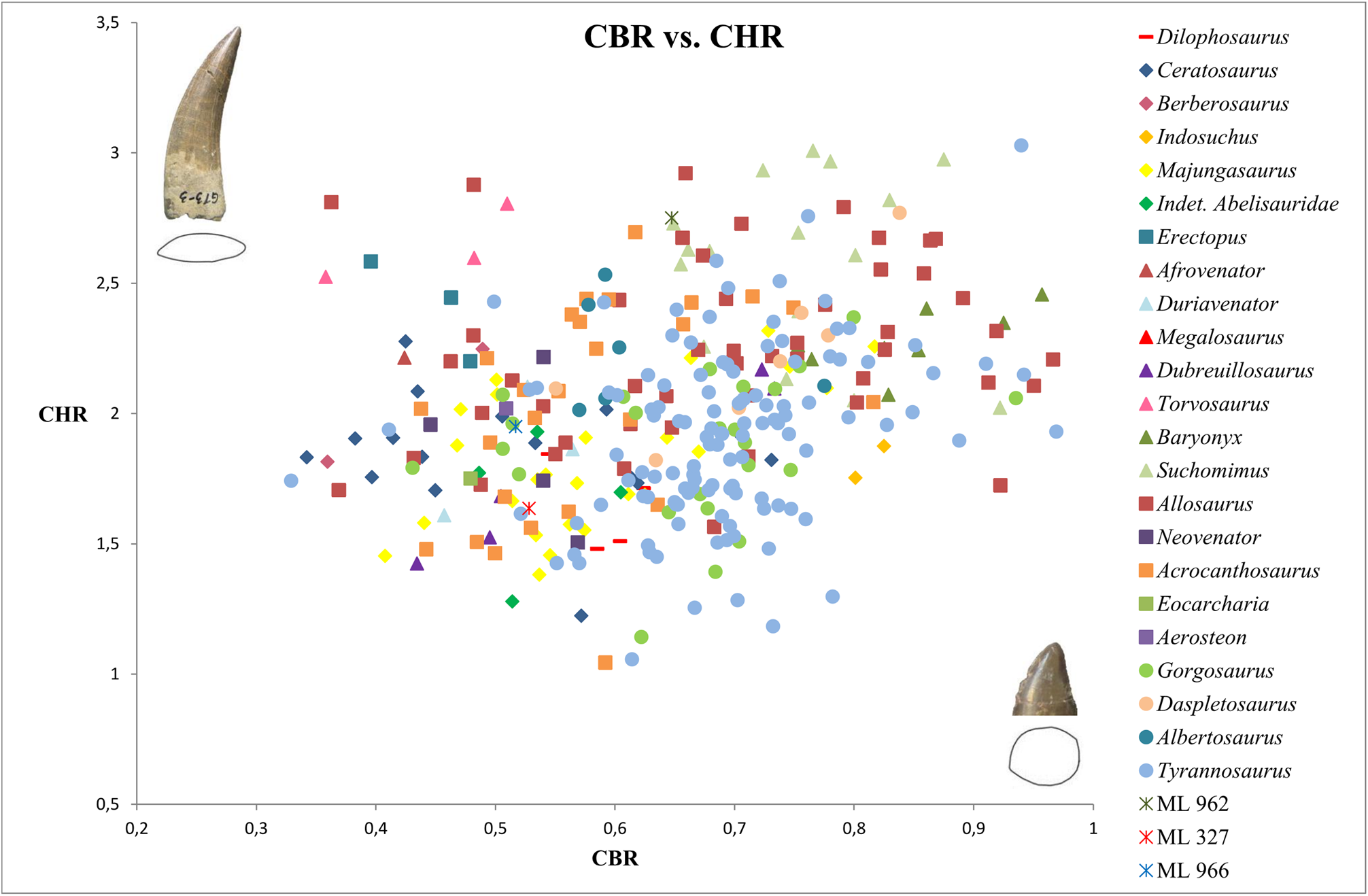
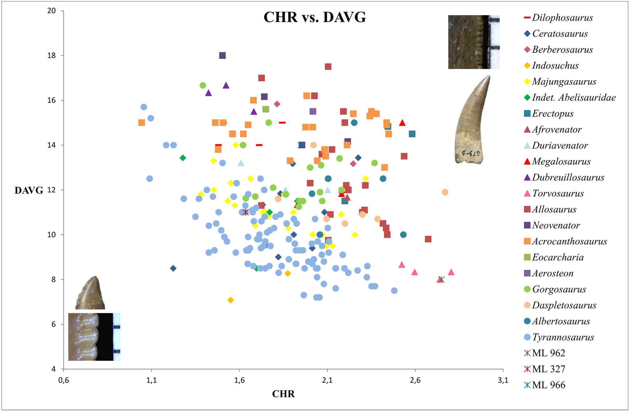
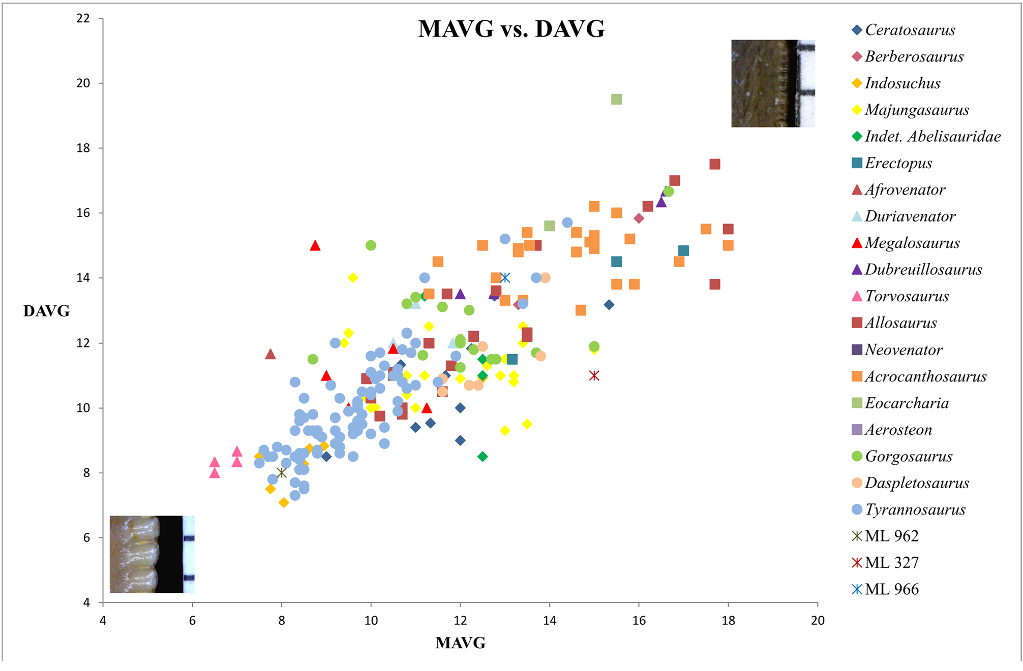
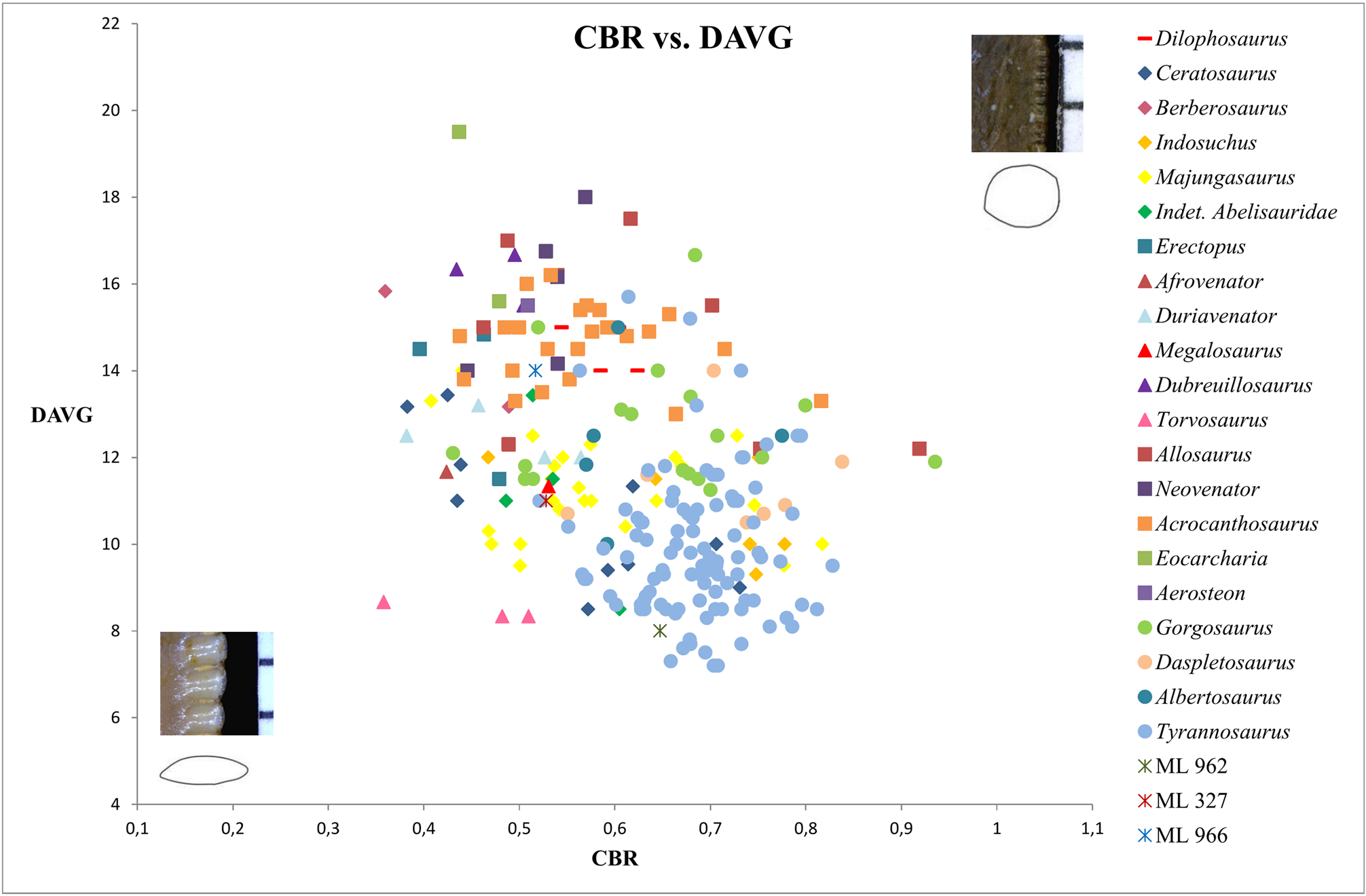
Both morphological and cladistic analyses support the identification of ML 962 to the taxon Torvosaurus . Bivariate plots of MAVG and DAVG ( Fig. 7 View FIGURE 7 ) show that ML 962 possesses the same number of denticles per five mm as Carcharodontosaurus , Tyrannosaurus and Indosuchus , and close values of denticles as Torvosaurus . However, bivariate plots of CHR and DAVG clearly illustrates the same values of ML 962 and Torvosaurus teeth ( Fig. 6 View FIGURE 6 ), on the opposite of bivariate graphs with CBR, as CBR values of ML 962 and Torvosaurus teeth are significantly different ( Figs. 5 View FIGURE 5 , 8 View FIGURE 8 ). This can be explained by the absence of mesial teeth of Torvosaurus in our dataset. As it has been mentioned previously, mesialmost teeth of many theropods are usually labiolingually thicker than lateral teeth, and this is clearly the case in Torvosaurus and Megalosauridae in which mesialmost teeth have an elliptical to rounded cross-section at the crown base instead of a lenticular outline typically present in the lateral teeth of these taxa. Following this observation, characters on mesialmost teeth were only coded in ML 962 in our data matrix.
The cladistic analysis performed on the datamatrix of dentition-based characters recovered ML 962 as a megalosaurid theropod, forming a polytomy with all members of this clade ( Fig. 1 View FIGURE 1 ). This lack of resolution can be explained by the absence of mesialmost teeth in Afrovenator and Megalosaurus , and the little information collected from mesialmost dentition of Eustreptospondylus , Duriavenator and Torvosaurus in our dataset. A similar position within the megalosaurid clade was found when the cladistic analysis was performed on the supermatrix, but ML 962 forms a polytomy with the megalosaurids Torvosaurus , Megalosaurus , Afrovenator and Duriavenator that bear large teeth ( Fig. 2 View FIGURE 2 , Appendix Fig. A10 View FIGURE 10 ).
Following the results of both cladistic and morphological analyses, we identify ML 962 as a mesial tooth, perhaps a dentary tooth, belonging to the species Torvosaurus tanneri . Material of Torvosaurus tanneri are not rare in the Kimmeridgian—Tithonian of Europe and North America and have been reported several times in the Lourinhã Formation previously ( Mateus & Antunes 2000b; Mateus 2005; Mateus et al. 2006). Therefore, this referral to Torvosaurus is consistent both stratigraphically and biogeographically.


No known copyright restrictions apply. See Agosti, D., Egloff, W., 2009. Taxonomic information exchange and copyright: the Plazi approach. BMC Research Notes 2009, 2:53 for further explanation.