
Lithobius (Sigibius) trichinocaput, Ma, Huiqin, Pei, Sujian, Li, Yaying & Shi, Baojun, 2012
|
publication ID |
https://doi.org/ 10.5281/zenodo.281530 |
|
DOI |
https://doi.org/10.5281/zenodo.3508492 |
|
persistent identifier |
https://treatment.plazi.org/id/03C387A3-BC0E-F415-FF2A-2C832F383388 |
|
treatment provided by |
Plazi |
|
scientific name |
Lithobius (Sigibius) trichinocaput |
| status |
sp. nov. |
Lithobius (Sigibius) trichinocaput sp. n.
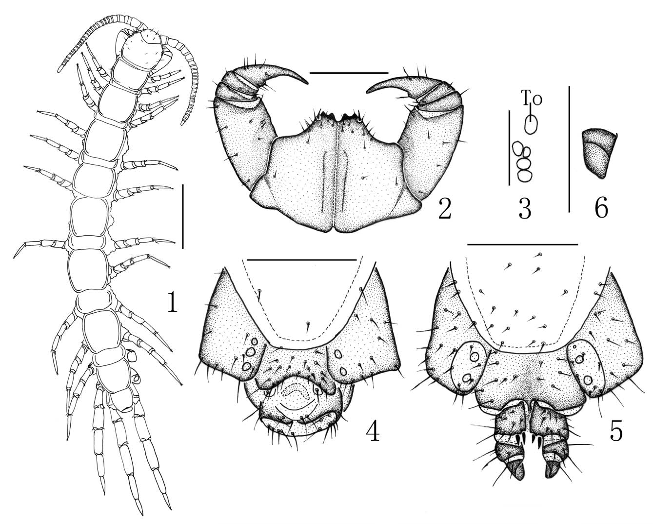
Material examined: Holotype: 3 ( Fig. 1 View FIGURES 1 – 6 ), body length 9.0 mm, cephalic plate 0.9 mm long, 0.9 mm broad, Xiaowutai National Natural Reserve, Yu County, Zhangjiakou City, Hebei Province, 39°54'N 115°00'E, 1236 m, 20 August 2005, leg. Zhi-sheng Zhang, Hui-qin Ma.
Paratypes: 4 ƤƤ, 2 3, same data as holotype.
Etymology: the specific name refers to the cephalic plate more obviously covered with setae than the other congeners.
Diagnosis: A Lithobius (Sigibius) species with body length up to 9.7 mm, antennae composed of up to 23–26 articles; 4 ocelli on each side, arranged in 2 irregular rows; Tömösváry’s organ moderately large, slightly larger than adjoining ocelli; 2+2 coxosternal teeth; porodonts moderately thick and strong, posterolateral to lateral tooth; posterior angles of all tergites without triangular projections; coxal pores 2–4, oval to round; female gonopods with 2+2 moderately large, coniform spurs; terminal claw simple; male gonopods short and small, with one long seta on terminal segment.
Description: Body length 7.6–9.7 mm, cephalic plate 0.55–0.96 mm long, 0.55–0.96 mm wide. Colour: basal antennal articles light grey to pale yellow-brown; tergites pale light grey to yellow-brown; cephalic plate, and T 15 pale yellow-brown with greyish hue; pleural region and SS pale grey; distal part of forcipules brown, basal and proximal parts of forcipules, forcipular coxosternite and S 15 yellow-brownish with greyish hue; all legs pale grey, tarsus of all legs yellow-brown.
Antennae: commonly 25+25 or 26+26 (one specimen with 23+23) articles; basal article longer than wide, second one markedly longer than wide, following articles gradually shortening, distal article up to 2.4–2.7 times as long as wide. Abundant setae on the antennal surface, less so on the basal articles, gradually increasing in density to about sixth or seventh article, then more or less constant.
Cephalic plate smooth, convex, pigment concentrated as close netlike veins, width equal to length; tiny setae emerging from pores and long setae scattered sparsely over the whole surface; frontal marginal ridge with shallow anterior median furrow ( Fig. 1 View FIGURES 1 – 6 ); lateral marginal ridge discontinuous, posterior margin straight or slightly convex, widening in the middle part.
Four oval to rounded ocelli on each side ( Fig. 3 View FIGURES 1 – 6 ) arranged in two irregular rows; the posterior one comparatively large; ocelli domed, translucent, usually darkly pigmented.
Tömösváry’s organ comparatively large ( Fig. 3 View FIGURES 1 – 6 -To), nearly rounded; lying far from adjoining ocelli, situated on the anterolateral margin of cephalic plate, visible larger than the adjoining ocelli.
Coxosternite subtrapezoidal ( Fig. 2 View FIGURES 1 – 6 ), anterior margin narrow, external sides lightly longer than internal side; median diastema moderately deep, V-shaped; anterior margin with 2+2 teeth (in one specimen 2+3); porodonts thick and stout, lying posterolateral to the lateral tooth ( Fig. 2 View FIGURES 1 – 6 ); some scattered setae on the ventral side of coxosternite.
All tergites smooth, without wrinkles, backside slightly hunched; T 1 posterolaterally narrower than anterolaterally, generally trapeziform, narrower than T 3; posterior margin of T 1 slightly convex or straight; posterior margin of TT 3, 5, 8, 10, 12 and 14 slightly concave, posterior margin ridge of TT 1, 3, 5, 8, 10, 12 and 14 discontinuous; all posterior angles generally rounded, without triangular projections; lateral margin ridge of all tergites continuous; tiny setae scattered very sparsely over the surface, 1–3 moderately long setae on anterior angles, 1–2 moderately long setae on the posterior margin of each tergite.
Posterior side of all sternites narrower than the anterior, generally trapeziform, comparatively smooth; setae inserted in pores scattered very sparsely on the surface, 2–3 slightly long setae on the surface of the anterior part of each sternite, 1–2 slightly long setae on the surface of the posterior part of each sternite.
Legs strong, tarsus 1–2 articulation indistinct on legs 1–13, well defined on legs 14 and 15. All legs with claws, fairly long, curved ventrad; anterior and posterior accessory spines on legs 1–13, the anterior one moderately long and slender, forming a small angle with the claw; the posterior one short and strong, forming a large angle with the claw; only posterior accessory spines on legs 14, no accessory spines on legs 15. Short to comparatively long setae scattered very sparsely over the surface of all segments of all legs, more setae scattered on the surface of tarsus; legs 14 and 15 slightly thicker and stronger than the other legs. In the female, tarsus 1 5.0–5.6 times as long as wide, tarsus 2 about 78%–82% length of tarsus on legs 15; tarsus 2 about 4.8–5.4 times as long as wide, tarsus 2 about 71%–77% length of tarsus on legs 15. Plectrotaxy as in table 1.
legs ventral dorsal
C Tr P F Ti C Tr P F Ti 1 - - p amp am - - mp ap a 2 - - p amp am - - mp ap a 3-10 - - mp amp am - - mp ap ap 11 - - (a)mp amp am - - mp (a)p ap 12 - - amp amp am a - mp p a 13 - m amp amp am a - mp p p 14 - m amp amp - a - mp p - 15 - m amp am - a - mp - -
Coxal pores 2–4, arranged in a row, round or slightly ovate, usually 2333, rarely 3343 or 2332. Coxal pore field set in a relatively shallow groove, the fringe of coxal pore-field with eminence, 5–7 short to moderately long setae scattered over the surface of the eminence.
Male S 15 anterolaterally broader than posterolaterally, generally trapeziform, posterior edge straight, short to long setae scattered very sparsely over the surface; sternites of genital segment nearly so long as wide, usually well sclerotised. Posterior margin of the sternite of the genital segment with a quite deep concavity between the gonopods, no bulge medially; comparatively long setae about evenly scattered on the ventral surface of the genital segment, few setae near S 15. Gonopods short and small, only a small bulge, with one long seta, apically slightly sclerotised ( Fig. 4 View FIGURES 1 – 6 ).
Female S 15 anterolaterally broader than posterolaterally, generally trapeziform, posteromedially straight, long setae scattered sparsely over the surface; sternite of genital segment usually well chitinised, wider than length; posterior margin of genital sternite deeply concave between the condyles of gonopods, except for a small, median, approximately coniform bulge; relatively long setae scattered over the ventral surface of the genital segment, few setae near S 15, regularly fringed with longer setae along the posterior margin. Gonopods: first article fairly broad, bearing 8–9 long setae arranged in about three rows; 2+2 moderately small, coniform spurs, inner spur smaller ( Fig. 5 View FIGURES 1 – 6 ); second article with 5 rather long setae, arranged in two irregular rows on its ventral side, one thick spicule on the external margin of the dorsal part of the second article; usually 1–2 long setae on the ventral surface of third article, two thick spicula on the external margin of the dorsal part of the third article; terminal claw simple ( Fig. 6 View FIGURES 1 – 6 ).
Habitat: The specimens were collected in Larix forest (at about 950-1000 m a.s.l.) in moist habitats under roadside stones or forest floor.
Remarks: L. (S.) trichinocaput sp. n. is morphologically close to L. (S.) siopius (Chamberlin & Wang, 1952) from Provo and Salt Lake City (Utah) (Chamberlin & Wang, 1952b), but could be well distinguished from latter by the presence of four ocelli on each side of cephalic plate rather than three ocelli as in the latter, terminal claw of female gonopods simple instead of bidentate, and the 15th dorsal plectrotaxy being 10200 other than 0 0 110 in the latter. It differs from L. (S.) trebinjanus Verhoeff, 1900 ( Eason, 1983) by having fewer antennal articles, different plectrotaxy and terminal claw of female gonopods simple (vs. a claw with a well-developed medial and a smaller lateral denticle in L. (S.) trebinjanus ).
On the other hand, Eason (1993) suggested that this subgenus is mainly distributed in the Mediterranean Region and Eastern Europe and apart from a few species know from Asia Minor, Transcaucasia and northern Iran, the subgenus is, hitherto, unknown in Asia, L. (S.) bullatus Eason, 1993 from Hong Kong is likely to have been introduced from Europe. While the new species is from Hebei Xiaowutaishan National Natural Reserve, Zhangjiakou City, Hebei Province, there has not been too much trade between Zhangjiakou City and Europe, moreover, no other comments about the zoogeographic meaning of this subgenus in East Asia, so we suggest the new species is likely to be native.
MAP 1. Map of distributions of Chinese Lithobius (Sigibius) species.
No known copyright restrictions apply. See Agosti, D., Egloff, W., 2009. Taxonomic information exchange and copyright: the Plazi approach. BMC Research Notes 2009, 2:53 for further explanation.