
Zeugophora cupka, Takemoto, 2019
|
publication ID |
https://doi.org/ 10.11646/zootaxa.4644.1.1 |
|
publication LSID |
lsid:zoobank.org:pub:02543A03-07E4-4F82-9372-CE85EBC99ABF |
|
persistent identifier |
https://treatment.plazi.org/id/03C38C25-407D-FD05-FF3B-FBB8FDA1A4DC |
|
treatment provided by |
Plazi |
|
scientific name |
Zeugophora cupka |
| status |
sp. nov. |
Zeugophora cupka View in CoL sp. nov.
[Japanese name: Okuezo-momobuto-hamushi]
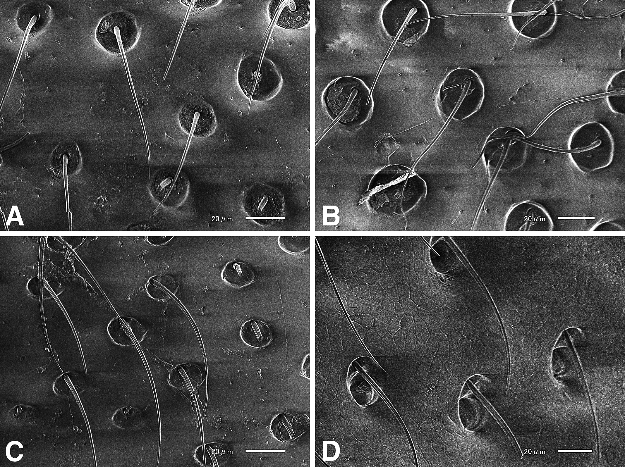
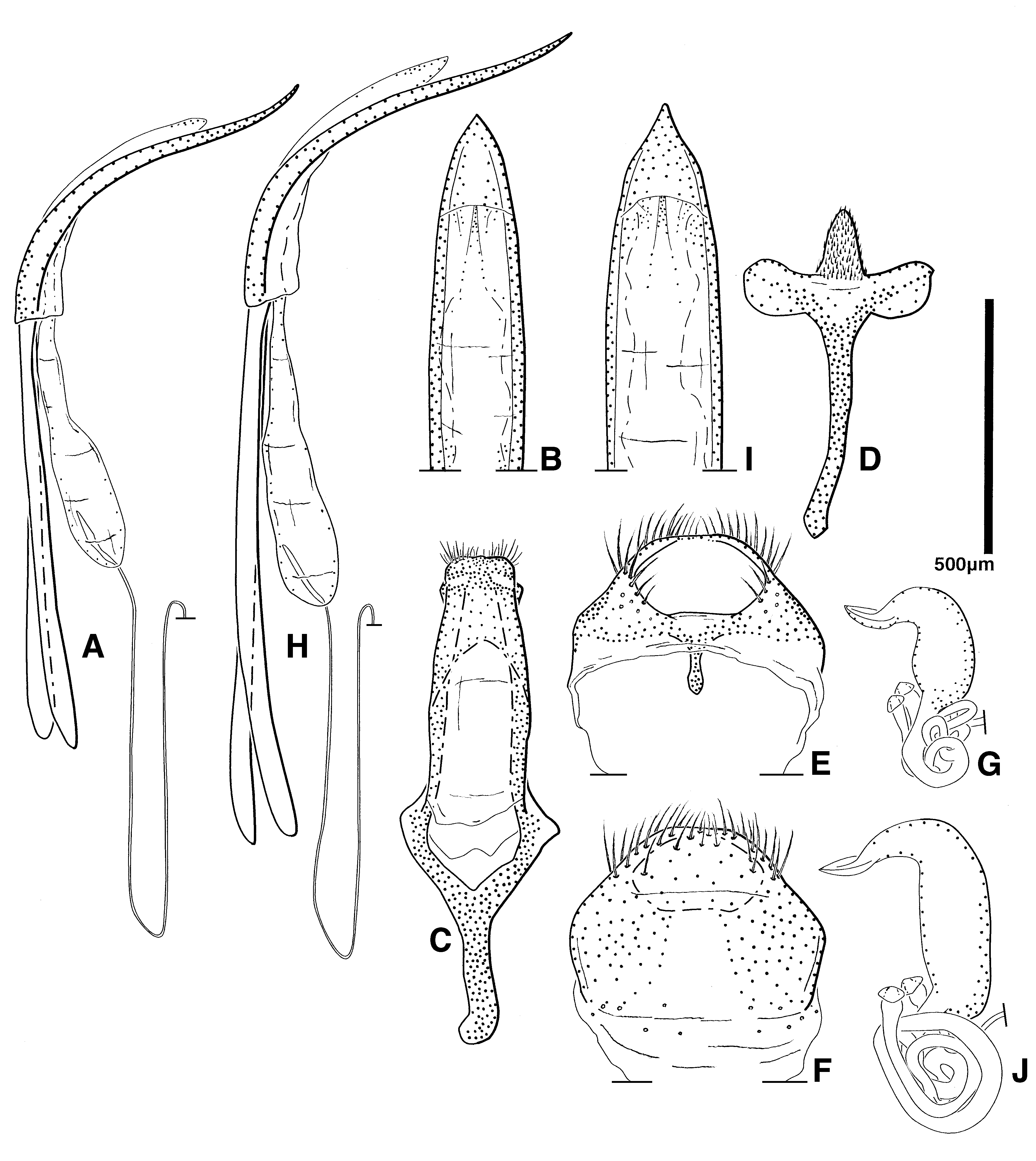
( Figs 44 View FIGURE 44 A–D; 45–48)
Zeugophora (Zeugophora) chujoi (mis det.): Kimoto, 1986b [ Japan: Hokkaido, Honshu].
Description. Measurement. Body length: PEL, male, 2.59–3.25 mm, female, 2.86–3.41 mm. Width: EW, male, 1.11–1.61 mm, female, 1.30–1.60 mm. Biometric data are given in Table 10.
Coloration. Surface shiny. Body fulvous. Head fulvous, blackened dorsally. Apex of mandible black. Antennae fulvous. Pronotum fulvous with black patch on central portion. Scutellum fulvous. Elytra fulvous with black patches on anterior corners and posterior halves of elytra, anterior and posterior patches sometimes connected. Ventral surface of head and prosternum fulvous, thorax and abdomen black. Leg fulvous except hind black coxa.
Habitus. Body oblong.
Head ( Fig. 46A View FIGURE 46 ) covered with sparse setiferous punctures; setae produced from inside punctures ( Fig. 47A View FIGURE 47 ); frontoclypeal suture bisinuate, middle portion narrow, about one third width of clypeus; labrum and clypeus convex and furnished with setae; length of clypeus longer than that of labrum. Anterior margin of clypeus weakly lined ( Fig. 46A View FIGURE 46 ). Canthus small, obtusely triangular with apex rounded, furnished with a few setiferous punctures. Apex of mandible bifurcate and furnished with one large tooth apically on inner margin; lateral angles furnished with setae ( Fig. 45D View FIGURE 45 ). Antenna weakly clavate; antennal segments short, except for first, third and fourth which are long and slender, eleventh pointed; first through fourth segments with setae, other segments with tiny dense setae ( Fig. 45C View FIGURE 45 ).
Pronotum ( Fig. 45A View FIGURE 45 , 46B View FIGURE 46 ) with blunt lateral tubercles, strongly tapered towards base; posterior marginal groove undulate, but lateral sides ambiguous; lateral basal angles weakly swollen; surface regularly convex, covered with coarse setiferous punctures ( Fig. 47B View FIGURE 47 ); distance between punctures 0.5–4.0 times their own diameter, sparser on posterior half of longitudinal median portion. Setae produced from inside punctures ( Fig. 47B View FIGURE 47 ).
Elytra long, leaving apex of pygidium exposed, moderately vaulted in lateral view; lateral sides subparallel; surface with reticulate coriaceous microsculpture, irregularly covered with dense setiferous punctures, punctures coarsely separated by 2–4 times their diameter; setae produced from anterior inner margin of puncture, posterior two-thirds of punctures deep ( Fig. 47D View FIGURE 47 ); sutural and lateral grooves complete from elytral base to apex; basal areas of elytra weakly convex on either side of suture; humerus protruding anteriorly; epipleuron narrow. Scutellum trapezoidal, with setae.
Disc of mesoventrite rugose; process short, furnished with setiferous punctures ( Fig. 45B View FIGURE 45 , 46C View FIGURE 46 ). Mesepisternum furnished with setiferous punctures, except near suture separating it from mesepimeron ( Fig. 45B View FIGURE 45 , 46C View FIGURE 46 ). Central area of metaventrite sparsely and lateral area densely covered with setiferous punctures ( Fig. 47C View FIGURE 47 ), setae produced anterior to shallow punctures ( Fig. 47C View FIGURE 47 ); posterior margin of mid coxal insertion markedly punctate. Surface of sternites with microsculpture, sparsely covered with setiferous punctures. In male, posterior margin of 7th sternite evenly curved ( Fig. 46D View FIGURE 46 ). In female, apex of pygidium concave; posterior margin of 7th sternite straight, but bisinuate on central portion ( Fig. 46E View FIGURE 46 ).
Legs furnished with setae; tibial setae becoming denser and longer apically. Hind femora thickened. Tibiae straight; mid and hind tibiae with sharply defined and finely crenulate carina along external face, apex with two spurs. Tarsi distinctly shorter than tibiae, basal three segments gradually widened apically.
Male genitalia as shown in Fig. 48 View FIGURE 48 A–F: 8th sternite furnished with setae, short, oblique anteriorly, membrane and sclerotized portion narrow ventrally, allowing for downward extension ( Fig. 48E, F View FIGURE 48 ); apex of spiculum three branched, central branch furnished with tiny setae ( Fig. 48D View FIGURE 48 ); median struts of median lobe approximately 1.5 times as long as median lobe ( Fig. 48A View FIGURE 48 ); apex of median lobe pointed ( Fig. 48B View FIGURE 48 ), lateral side flattened ( Fig. 48A View FIGURE 48 ); paramere well developed ( Fig. 48C View FIGURE 48 ).
Spermatheca as shown in Fig. 48G View FIGURE 48 .
Etymology. The species name, cupka (cúpka) means "east" in the Ainu language of Hokkaido, Japan.
Host plant. Populus suaveolens Fisch [Japanese name: Doronoki].
Remarks. External traits of Zeugophora cupka are somewhat similar to Z. turneri Power, 1863 which is distributed in northern, north-central, and eastern Europe to Siberia and Mongolia. I examined external traits and male and female genitalia of both species. They can be distinguished from the latter by the following characters: 1) body coloration; Z. turneri is fully testaceous while Z. cupka has some color variation as described above; 2) shape of median lobe; in Z. turneri , median lobe is wider and apex is narrower than in Z. cupka ( Fig. 48B, I View FIGURE 48 ); and 3) shape of spermatheca; in Z. turneri , the shape of the spermathecal capsule is vertically long, while that of Z. cupka is short and the posterior portion is swollen ( Fig. 48G, J View FIGURE 48 ).
Types. Holotype ♀ ( TTPC): " Kamikawa , Sôunkyô, Hokkaidô, 26.VI.2018, T. Takemoto leg.", "18-tt-259, ex. by T. Takemoto, SEHU, Japan." . Paratypes: Japan. [Hokkaido] Tokachi Dist .: 1 ex., Shintoku, Tomuraushi, 18– 23.IX.2013, Y. Hirano leg. ( TTPC) ; 1 ex., Kamishihoro, Tokachi-mitsumata, 21.VII.2018, S. Urabe leg. ( TTPC) .
Kamikawa Dist.: 3 exs., Kamikawa, Sôunkyô, 12. VI .2018, T. Takemoto leg. ( TTPC), 20 exs., same data but differ- ent date and collector, 23. VI .2018, S. Urabe leg. ( TTPC), 32 exs., same data but different date, 26. VI .2018 ( TTPC) , 4 exs., same data but different date and collector, 11.VII.2018, Y. Okita leg. ( TTPC). Okhotsk Dist .: 2 exs., Ru- beshibe, Kinka-tôge, 30. VI .2018, S. Urabe leg. ( TTPC).
Distribution. Japan: Hokkaido.


| VI |
Mykotektet, National Veterinary Institute |
No known copyright restrictions apply. See Agosti, D., Egloff, W., 2009. Taxonomic information exchange and copyright: the Plazi approach. BMC Research Notes 2009, 2:53 for further explanation.