
Triceratops, Marsh, 1889
|
publication ID |
https://doi.org/ 10.1080/02724634.1996.10011297 |
|
DOI |
https://doi.org/10.5281/zenodo.3812737 |
|
persistent identifier |
https://treatment.plazi.org/id/03C3CB50-FF9B-3443-F9FE-170CF816FBF9 |
|
treatment provided by |
Jeremy |
|
scientific name |
Triceratops |
| status |
|
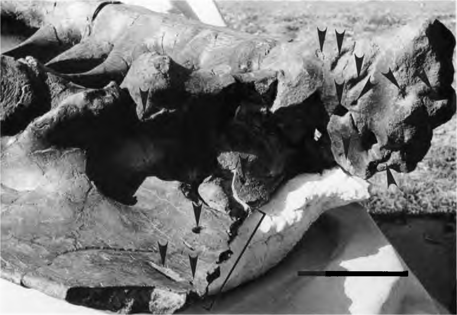
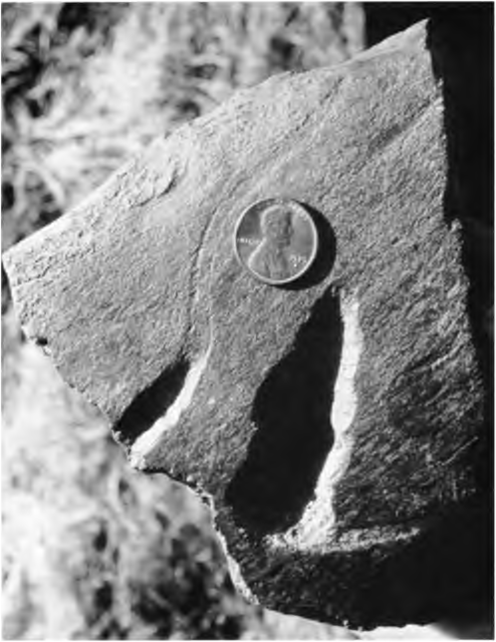
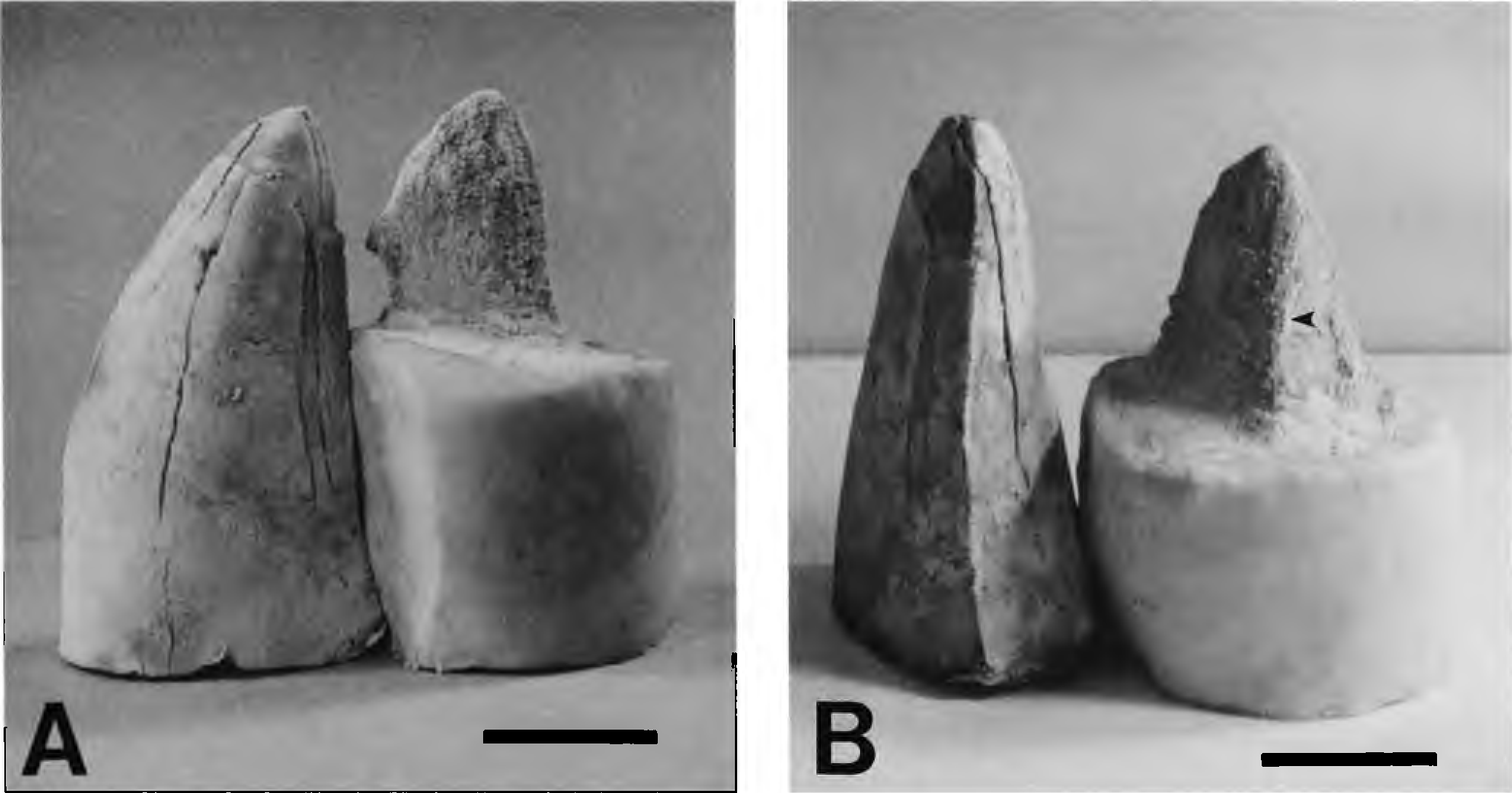
Dozens of T. rex tooth marks appear on a Triceratops sp. pelvis (sacrum and left ilium) that is housed at the Museum of the Rockies, Bozeman, Montana (MOR 799). This specimen was found by K. H. Olson in the Late Maastrichtian Hell Creek Formation of east-central Montana . The pelvis bears 58 definitive bite marks and 22 possible others ( Figs. 1 View FIGURE 1 , 2 View FIGURE 2 ). Many bite marks overlay one another, making an exact count impossible. We suggest that the tooth marks were produced by an adult T. rex because of their large size (punctures up to 2.5 cm in width and 3.7 cm in depth), their cross-sectional areas are rounded (unlike the more elliptical punctures made by most non-tyrannosaurid theropods whose teeth had lenticular cross-sectional areas: see Farlow et al., 1991), their spacing is wide (>4 cm), and some bones bear coarse serration marks on their periosteal surfaces. Additionally, a cast of one of the deeper bites matches the morphology of an adult T. rex lateral tooth (sensu Farlow et al., 1991), including its carina ( Fig. 3A, B View FIGURE 3 ). Finally, no other known carnivorous animals present in the Hell Creek fauna were large enough to have inflicted comparable damage.
At least thirty-nine puncture marks are located on the ventral and both lateral surfaces of the Triceratops sacrum, showing that it was overturned either prior to or during feeding.
Most bites occur on the first sacral vertebra ( Fig. 1 View FIGURE 1 ). It appears the tyrannosaur(s) may have detached the pelvis from the torso by repetitively biting into this centrum, as it has been nearly bitten in half. The majority of these bite marks are in the form of deep localized punctures and appear to have been made by a tyrannosaur’s larger anterior lateral teeth. A few elongate bite furrows from shallower biting also appear. The largest is located on the transverse process of the third sacral vertebra and is 1.0 cm in depth and 11.8 cm long. Between the transverse processes of two adjacent sacral vertebrae (second and third) are four faint vertically inclined furrows that appear to be nipping marks made by a tyrannosaur’s premaxillary teeth. (Unfortunately they show low relief and therefore are not figured here.)
The left ilium of the Triceratops bears at least 19 bite marks on both its ventral and dorsal surfaces ( Figs. 1 View FIGURE 1 , 2 View FIGURE 2 ). The bite marks are distributed around the periphery of the iliac crest. Approximately one-sixth of the anterior end of the ilium was bitten off by repetitive bites concentrated in this region. The majority of these bites are furrow-shaped and were likely inflicted by a tyrannosaur’s larger anterior lateral teeth. The longest furrow is 0.9 cm in depth and 10.1 cm long.
No known copyright restrictions apply. See Agosti, D., Egloff, W., 2009. Taxonomic information exchange and copyright: the Plazi approach. BMC Research Notes 2009, 2:53 for further explanation.