
Mesanophrys cf. carcini Small & Lynn in Aescht, 2001
|
publication ID |
https://doi.org/10.5852/ejt.2016.249 |
|
publication LSID |
urn:lsid:zoobank.org:pub:43278833-B695-4375-B1BD-E98C28A9E50E |
|
DOI |
https://doi.org/10.5281/zenodo.5631541 |
|
persistent identifier |
https://treatment.plazi.org/id/03C487DB-6859-FFB9-5482-FB7F8BE7321D |
|
treatment provided by |
Plazi |
|
scientific name |
Mesanophrys cf. carcini Small & Lynn in Aescht, 2001 |
| status |
|
Mesanophrys cf. carcini Small & Lynn in Aescht, 2001
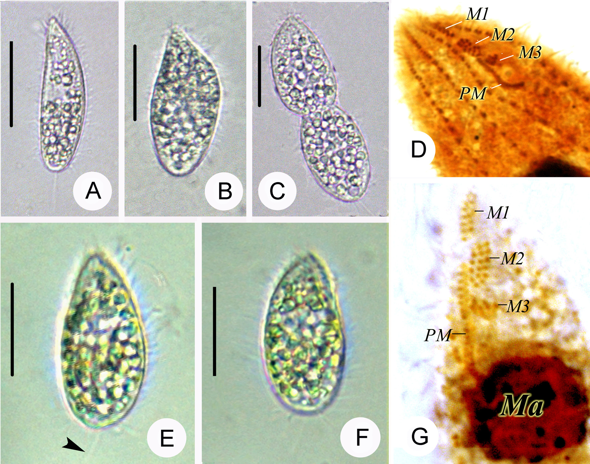
Fig. 2A–C View Fig. 2 ; Table 1 View Table 1
Description
Description based on Alabama population: body ( in vivo) 35–55 × 15–25 μm, spindle-shaped ( Fig. 2A View Fig. 2 ) or pyriform ( Fig. 2B View Fig. 2 ), having sharply pointed anterior end and narrowly rounded caudal end ( Fig. 2A View Fig. 2 ); variability in shape likely attributable to nutritional conditions ( Fig. 2A–B View Fig. 2 ) or division ( Fig. 2C View Fig. 2 ). Buccal field 10–15 μm in length or 25–30% of body length. Somatic cilia distributed in a dense lateral field, 5–8 μm long ( Fig. 2A View Fig. 2 ). Extrusomes undetectable ( Fig. 2A–C View Fig. 2 ). Cytoplasm transparent or grey, granulated; cytoplasmic granules 3–5 μm long, 1–1.5 μm wide, refractive. Macronucleus single, large, 8–13 μm long or 20–30% of body length, 8–12 μm wide or 40–50% of body width, spheroid, centrally located. One micronucleus attached to macronucleus. Locomotion by moving on substrate or swimming in water. Somatic cilia 7 to 10 μm long, ten somatic kineties composed of dikinetids in anterior ⅔ of cell. Membranelle 1 (M1) slightly separated from apex of anterior end, comprising two rows of kinetids each with seven to nine basal bodies ( Fig. 2D View Fig. 2 ). M2 bearing three to five longitudinal rows of cilia, with each longitudinal row having six to eight basal bodies ( Fig. 2D View Fig. 2 ). M3 posterior to M2, comprising three rows of kinetids ( Fig. 2D View Fig. 2 ).
No known copyright restrictions apply. See Agosti, D., Egloff, W., 2009. Taxonomic information exchange and copyright: the Plazi approach. BMC Research Notes 2009, 2:53 for further explanation.
|
Kingdom |
|
|
Phylum |
|
|
Class |
|
|
Order |
|
|
Family |
|
|
Genus |