
Uronema marinum Dujardin, 1841
|
publication ID |
https://doi.org/10.5852/ejt.2016.249 |
|
publication LSID |
urn:lsid:zoobank.org:pub:43278833-B695-4375-B1BD-E98C28A9E50E |
|
DOI |
https://doi.org/10.5281/zenodo.5631551 |
|
persistent identifier |
https://treatment.plazi.org/id/03C487DB-685C-FFB3-571B-FEF388F03772 |
|
treatment provided by |
Plazi |
|
scientific name |
Uronema marinum Dujardin, 1841 |
| status |
|
Uronema marinum Dujardin, 1841
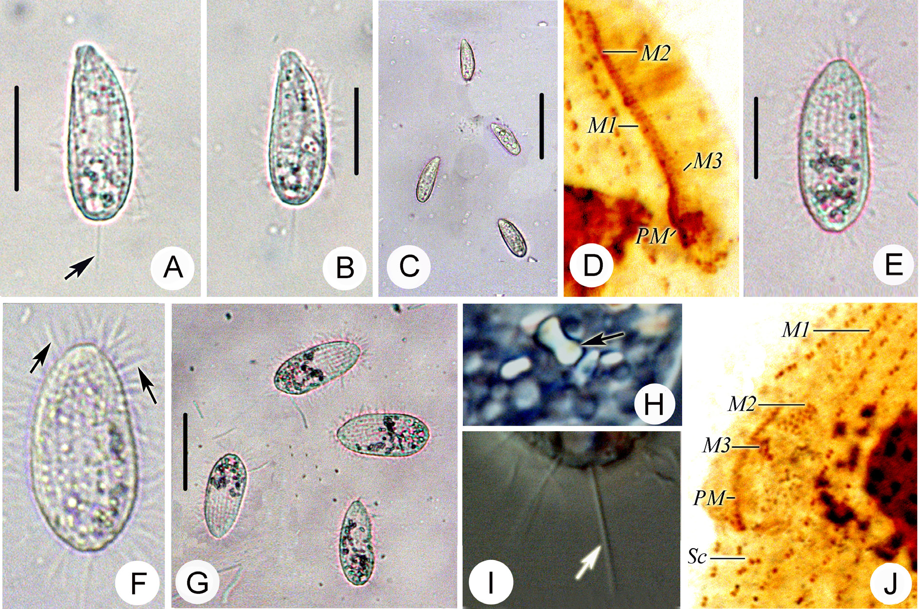
Fig. 4 View Fig. 4 F–I; Table 1 View Table 1
Description
Description based on Alabama population: Size in vivo 10–25 × 6–10 μm, elongate-elliptical in outline ( Fig. 4 View Fig. 4 F). Anterior end flat, with an apical plate, dorsal posterior area slightly rounded ( Fig. 4 View Fig. 4 F–H). Buccal field 50% of body length. Pellicle smooth ( Fig. 4 View Fig. 4 F–I). Extrusomes approximately 1 µm long, rodshaped, closely beneath pellicle. One large globular to ellipsoidal macronucleus centrally located with numerous tiny (approximately 1×1 μm) irregularly-shaped peripheral nucleoli ( Fig. 4 View Fig. 4 F–I). Contractile vacuole about 3 μm in diameter at posterior end of cell ( Fig. 4 View Fig. 4 H). Movement unremarkable, either swimming continuously or resting on the bottom. Twelve to 13 ciliary rows. Buccal apparatus typical of genus: M1 positioned near apical plate and composed of 5–7 kinetosomes; M2 slightly longer than M1; M3 very short ( Fig. 4 View Fig. 4 I). PM on right of shallow buccal cavity, composed zigzaging row of basal bodies, extending anteriorly to middle of M2 ( Fig. 3 View Fig. 3 I).
No known copyright restrictions apply. See Agosti, D., Egloff, W., 2009. Taxonomic information exchange and copyright: the Plazi approach. BMC Research Notes 2009, 2:53 for further explanation.
|
Kingdom |
|
|
Phylum |
|
|
Class |
|
|
Order |
|
|
Family |
|
|
Genus |