
Uronemita filificum Kahl, 1931
|
publication ID |
https://doi.org/10.5852/ejt.2016.249 |
|
publication LSID |
urn:lsid:zoobank.org:pub:43278833-B695-4375-B1BD-E98C28A9E50E |
|
DOI |
https://doi.org/10.5281/zenodo.5631549 |
|
persistent identifier |
https://treatment.plazi.org/id/03C487DB-685D-FFBD-570E-FEF38E963617 |
|
treatment provided by |
Plazi |
|
scientific name |
Uronemita filificum Kahl, 1931 |
| status |
|
Uronemita filificum Kahl, 1931
Fig. 4A–E View Fig. 4 ; Table 1 View Table 1
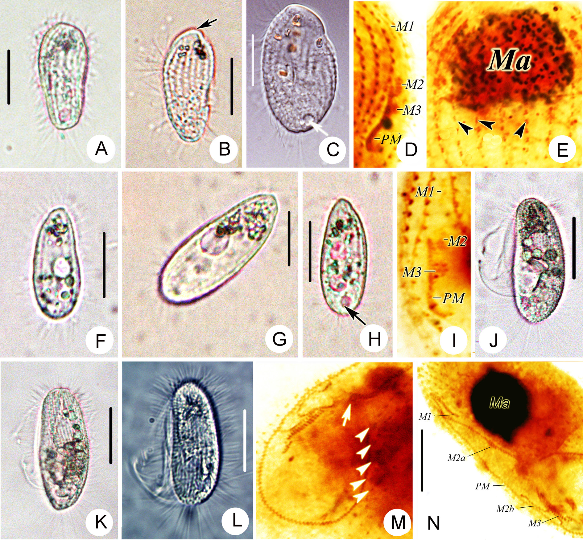
Description
Description based on Alabama population: body 30–45 × 15–20 μm in vivo, inverted pear-shaped with large, conspicuous apical plate ( Fig 4A–B View Fig. 4 ). Dorsal side conspicuously convex. Length of buccal field 60% of body. Extrusomes appromately 2 µm long, rod-shaped, closely beneath pellicle. Cytoplasm colourless to grayish, containing several (ca 3 μm across) food vacuoles and dumbbell-shaped crystals (ca 1–2 µm long) often concentrated in anterior end of body ( Fig. 4C View Fig. 4 ). Single macronucleus larg ( Fig. 4E View Fig. 4 ). Locomotion by swimming aimlessly, sometimes rotates while attached to substratum by caudal cilium. Eighteen or 19 somatic kineties, anterior third of each composed of dikinetids ( Fig. 4E View Fig. 4 ). M1 single-rowed with five or six kinetosomes ( Fig. 4D View Fig. 4 ); M2 three-rowed; M3 smaller and close to M2 ( Fig. 4D View Fig. 4 ). PM on right of shallow buccal cavity, with zigzag row of basal bodies, extending anteriorly to the middle of M2. Scutica consisting of three or four basal body pairs ( Fig. 4D View Fig. 4 ).
No known copyright restrictions apply. See Agosti, D., Egloff, W., 2009. Taxonomic information exchange and copyright: the Plazi approach. BMC Research Notes 2009, 2:53 for further explanation.
|
Kingdom |
|
|
Phylum |
|
|
Class |
|
|
Order |
|
|
Family |
|
|
Genus |