
Cymbasoma dakini, Suárez-Morales, Eduardo & Mckinnon, David, 2016
|
publication ID |
https://doi.org/10.11646/zootaxa.4102.1.1 |
|
publication LSID |
lsid:zoobank.org:pub:9A7BA798-AA7C-4CAA-B42C-1E260CA573E4 |
|
DOI |
https://doi.org/10.5281/zenodo.6091289 |
|
persistent identifier |
https://treatment.plazi.org/id/03C4CA6D-D514-FFB1-FF12-544B933B2868 |
|
treatment provided by |
Plazi |
|
scientific name |
Cymbasoma dakini |
| status |
sp. nov. |
Cymbasoma dakini sp. nov
( Figs 5–8 View FIGURE 5 View FIGURE 6 View FIGURE 7 View FIGURE 8 )
Material examined. Adult female holotype and two paratype females from Corinella, Western Port Bay, Victoria, Australia ( 38°23.115’ S, 145°25.371’ E). Holotype partially, ethanol-preserved; dissected parts mounted on slide in glycerine, sealed with Entellan®. Undissected paratypes (2) each mounted on slides in glycerine, sealed as holotype. Date of collection: 17th June 1985. Slides deposited in the collection of MTQ, Australia (cat. MTQ W34372, MTQ W34373, MTQ W34374, respectively).
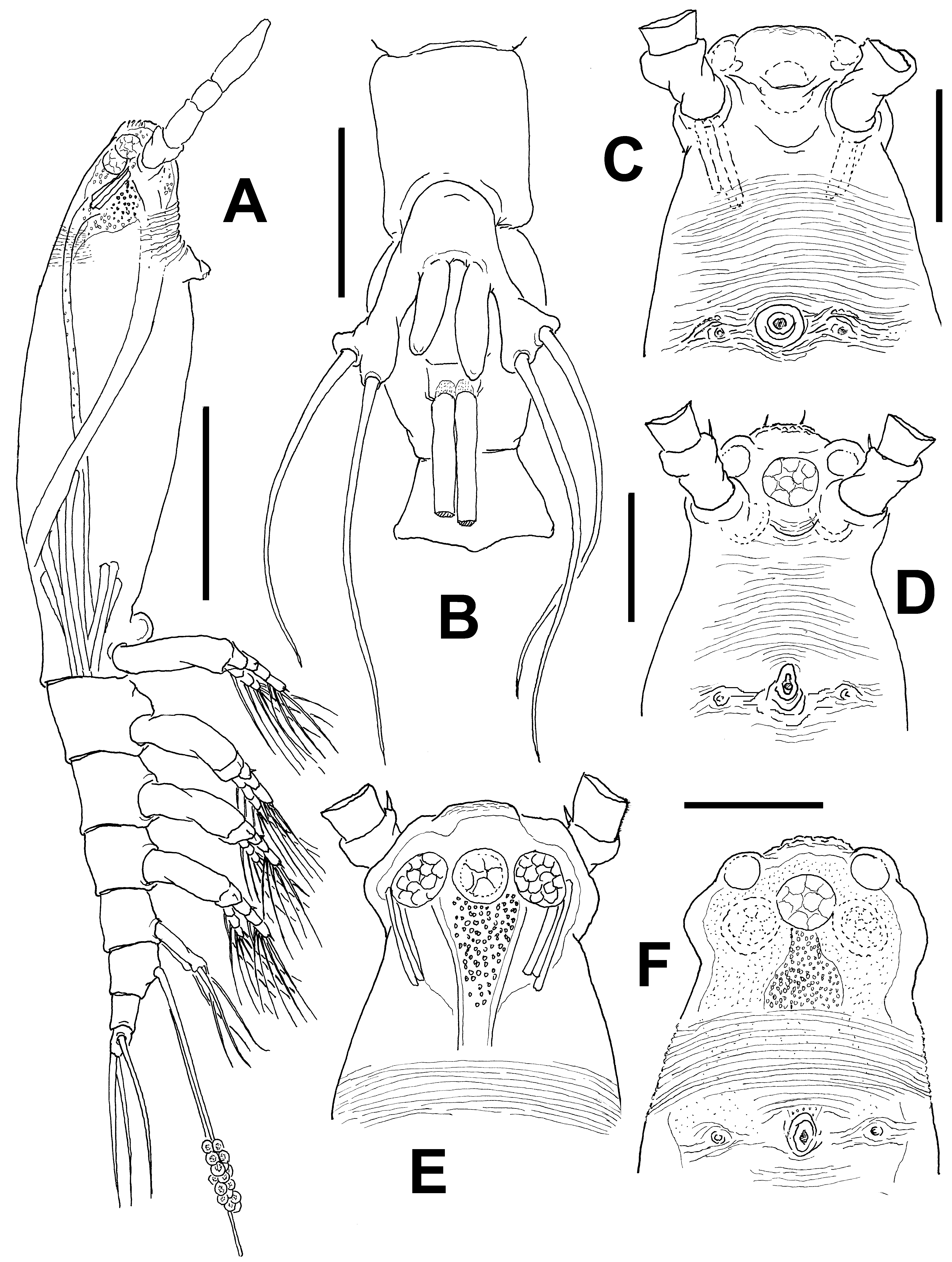
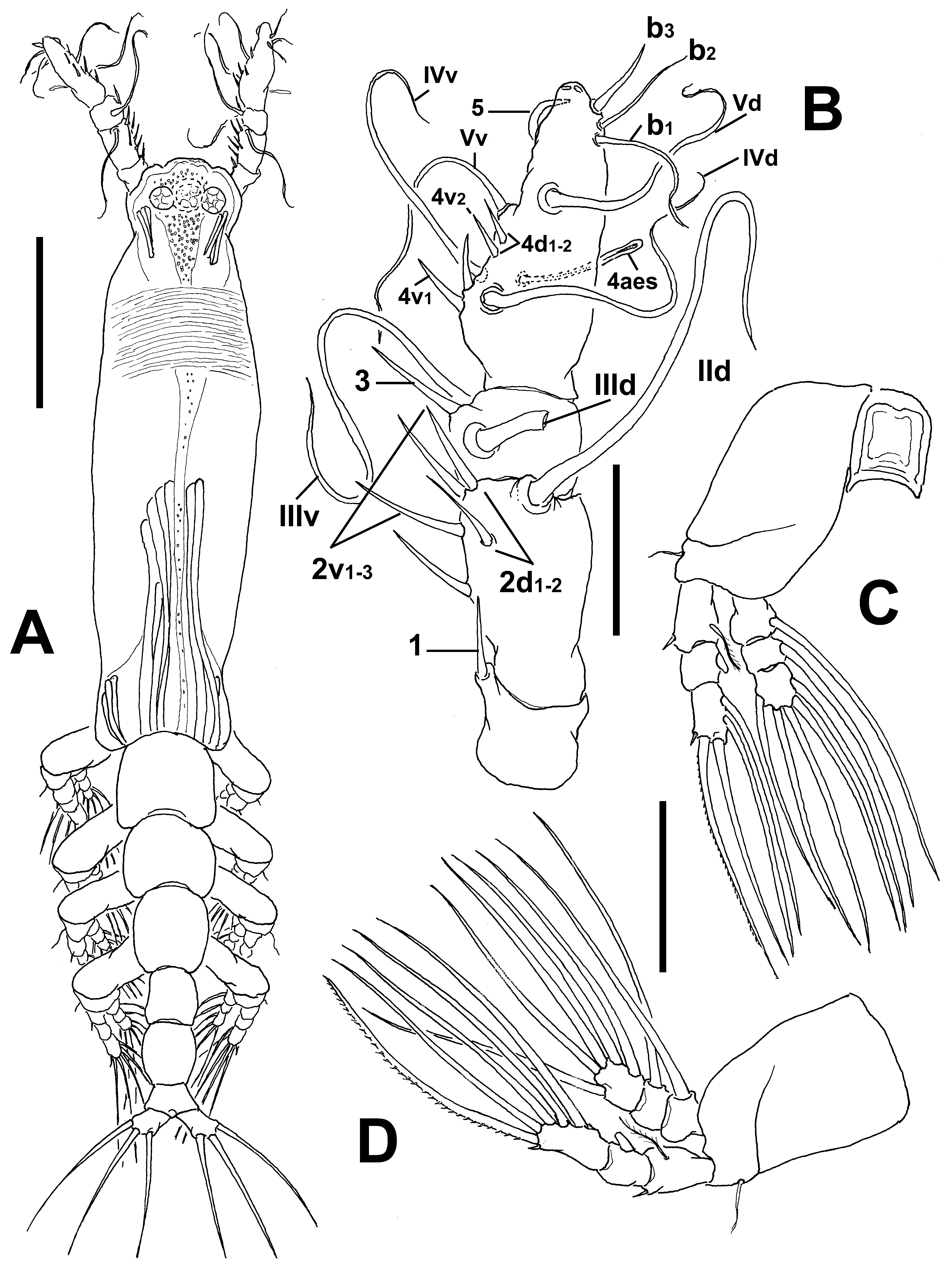
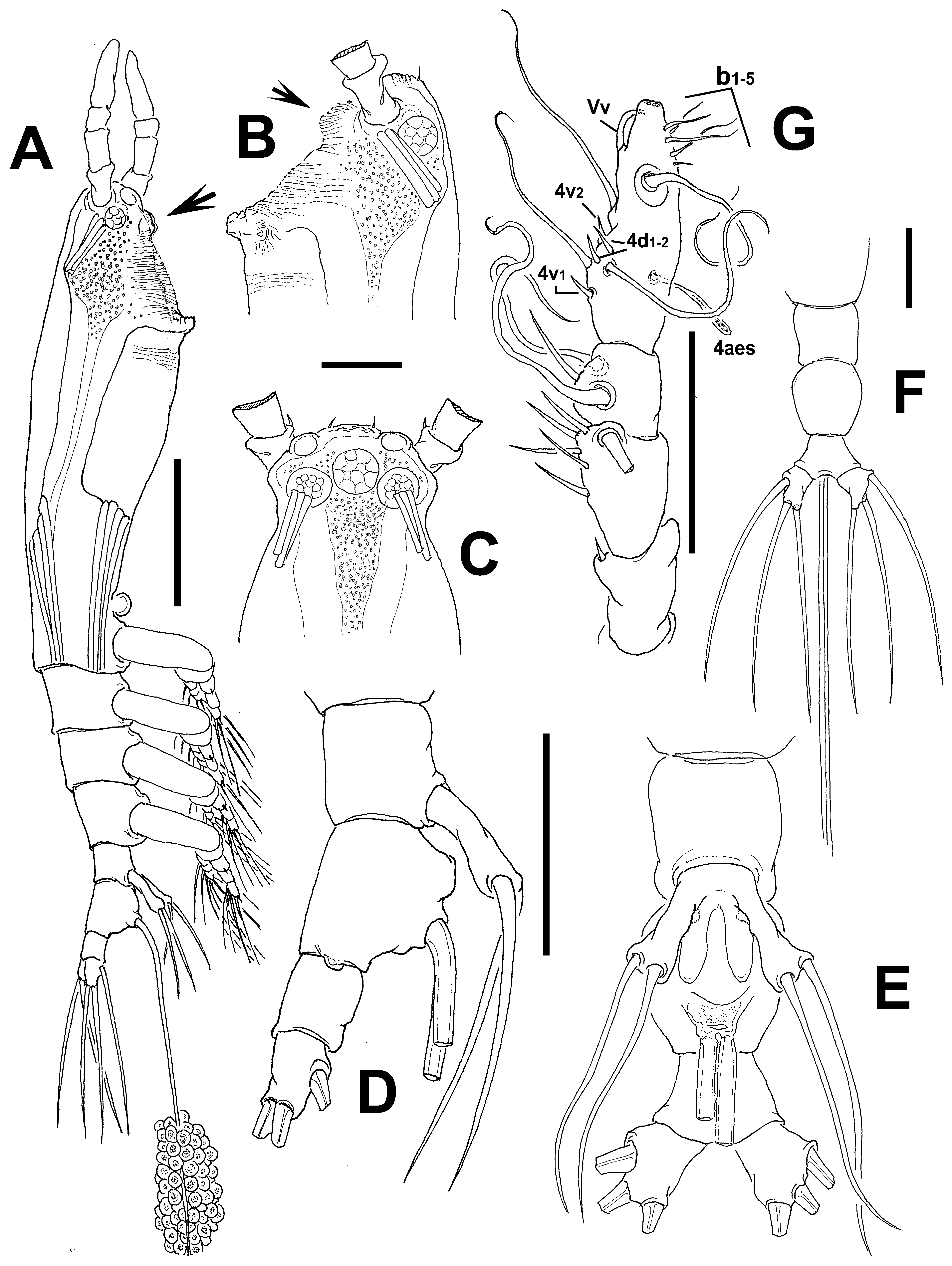
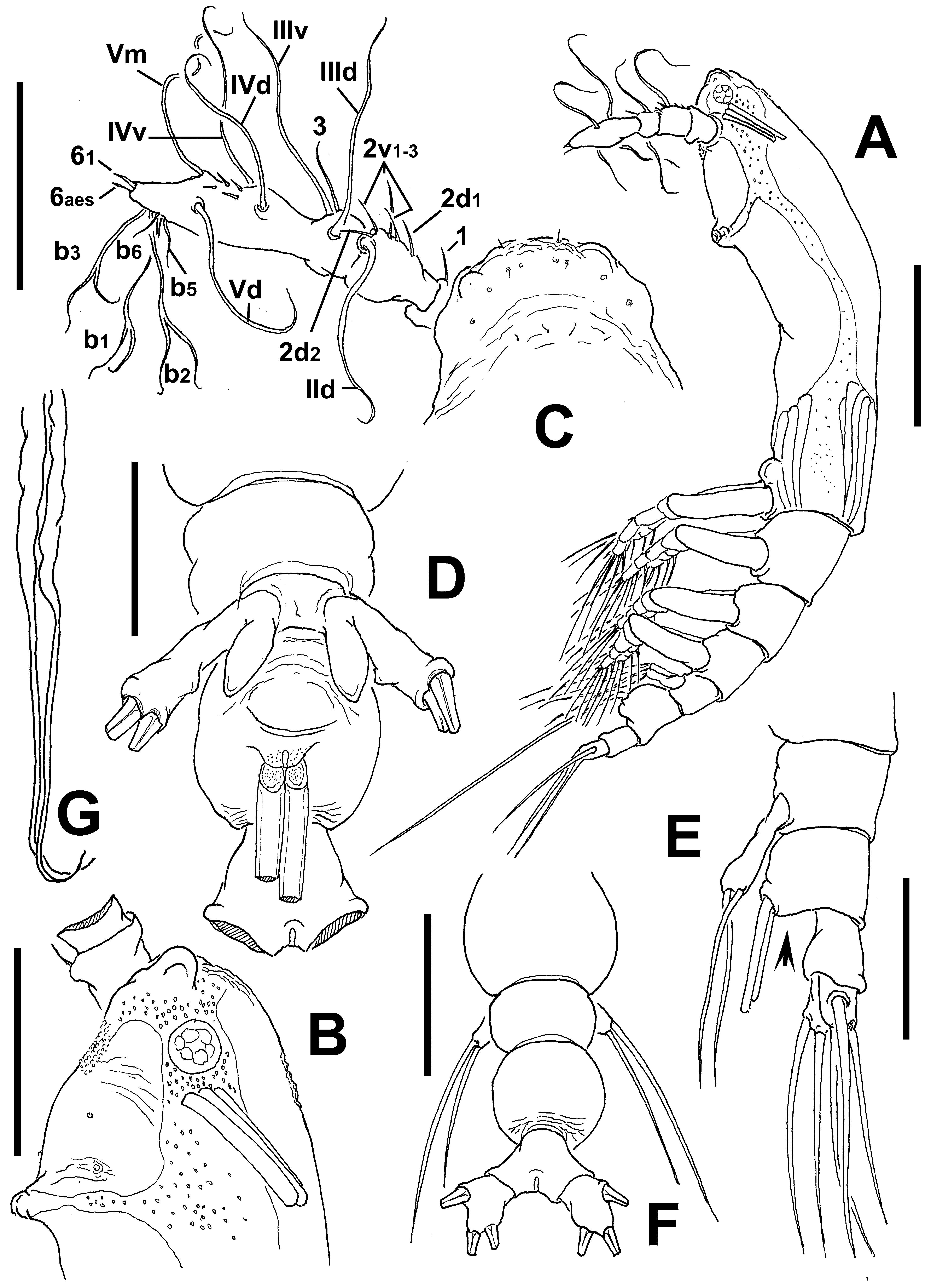
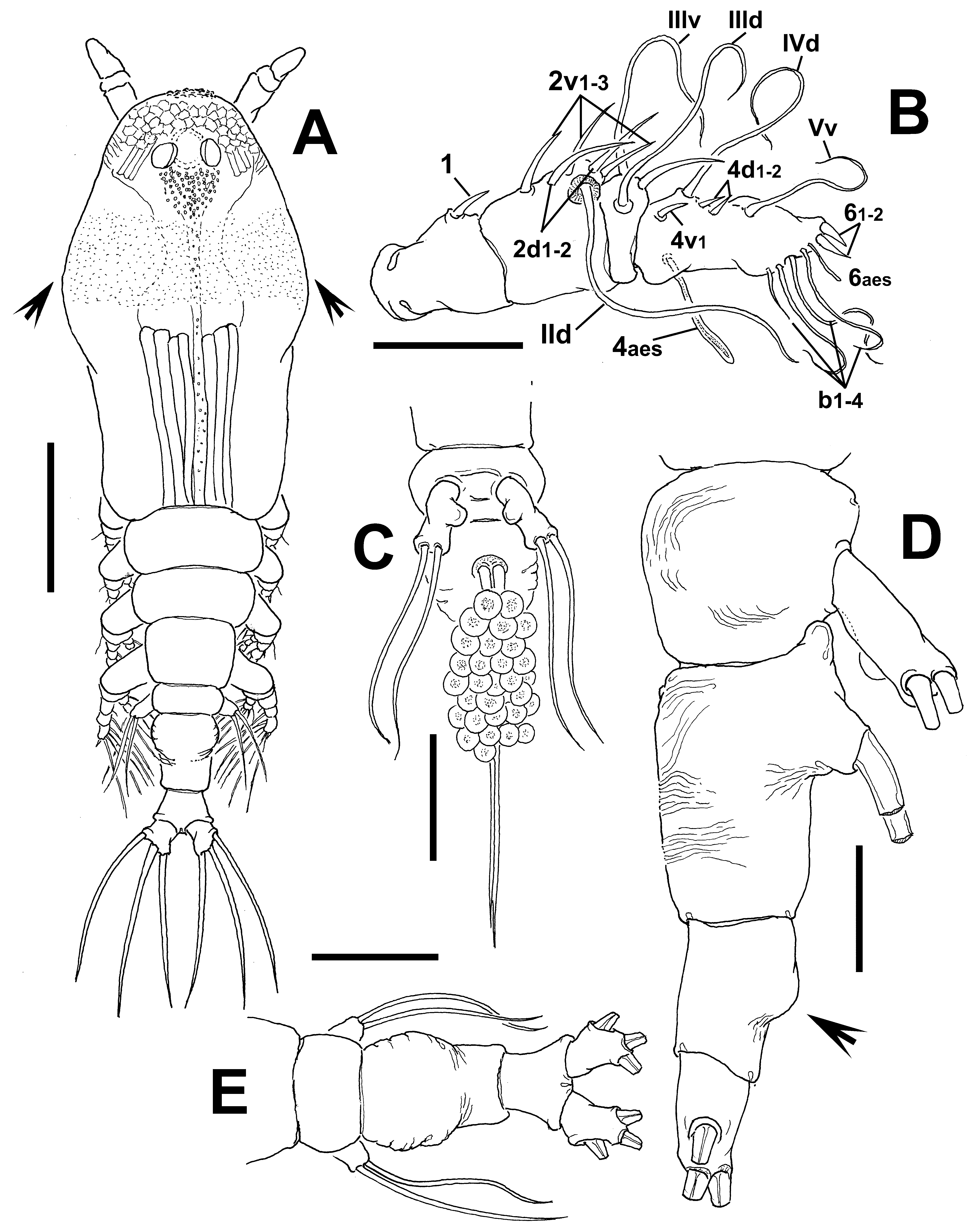
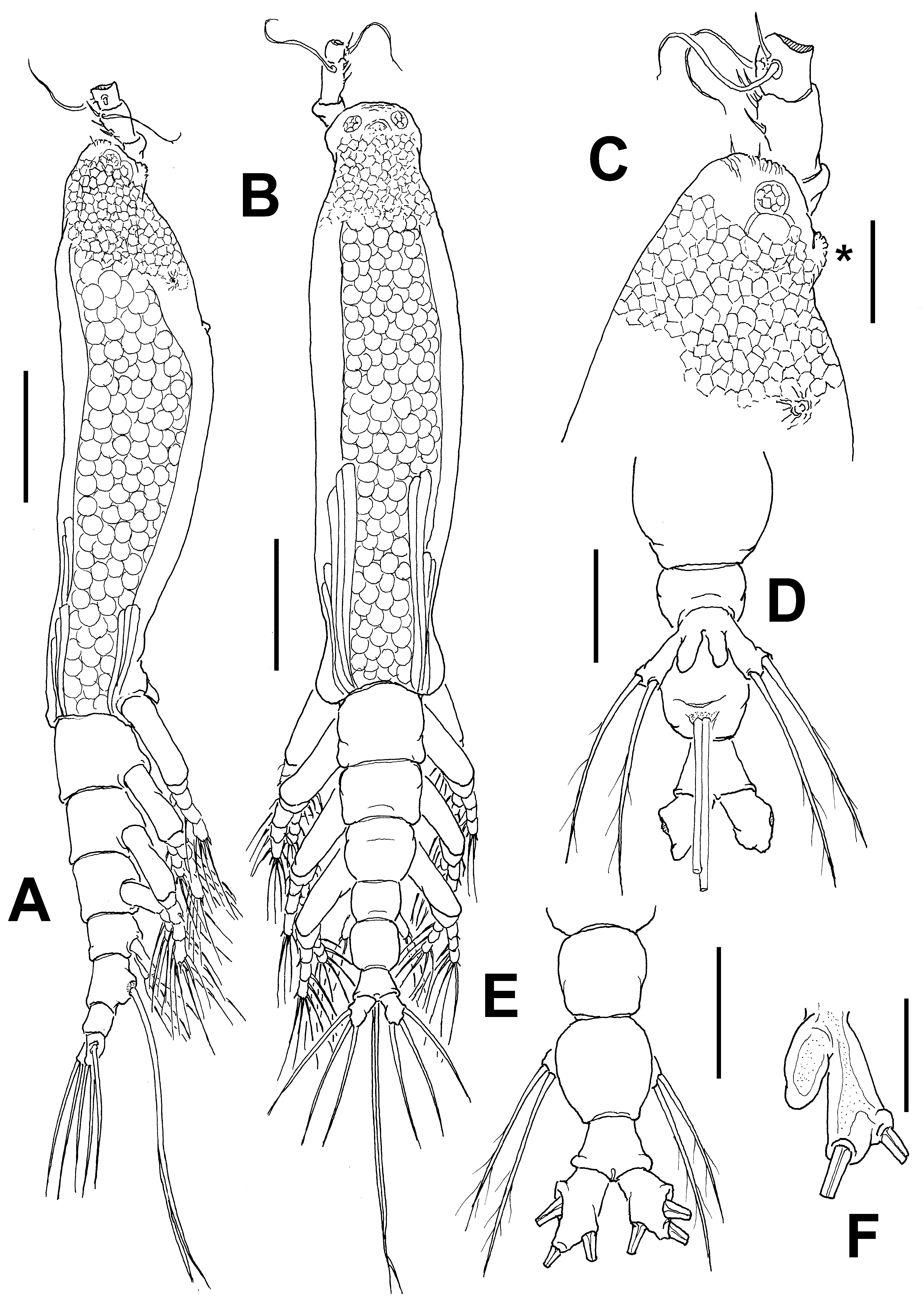
Description of adult female. Body moderately elongate, slender ( Figs 5 View FIGURE 5 A, 6A, 7A); body length of holotype female 1.15 mm; size range 1.09–1.15 mm, size average ( n =3) 1.13± 0.02 mm. Cephalothorax 0.64–0.67 mm long, representing 62.1–63.2 % of total body length. Midventral oral papilla located at 25% of cephalothorax length. Pair of relatively large ocelli present, pigment cups moderately developed, medially conjoined, weakly pigmented; ventral cup and lateral cups equally sized ( Figs 5 View FIGURE 5 E, F, 7C). Cephalic area with flat “forehead”, ornamented with shallow transverse striations ( Figs 5 View FIGURE 5 C–F, 7C) with pair of sensilla ( Fig. 5 View FIGURE 5 C). Symmetrical rounded protuberances at both sides of flat forehead. Low ventral rounded protuberance ornamented with transverse striae (arrowed in Figs 7 View FIGURE 7 A, B) in one specimen, protuberance not observed in the other specimens ( Fig. 5 View FIGURE 5 A). Additional cephalic cuticular ornamentation including transverse, shallow cuticular ridges overlying posterior surface of oral region on dorsal surface. Ventral surface with additional features including: 1) transverse striations between the antennulary bases and oral papilla, lighter striation on post-oral surface; 2) pair of symmetrical nipple-like processes on anterior ventral surface at each side of oral area ( Figs 5 View FIGURE 5 C, D, F, 7B).
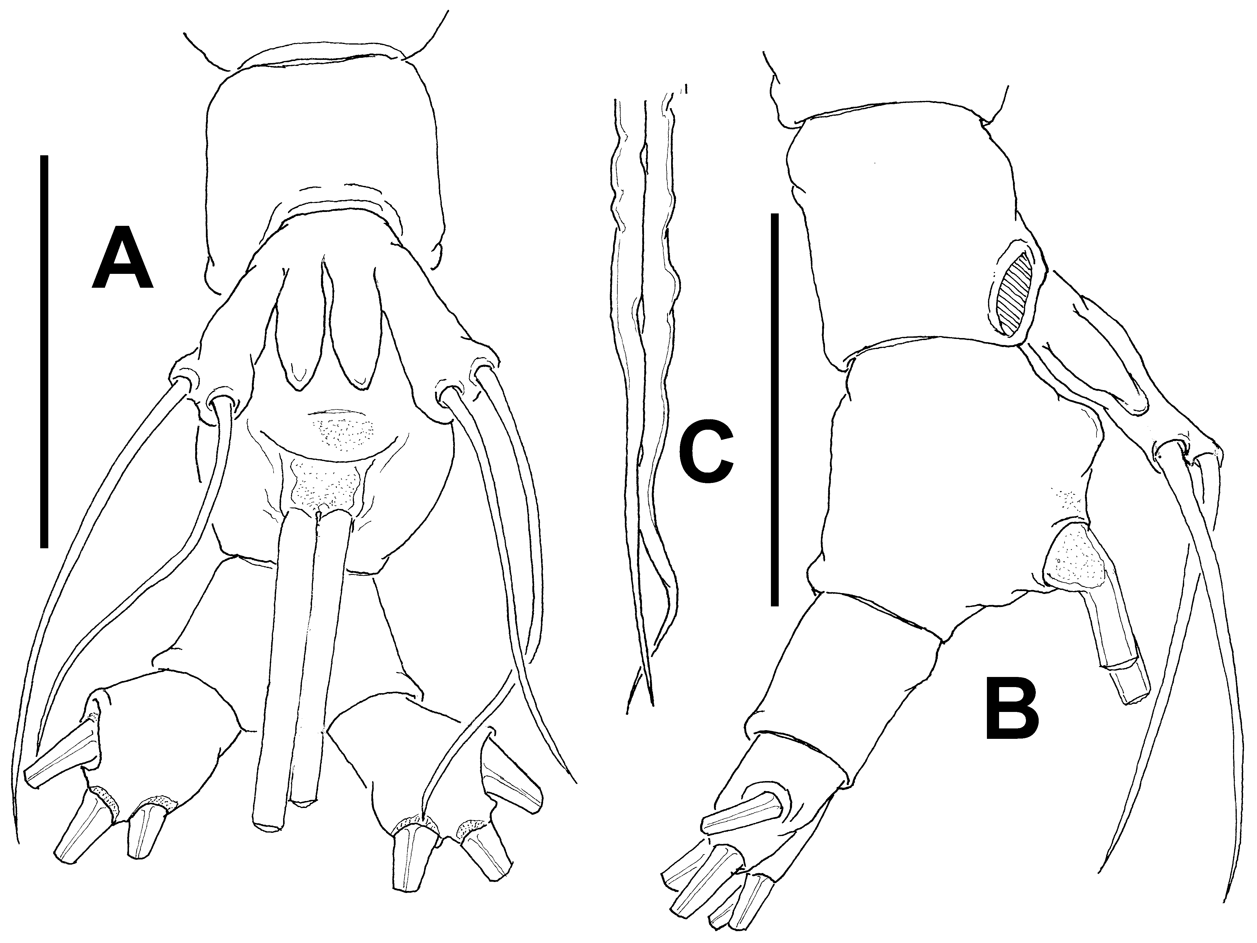
Urosome consisting of fifth pedigerous somite, genital double-somite and anal somite, together representing 15% of total body length. Relative lengths of urosomites (fifth pedigerous, genital double and free somite) 37.5: 43.5: 19 = 100, respectively. Genital double-somite with smooth dorsal and ventral surfaces ( Figs 6 View FIGURE 6 A, 7D, 8B); with low ventral process on anterior margin ( Figs 7 View FIGURE 7 D, 8B). Caudal ramus subrectangular, 1.2 times longer than wide, armed with three subequally long, sparsely setulated caudal setae ( Fig. 7 View FIGURE 7 E, F). Ovigerous spines paired, relatively short, 45–50 % of total body length ( 0.51–0.57 mm) ( Figs 5 View FIGURE 5 A, 7A). Spines basally separated, slender, straight at their base and along shaft, without distal expansions and tapering apically, one spine slightly shorter; spines 0.35–0.40 mm long in two specimens ( Figs 5 View FIGURE 5 A, 7A).
Antennule length 0.20–0.23 mm, representing about 15.9–21.2 % of total body length and 30–33.5 % of cephalothorax length in the three specimens examined; 4-segmented. Relative length of distal antennulary segment as: 40.2, 42.5, and 48 in each of the three specimens examined. In terms of the pattern described by Grygier & Ohtsuka (1995) for female monstrilloid antennulary armature, setae (Roman numerals) and spines (Arabic numerals), stout, spiniform element 1 present on first segment; elements on second segment: 2d1-2, 2v 1-3, and IId. Third segment with stout, long element 3 plus elements IIId and IIIv. Segment 4 bearing elements 4d1,2, 4v 1-2, element 4v 3 not observed; setae IVd, IVv, Vd, Vv present. Element 5 spiniform, strongly curved inwards. Subterminal elements b1-3 present, unbranched, short; elements 61 and 62 not present in specimens, but apical sockets were observed ( Figs 6 View FIGURE 6 B, 7G).
Incorporated first pedigerous somite and succeeding three free pedigerous somites each bearing a pair of biramous legs. Pedigerous somites 2–4, together accounting for 23.5, 24.5, 24.2 % of total length in dorsal view in each of the specimens examined. Legs 1–4 slightly increasing in size posteriorly. Intercoxal sclerites of legs 1–4 subrectangular, widest at base, tapering distally, surface and posterior margin smooth ( Fig. 6 View FIGURE 6 C). Bases of legs 1–4 articulating with large, rectangular coxa along oblique line; with hair-like lateral seta; on leg 3, this seta about 2.2 times longer, slightly setulated from proximal half and slightly thicker than those on the other legs. Endopods and exopods of legs 1–4 triarticulated. Ramal setae all biserially plumose except spiniform outer seta on exopodal segments 1 and 3, and inner seta of first exopodal segment, these latter being short, slender, and sparsely setulated. Spine on distal exopodal segment of right leg 4 noticeably short ( Fig. 6 View FIGURE 6 D). Also, outermost apical exopodal setae of legs 1–4 with inner margin naked, outer margin spinulose.
Armature formula of legs 1–4:
Fifth legs medially conjoined, bilobate, inner (endopodal) lobe elongate, digitiform unarmed, rounded distally, reaching about ¾ the length of outer lobe. Outer (exopodal) lobe elongate, slender, armed with two subequally long setae on distal position ( Figs 5 View FIGURE 5 B, 7E, 8A). Male: unknown.
Type locality. Corinella, Western Port Bay, Victoria, Australia ( 38°23.115’S, 145°25.371’E).
Etymology. The species is named after William J. Dakin, first author of one of the most influential contributions on Australian marine zooplankton ( Dakin & Colefax 1940), which includes the earliest records of Australian monstrilloids.
Diagnosis. Cymbasoma with third antennulary segment representing more than 40% of antennule length, with dorsal and ventral fringes of cuticular striation on cephalic area. Paired cephalic protuberances on both sides of flat forehead area. Fifth leg with two setae on elongate outer lobe, inner lobes digitiform, unarmed, slightly asymmetrical. Anal somite not constricted. Ovigerous spines representing 45–55% of total body length.
Remarks. This species belongs to a group of Australian Cymbasoma in which the female fifth leg bears two setae on the outer lobe and a naked inner lobe. This is an unusual character among species of Cymbasoma . It is known only in C. agoense Sekiguchi, 1982 from Japan and in some of the Australian species included in this work. In some cases like in C. nicolettae Suárez-Morales, 2002 or C. thompsonii ( cf. Sars 1921), the innermost seta is very small, slender and could be overlooked during casual observation, but this is not the case in the Australian species. Two setae on the fifth leg are known also in a few species of Monstrilla , like M. leucopis Sars, 1921 (Suárez-Morales 2010) , M. helgolandica Claus, 1863 or M. hamatapex Grygier & Ohtsuka, 1995 . The new species differs from its congeners sharing this relevant character in having a conspicuous dorsal field of wrinkles on the cephalic area, and a long fifth leg inner lobe, barely reaching the distal margin of the outer lobe. This lobe is relatively short and robust in C. agoense ( cf. Sekiguchi 1982: fig. 6F) and the body shape and proportions are clearly different in these two species. It differs from the other Australian congeners in the presence of a narrow, long digitiform inner lobe, an unconstricted anal somite, a genital double-somite with rounded lateral margins and a low anteroventral protuberance. The inner lobe of the fifth leg is clearly shorter in Cymbasoma sp. ( Fig. 70 View FIGURE 70 D) and also in C. lourdesae sp. nov. ( Fig. 24 View FIGURE 24 D) and in C. tharawalorum sp. nov. ( Fig. 65 View FIGURE 65 C). The genital double-somite has a different shape in C. lourdesae ( Fig. 24 View FIGURE 24 E) and in C. tharawalorum ( Fig. 65 View FIGURE 65 D). Also, the antennulary segments 3– 4 are fused in C. lourdesae ( Fig. 24 View FIGURE 24 C) whereas they are separated in the new species C. dakini . In C. tharawalorum the antennules are distinctly compressed ( Fig. 65 View FIGURE 65 B), thus differing from the antennule of C. dakini sp.nov.
No known copyright restrictions apply. See Agosti, D., Egloff, W., 2009. Taxonomic information exchange and copyright: the Plazi approach. BMC Research Notes 2009, 2:53 for further explanation.
|
Kingdom |
|
|
Phylum |
|
|
Class |
|
|
Order |
|
|
Family |
|
|
Genus |