
Cymbasoma lenticula, Suárez-Morales, Eduardo & Mckinnon, David, 2016
|
publication ID |
https://doi.org/10.11646/zootaxa.4102.1.1 |
|
publication LSID |
lsid:zoobank.org:pub:9A7BA798-AA7C-4CAA-B42C-1E260CA573E4 |
|
DOI |
https://doi.org/10.5281/zenodo.6091311 |
|
persistent identifier |
https://treatment.plazi.org/id/03C4CA6D-D520-FFE4-FF12-507895142AFB |
|
treatment provided by |
Plazi |
|
scientific name |
Cymbasoma lenticula |
| status |
sp. nov. |
Cymbasoma lenticula sp. nov.
( Figs 38–41 View FIGURE 38 View FIGURE 39 View FIGURE 40 View FIGURE 41 )
Material examined. Holotype: adult female from Western Port Bay (Station G1 of Kimmerer & McKinnon 1985), Victoria, Australia ( 38°31.306’ S, 145°4.858’ E), partially dissected, mounted on 2 slides in glycerine, sealed with Entellan®. Date of collection: 29th November 1983. Slides deposited in the collection of MTQ, Australia (cat. MTQ W34394). Allotype adult male from Warneet, Western Port Bay, Victoria, Australia ( 38°13.200’ S, 145°18.758’ E), partially dissected, mounted on 2 slides in glycerine, sealed with Entellan®. Date of collection: 12th June 1984. Vial and slides deposited in the collection of MTQ, Australia (cat. MTQ W34395).
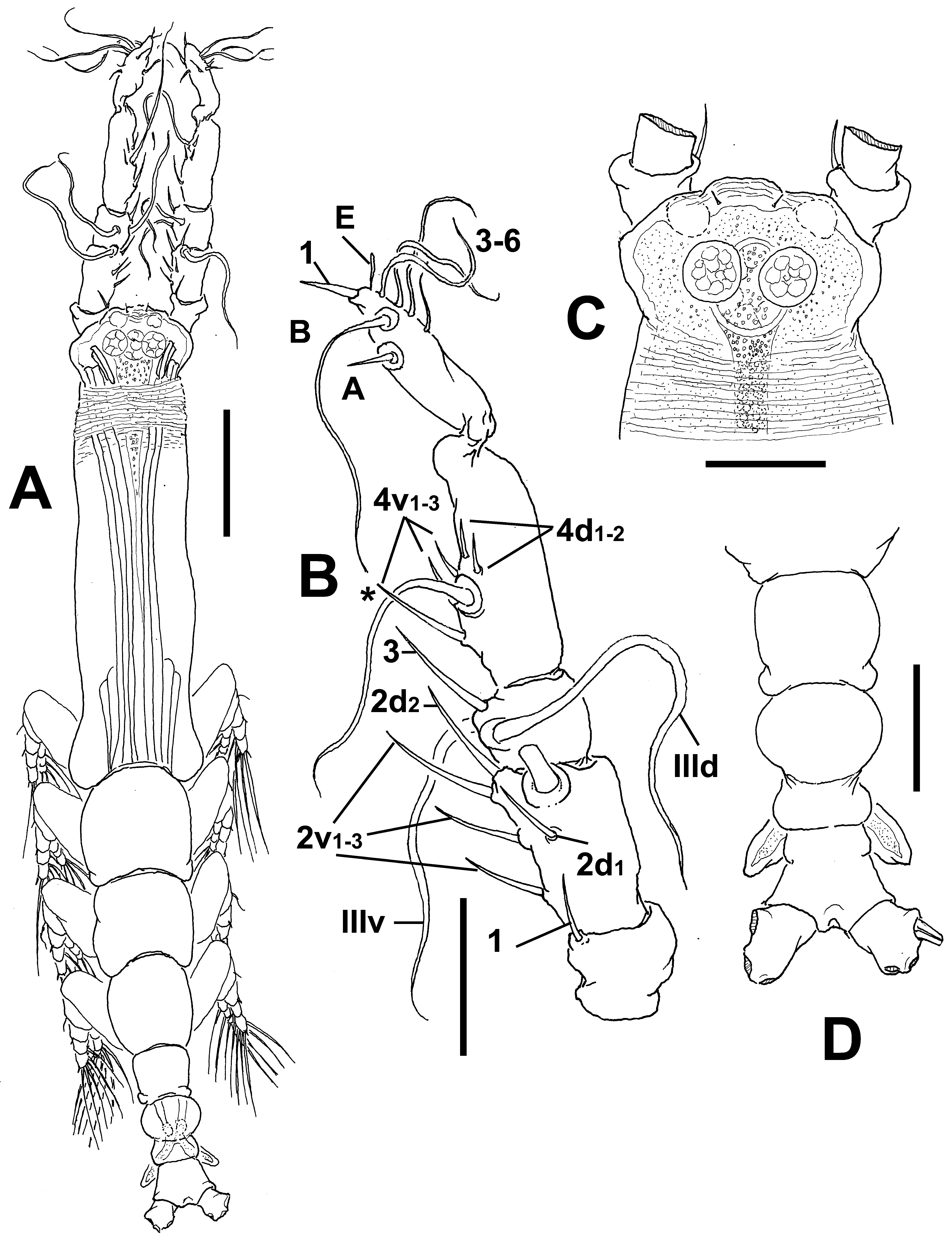
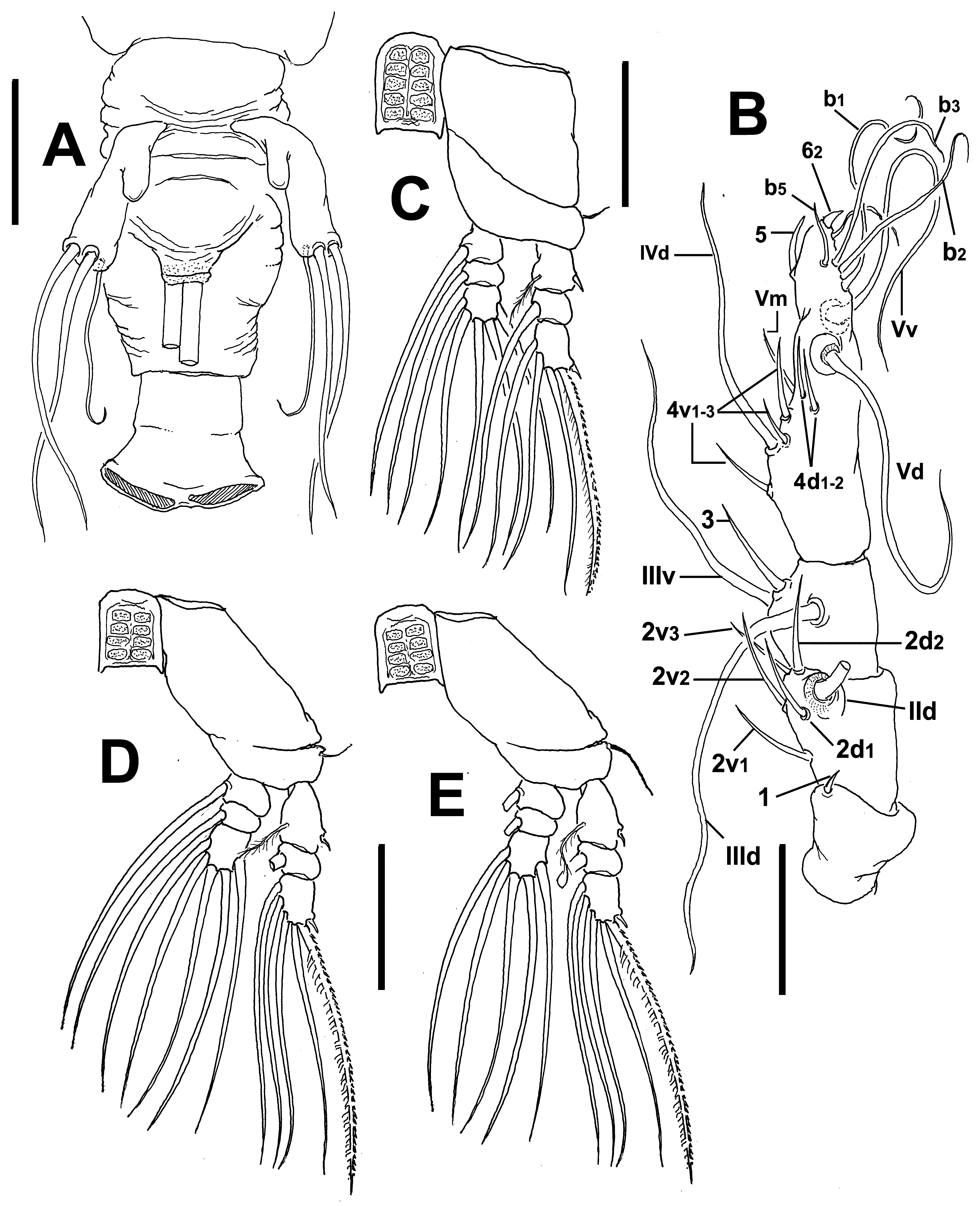
Description of adult female. Body relatively slender ( Fig. 38 View FIGURE 38 A); body length of holotype female 1.22 mm. Cephalothorax approximately 0.71 mm long, representing 58% of total body length. Midventral oral papilla moderately protuberant, located at 26% of cephalothorax length. Pair of relatively small ocelli present, pigment cups medially separated, unpigmented; ventral cup slightly larger than lateral cups ( Fig. 38 View FIGURE 38 B). Anteriormost cephalic area narrower than rest of cephalothorax, with slightly produced "forehead”. Cephalic frontal area with pattern of transverse striations flanked by pair of rounded lens-like anteriorly directed protuberances ( Fig. 38 View FIGURE 38 B, D, E), frontal sensilla absent. Single low medial protuberance present on ventral surface between antennule bases and nipple-like processes ( Fig. 38 View FIGURE 38 C). Dorsal surface of cephalothorax smooth except for field of wrinkles (arrowed in Fig. 38 View FIGURE 38 C), ventral surface with field of transverse cuticular wrinkles at perioral area ( Fig. 38 View FIGURE 38 D). Ventral surface also with 1) pair of symmetrical, crescent-shaped processes on anterior ventral surface located near bases of antennules, with no adjacent striae ( Fig. 38 View FIGURE 38 D); 2) pair of nipple-like processes with concentric and transverse striae.
Urosome consisting of fifth pedigerous somite, genital double-somite and anal somite, together representing 16.5% of total body length. Relative lengths of urosomites (fifth pedigerous, genital double and free anal somites) 26.8: 38.3: 34.9 = 100, respectively ( Figs. 38 View FIGURE 38 F, G). Lateral margins of fifth pedigerous somite straight, with deep lateral corrugation. Genital double-somite longest of urosome, with lateral margins corrugated ( Fig. 38 View FIGURE 38 G), anterior half swollen, tapering posteriorly into straight margins ( Fig. 39 View FIGURE 39 A); anteroventral process remarkably strong, reaching insertion of fifth leg ( Fig. 38 View FIGURE 38 F, H). Ovigerous spines paired, basally separated, slender, straight, broken off in specimen. Anal somite almost as long as genital double-somite, with medial constriction and faint suture ( Fig. 38 View FIGURE 38 G). Caudal ramus short, subquadrate, about 1.2 times as long as wide, armed with three caudal setae.
Antennule length 0.25 mm, representing about 21% of total body length and 34% of cephalothorax length, 4- segmented. Relative length of distal antennulary segment 51%. In terms of pattern by Grygier & Ohtsuka (1995), element 1 present on first segment; on second segment: long, slender elements 2d1-2, 2v 1-3, and IId. Third segment with element 3 being stout, spiniform, elements IIId and IIIv setiform, of normal aspect. Segment 4 bearing elements 4d1,2, 4v 1-3; setae IVd, Vd, Vv, Vm (short, spiniform), and 4aes present. Element 5 spiniform, curved. Subterminal elements b1-3,5 present, unbranched, element 62 present, other apical elements not observed, probably broken off ( Fig. 39 View FIGURE 39 B).
Incorporated first pedigerous somite and succeeding three free pedigerous somites each bearing a pair of biramous legs. Pedigerous somites 2–4, together accounting for 23% of total body length. Legs 1–4 slightly increasing in size posteriorly. Intercoxal sclerites of legs 1–4 subrectangular, with patches of spinules, posterior margin straight, smooth. Bases of legs 1–4 articulating with large, rectangular coxa along oblique line; with hairlike lateral seta ( Fig. 39 View FIGURE 39 C–E); on leg 3, this seta about 2.5 times longer, thicker than those on other legs ( Fig. 39 View FIGURE 39 E).
Endopods and exopods of legs 1–4 triarticulated. Ramal setae all biserially plumose except spiniform outer seta on exopodal segments 1 and 3, and inner seta of first exopodal segment, these latter being short, slender. Outermost distal spines on third exopodal segment of legs 1–4 short, 0.25 times as long as segment. Outermost apical exopodal setae of legs 1–4 with inner margin setulose, outer margin spinulose.
Armature formula of legs 1–4:
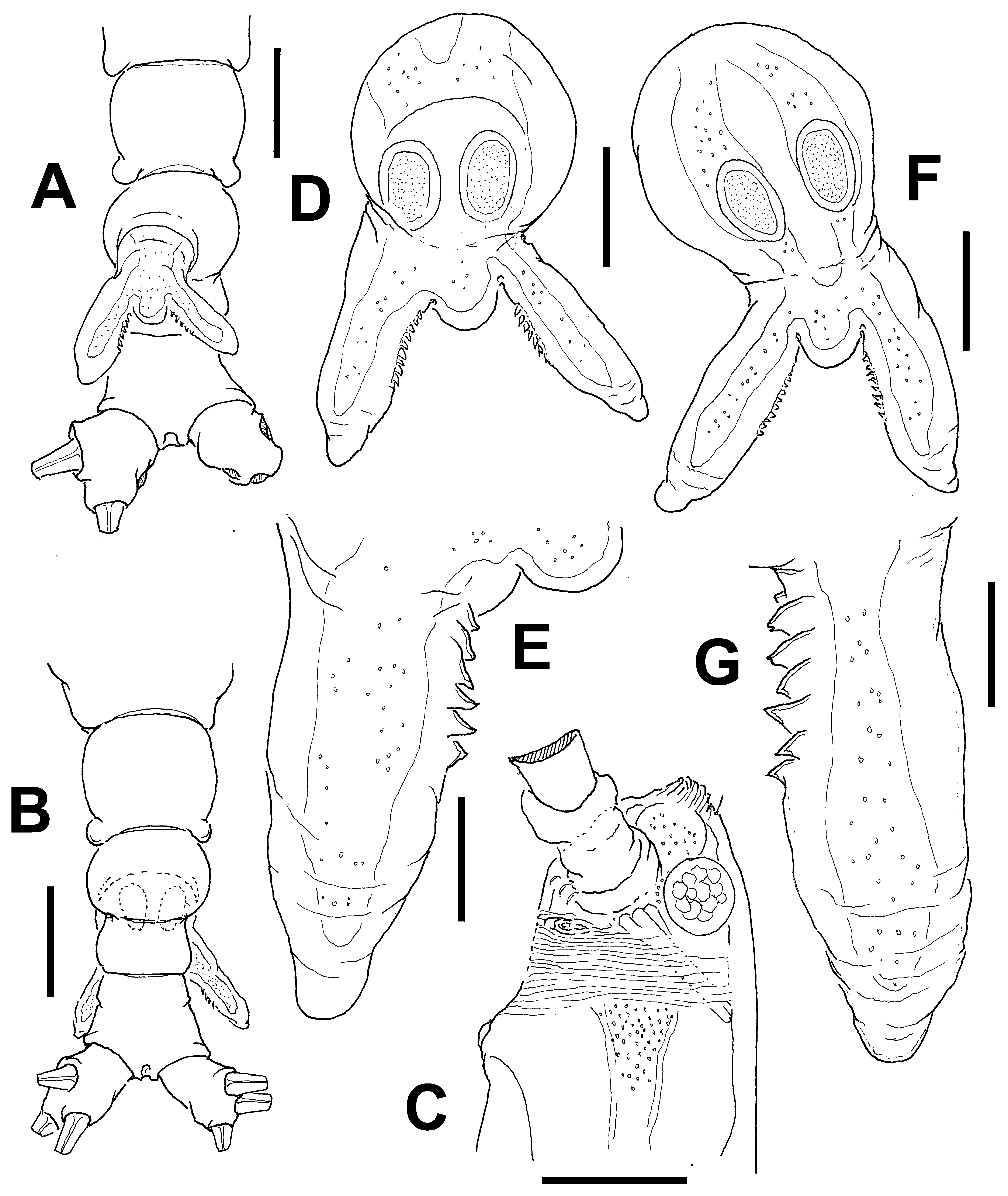
Fifth legs medially separate, bilobate, outer (exopodal) lobe elongate, distally truncate.
Outer lobe armed with three subdistal setae, innermost seta being shorter and narrower than other two ( Fig. 39 View FIGURE 39 A). Inner lobe thumb-shaped, noticeably shorter than outer lobe, unarmed, arising proximally from exopodal lobe, not reaching half its length ( Fig. 39 View FIGURE 39 A).
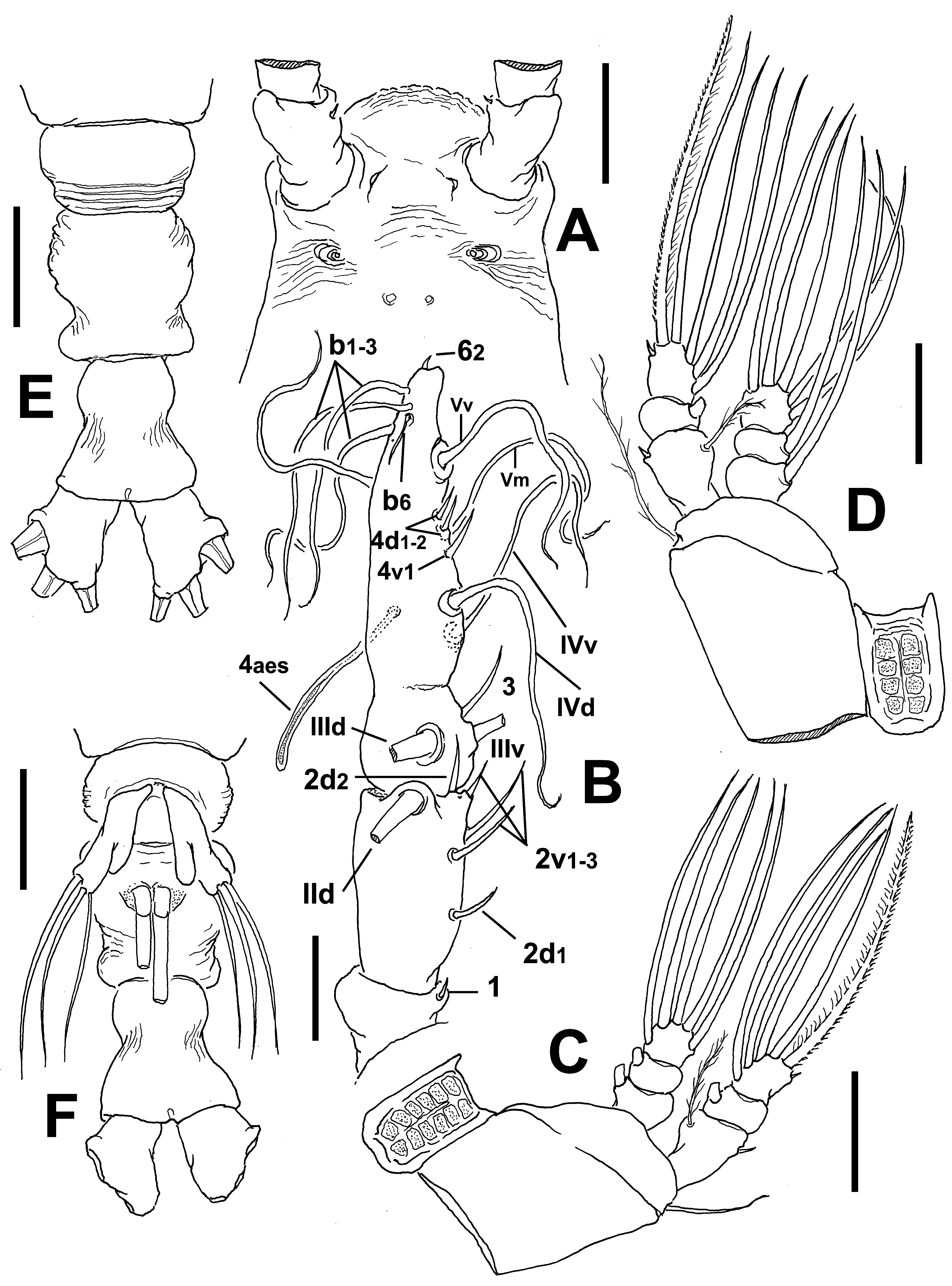
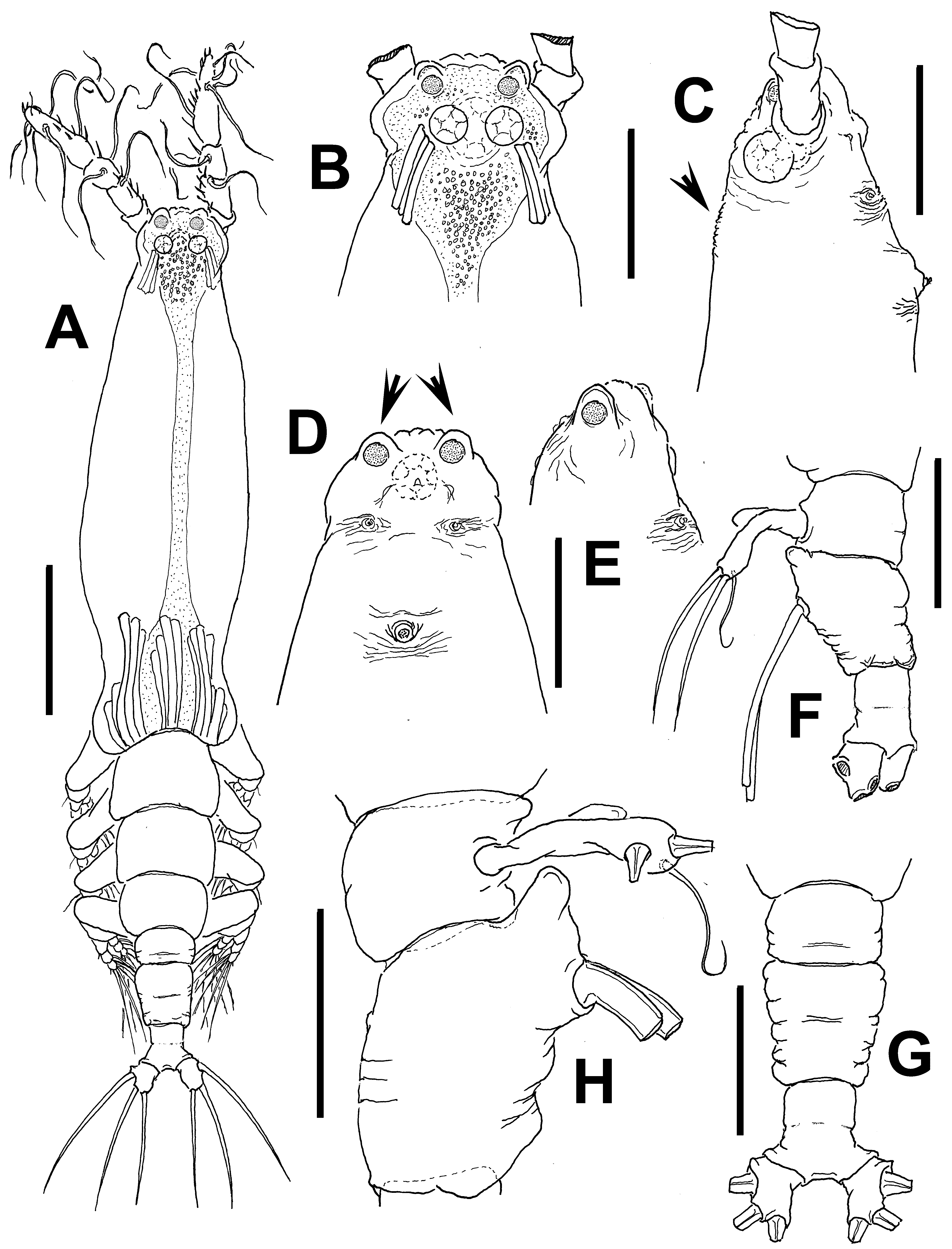
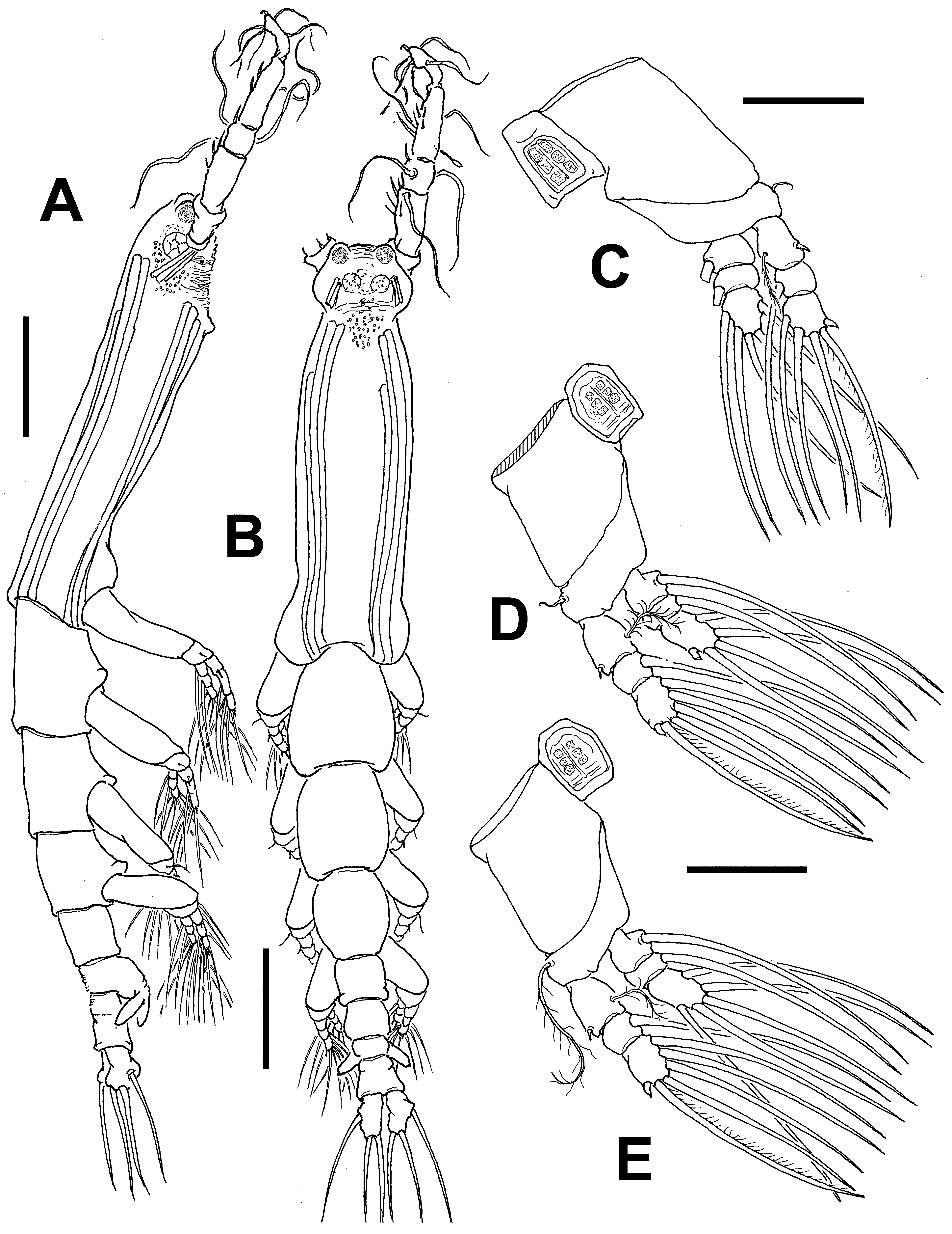
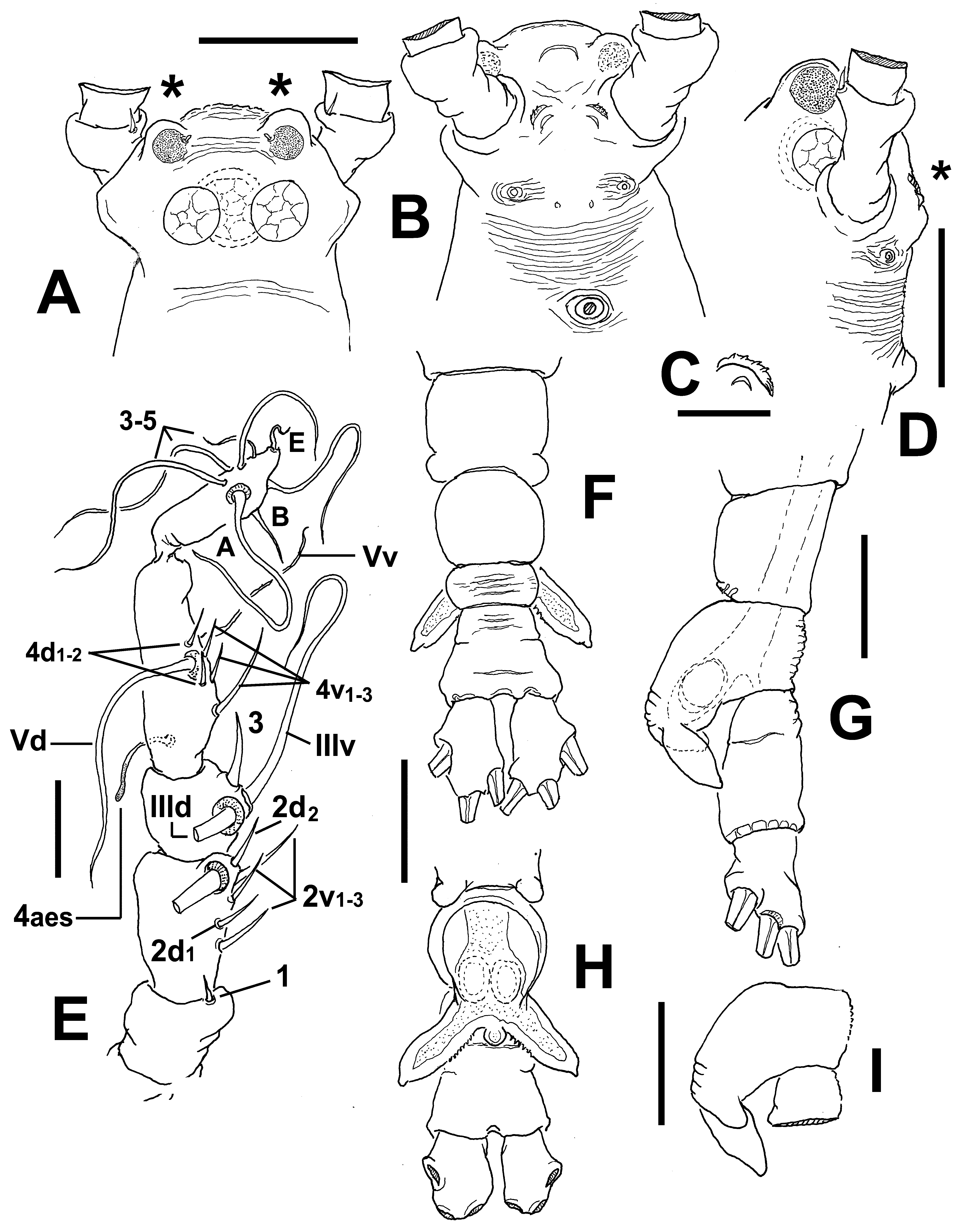
Description of male. Total body length 1.47 mm. Cephalothorax 0.68 mm long, representing 46% of total body length ( Fig. 40 View FIGURE 40 A, B). Midventral oral papilla moderately developed, located at 27% of cephalothorax length ( Figs 40 View FIGURE 40 B, 41D). Cephalic region protuberant bilaterally in dorsal and ventral views ( Fig. 41 View FIGURE 41 A, B). Cephalic frontal area as in female, with pattern of few transverse striations flanked by pair of rounded, lens-like anteriorly directed protuberances (asterisks in Figs. 41 View FIGURE 41 A); frontal sensilla absent. Pair of dorsal ocelli present, weakly developed; pigment cups medium-sized. Ocelli faintly pigmented. Forehead area moderately produced anteriorly. Ventral protuberant process as in female, but slightly smaller (asterisk in Fig. 41 View FIGURE 41 D). Other cuticular ventral processes including pair of crescent- shaped minute processes ( Fig. 41 View FIGURE 41 C) and nipple-like processes, the latter with adjacent wrinkles.
Fifth pedigerous somite with smooth dorsal surface ( Fig. 41 View FIGURE 41 G). Genital somite with striated dorsal surface. Genital complex of type II ( Suárez-Morales & McKinnon 2014), represented by pair of mammiliform, moderately divergent genital lappets with a few spinules on proximal inner margin ( Fig. 41 View FIGURE 41 F, H); lappets symmetrical and smooth, reaching to halfway of anal somite. Rounded, small protuberant medial process present at common basal joint of lappets ( Fig. 41 View FIGURE 41 H). Anal somite as in female, weakly constricted, with suture, about 1.5 times as long as preanal somite ( Fig. 41 View FIGURE 41 F). Caudal rami as long as anal somite, subrectangular, 1.6 times as long as wide ( Fig. 41 View FIGURE 41 F). Each ramus with three setae.
Antennulary length 0.27 mm. Antennules representing 30% of total body length, and 56% of cephalothorax length. Antennule 5-segmented, all segments separated, with segment 5 distal to geniculation ( Fig. 41 View FIGURE 41 E). Setal element 1 on first segment short, spiniform, as in female. Antennulary elements 2v 1-3, 2d1,2, and IId present on second segment. Setal elements IIId, IIIv, and 3 present on third segment; element 3 slender, weakly curved. Fourth segment with elements 4d 1-2, 4v 1– 3, element 4v 1 being longest of group. Fifth segment with 3 “b”-group setae, elements b1-3 long, distally unbranched, as in female. According to Huys et al. (2007) setal nomenclature of the distal segment, elements A, B, E and 3–5 present.
Pedigerous somites 2–4 noticeably elongate, together accounting for 38% of total body length in dorsal view. Coxae of each pair unarmed, joined by intercoxal sclerite which is slightly longer than wide and ornamented with spinulose patches, as in female. Legs 1–4 as in female except for naked outer margin of apical outermost seta on third exopodal segments of legs 1–4 ( Fig. 40 View FIGURE 40 C–E).
Type locality. Western Port Bay, Victoria, Australia ( 38°31.306’ S, 145°4.858’ E).
Etymology. From the Latin noun lenticula (= small lens); the name in apposition refers to the paired lensshaped structures on the cephalic region in both sexes of this species.
Diagnosis. Cymbasoma with cephalic area with two rounded, anteriorly projected lens-like processes flanking corrugate frontal area. Low medial cephalic protuberance on ventral surface between rounded processes and nipple-like processes. Genital double-somite with strongly developed antero-ventral protuberance, lateral margins corrugated. Anal somite almost as long as genital double-somite, with weak constriction and faint suture. Fifth leg with short inner lobe arising proximally, not reaching to half of inner margin of exopodal lobe; innermost exopodal seta shorter and thinner than the other two. Male with similar lens-like processes on cephalic region, anal somite with weak suture, genital complex of type II with mammiliform genital lappets and small medial rounded process at insertion of lappets.
Remarks. Male and female specimens were assigned to the same species on two of the criteria to match both sexes of the same species, their co-occurrence in the same sample/area (Western Port Bay, Victoria) and their sharing of peculiar, distinctive morphological features ( Grygier & Ohtsuka 2008; Suárez-Morales 2011, 2015). The male and female specimens of C. lenticula sp. nov. possess the peculiar lens-like cephalic processes, a low ventral protuberance on the cephalic region ( Figs. 38 View FIGURE 38 C, 41D), a constricted anal somite ( Figs. 38 View FIGURE 38 G, 41F, H), identical legs including ornamented intercoxal sclerites, and similar details of the antennulary armature, including a remarkably small element 1and unbranched “b” setae on the last segment. This species can be distinguished from its known congeners by a combination of characters including, 1) a short, well-defined female fifth leg inner lobe arising proximally and not reaching to half of the inner margin of the outer lobe; this structure of the fifth leg has not been observed in other species of the genus; 2) female fifth leg innermost exopodal seta shorter and thinner than the other two; 3) anal somite with a weak constriction; 4) two rounded lens-like processes flanking wrinkled frontal area in the cephalic area in both the male and the female; 5) strongly developed anteroventral process on the female genital double-somite. In the new species the fifth leg inner lobe is relatively shorter and the outer lobe longer ( Fig. 39 View FIGURE 39 A) than in C. constrictum ( Fig. 37 View FIGURE 37 F). The proportional length of the anal somite with respect to the genital double-somite is similar to that in C. striifrons ( Chang 2012: fig. 2A), but these two species differ in several other characters. In the new species the anal somite is constricted, a character absent in C. striifrons ; also, the fringe of striae present on the body of C. striifrons ( Chang 2012: fig. 1A) is absent in the C. lenticula sp. nov. The antennules are relatively longer in the new species (21% of total length) than they are in C. striifrons (16%) while the distal antennulary segment is remarkably short in the latter ( Chang 2012: fig. 2B), thus diverging from the pattern observed in the antennules of the new species ( Fig. 37 View FIGURE 37 B). Also, the fifth leg inner lobe is poorly developed in C. striifrons ( Chang 2012: fig. 2C) and short, but well-defined in the new species. Yet another species with a short fifth leg inner lobe arising proximally and not reaching to half of the outer lobe is C. rigidum Thompson, 1888 , from off the Nicobar Islands as reported by Sewell (1949). However, this species probably does not belong to Cymbasoma since Sewell’s (1949) illustrations show an additional, completely separated somite between the genital double- and the anal somites plus three caudal setae, an unlikely combination in the genus. It is probable that a small fourth caudal seta was overlooked by Sewell (1949). If this is confirmed, the Nicobar material would closely resemble Sars’ (1921) record and illustrations of a female C. rigidum , showing a deeply constricted anal somite and four caudal setae. Sewell’s specimens should be re-examined because they seem to differ from the original description by Thompson (1888) showing a non-constricted anal somite and three caudal setae. The male differs from the other males by the presence of the distinctive lens-like processes, the elongated pedigerous somites 2–4, three caudal setae, and genital lappets with an inner row of small spines. It shares with C. bullatum a similar genital complex but in C. bullatum the genital lappets are smooth and the medial rounded protuberance between the lappets is relatively larger than in the new species. It differs from the male of C. annulocolle in several subtle characters, including the lack of a fringe of striae, and the smaller, ball-shaped medial process at the insertion of genital lappets ( Fig. 38 View FIGURE 38 H), which diverges from the wide, rounded medial process in C. annulocolle ( Fig. 17 View FIGURE 17 D, F). The antennulary elements 1 and 3 and also all those of the 2v-d group on the second segment are clearly longer in C. annulocolle ( Fig. 15 View FIGURE 15 B) than in C. lenticula ( Fig. 41 View FIGURE 41 E).
No known copyright restrictions apply. See Agosti, D., Egloff, W., 2009. Taxonomic information exchange and copyright: the Plazi approach. BMC Research Notes 2009, 2:53 for further explanation.
|
Kingdom |
|
|
Phylum |
|
|
Class |
|
|
Order |
|
|
Family |
|
|
Genus |