
Cymbasoma apicale, Suárez-Morales, Eduardo & Mckinnon, David, 2016
|
publication ID |
https://doi.org/10.11646/zootaxa.4102.1.1 |
|
publication LSID |
lsid:zoobank.org:pub:9A7BA798-AA7C-4CAA-B42C-1E260CA573E4 |
|
DOI |
https://doi.org/10.5281/zenodo.6091307 |
|
persistent identifier |
https://treatment.plazi.org/id/03C4CA6D-D528-FF9A-FF12-579D95F92B63 |
|
treatment provided by |
Plazi |
|
scientific name |
Cymbasoma apicale |
| status |
sp. nov. |
Cymbasoma apicale sp. nov.
( Figs 34 View FIGURE 34 , 35 View FIGURE 35 )
Material examined. Holotype: adult female from near St Kilda, Port Phillip Bay, Victoria, Australia ( 37°53.466’ S, 144°55.687’ E; Station K of Kimmerer & McKinnon 1985), partially dissected, ethanol-preserved; dissected parts mounted on slide in glycerine, sealed with Entellan®. Date of collection: 23rd January 1984. Slides deposited in the collection of MTQ, Australia (cat. MTQ W34392).
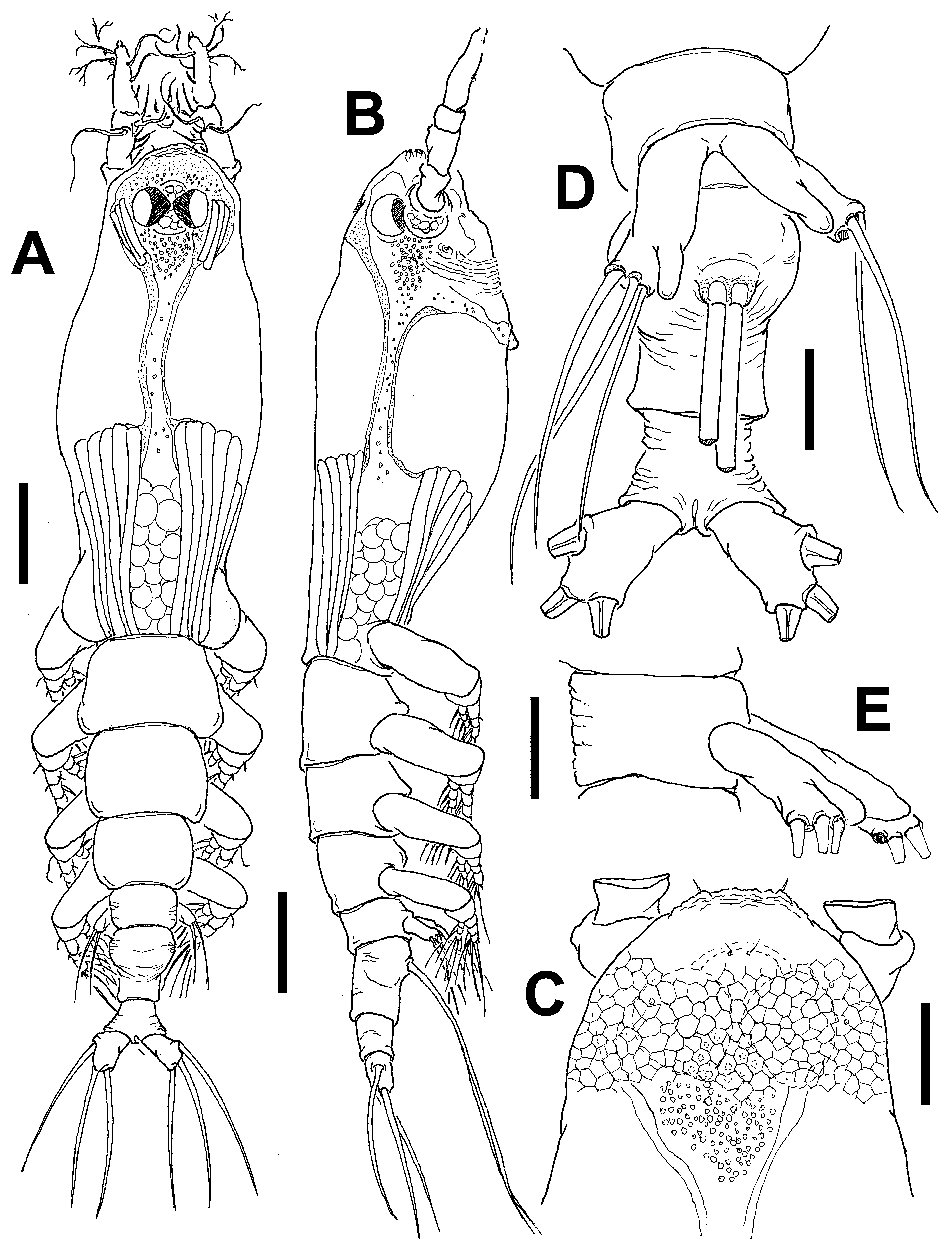
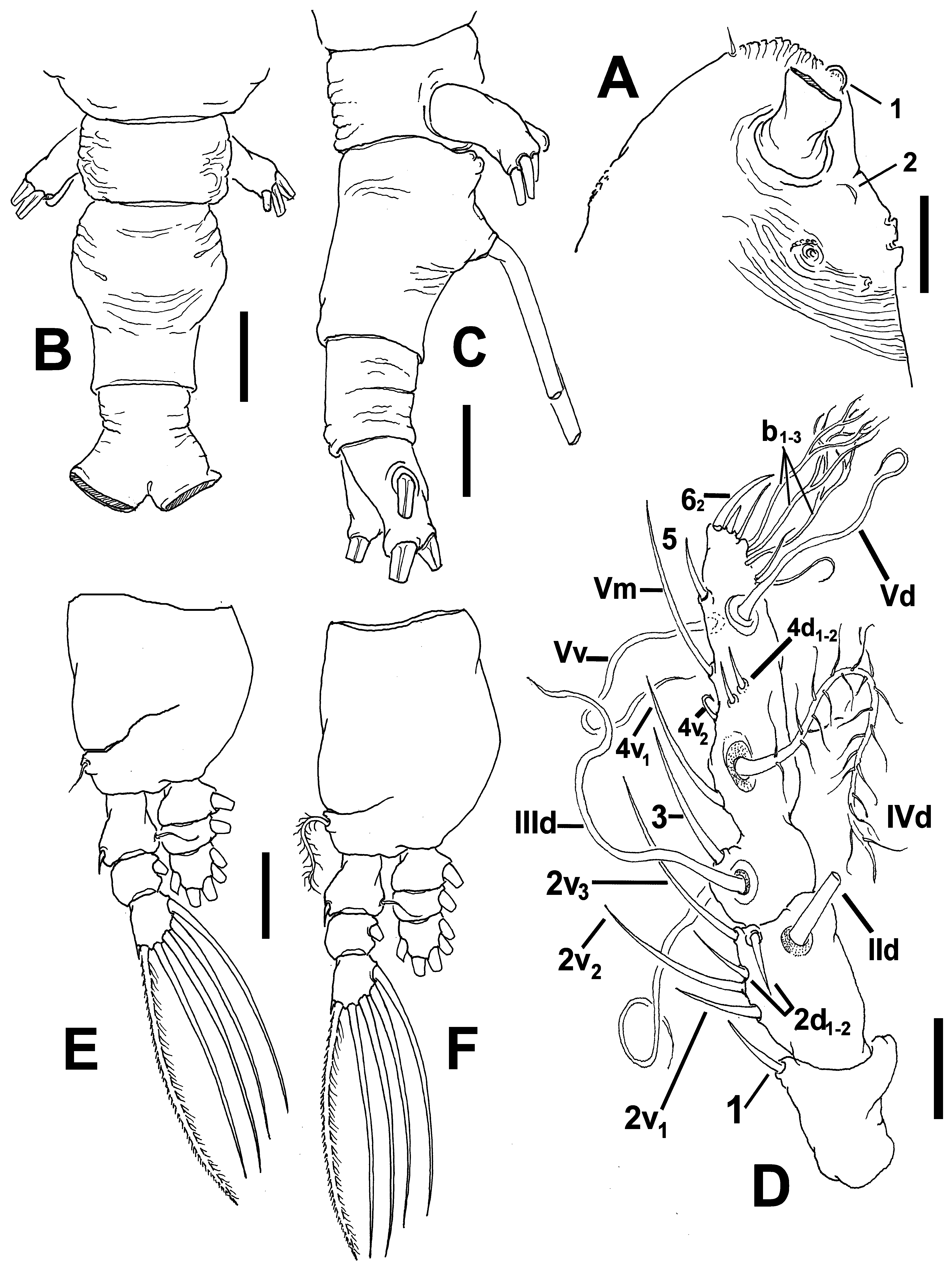
Description of adult female. Body robust, relatively wide in dorsal view ( Fig. 34 View FIGURE 34 A, B); body length of holotype female 1.82 mm. Cephalothorax approximately 0.9 mm long, representing about 50% of total body length. Midventral oral papilla moderately protuberant, located at 30% of cephalothorax length. Pair of relatively large ocelli present, pigment cups well developed, medially conjoined, intensely pigmented at inner half; ventral cup larger than lateral cups ( Fig. 34 View FIGURE 34 A). Cephalic area wide, with rounded, slightly produced "forehead”. Cephalic frontal area wide, ornamented with pattern of transverse striations ( Fig. 34 View FIGURE 34 C), pair of short sensilla present and single medial globose protuberance (marked as # 1 in Fig. 35 View FIGURE 35 A). Dorsal surface of cephalothorax smooth except for field of shallow reticulation overlying ocellar area ( Fig. 34 View FIGURE 34 C), ventral surface with field of transverse cuticular wrinkles at preoral area. Ventral surface also with 1) pair of symmetrical, crescent-shaped processes on anterior ventral surface located near bases of antennules, with no adjacent striae (marked as # 2 in Fig. 35 View FIGURE 35 A); 2) pair of nipple-like processes with concentric pattern of striae.
Urosome consisting of fifth pedigerous somite, genital double-somite and anal somite, together representing 17% of total body length. Relative lengths of urosomites (fifth pedigerous, genital double- and free anal somites) 25.7: 48.5: 25.8 = 100, respectively. Lateral margins of fifth pedigerous somite straight, with transverse wrinkles on dorsal and lateral surfaces. Genital double-somite longest of urosome, with ventral and dorsal surfaces ornamented with wrinkles ( Fig. 34 View FIGURE 34 D, E, 35B, C), anterior half swollen, posterior half with straight margins. Ovigerous spines paired, basally separated, slender, straight at their bases ( Fig. 34 View FIGURE 34 D), relatively short ( 0.63 mm), approximately 33% of total body length. Anal somite without clear medial constriction but with deep transverse wrinkles along lateral margins, reaching ventral surface ( Fig. 35 View FIGURE 35 B). Caudal ramus subrectangular, about 1.6 times as long as wide, armed with three caudal setae.
Antennule length 0.38 mm, representing about 21% of total body length and 36% of cephalothorax length, 4- segmented. Relative length of distal antennulary segment 46.5%. In terms of pattern described by Grygier & Ohtsuka (1995) for female monstrilloid antennulary armature, moderately long, slender element 1 present on first segment; elements on second segment: 2d1-2, 2v 1-3, and IId. Elements 2v 1-2 remarkably long, the latter reaching proximal 1/3 of fourth antennulary segment ( Fig. 35 View FIGURE 35 D). Third segment with element 3 being long, spiniform, elements IIId and IIIv setiform, of normal aspect. Segment 4 bearing short elements 4d1,2 and 4 v1-2, the former long, the latter short, hook-like; element 4v 3 not observed. Setae IVd, Vd, Vv, Vm present. Element 5 present, straight, spiniform. Subterminal elements b1-3 present, distally branched, elements b6 and b5 slender. Elements 61 and 62 present, the latter being remarkably long ( Fig. 35 View FIGURE 35 D).
Incorporated first pedigerous somite and succeeding three free pedigerous somites each bearing a pair of biramous legs. Pedigerous somites 2–4, together accounting for 27% of total body length. Intercoxal sclerites of legs 1–4 subrectangular, surface and posterior margin smooth. Bases of legs 1–4 with hair-like lateral seta ( Fig. 35 View FIGURE 35 E, F); on leg 3, this seta about three times longer, thicker than those on other legs ( Fig. 35 View FIGURE 35 F). Endopods and exopods of legs 1–4 triarticulated. Ramal setae all biserially plumose except spiniform outer seta on exopodal segments 1 and 3, and inner seta of first exopodal segment, these latter being short, slender. Outermost distal spines on third exopodal segment of legs 1–4 short, 0.2 times as long as segment. Outermost apical exopodal setae of legs 1–4 with inner margin setulose, outer margin spinulose.
Armature formula of legs 1–4:
Fifth legs medially conjoined, bilobate, outer (exopodal) lobe subrectangular, distally truncate. Outer lobe armed with three subdistal setae ( Fig. 34 View FIGURE 34 D). Inner lobe thumb-shaped, noticeably shorter than outer lobe, unarmed, arising subdistally from exopodal lobe and reaching its distal margin ( Fig. 34 View FIGURE 34 E, 35C).
Male: unknown.
Type locality. Port Phillip Bay, Victoria, Australia ( 37°53.466’ S, 144°55.687’ E).
Etymology. The specific name, an adjective derived from the Latin apicalis (= apical), refers to the position of the inner lobe of the female fifth leg, which arises very near the apex of the outer lobe.
Diagnosis. Cymbasoma with relatively short cephalothorax, representing 50% of total body length, third antennulary segment representing 42% of antennule length, with elements 2v 2-3 and 62 remarkably long. Cephalic region with pair of crescent-like processes on ventral surface and small medial ball-like protuberance in same position. Eyes well-developed, intensely pigmented at inner half. Cephalothorax with dorsal field of reticulation covering part of cephalic region. Genital double-somite longest of urosome, with rounded proximal half, distal half with straight margins. Anal somite with corrugate lateral margins. Fifth leg with elongate, outer lobe with three distal setae, inner lobe distinctly arising subdistally, narrow, short, reaching distal end of outer lobe, unarmed. Ovigerous spines short, 33% of total body length.
Remarks. This species has a fifth leg with a short, thumb-like inner lobe arising from the distal half of the outer exopodal lobe and reaching its distal end, thus diverging from the pattern of other Australian species of Cymbasoma with a long, digitiform lobe arising basally, like C. annulocolle , C. bidentatum and C. markhasevae . It most closely resembles C. davisi from Florida ( Suárez-Morales & Pilz 2008) which shares with the new species the same type of fifth leg, a ventral field of transverse wrinkles on the preoral area, and a similar body shape and proportions except for a narrower cephalic area (both dorsally and laterally) in the new species. The preoral area of C. davisi is strongly concave even in different specimens ( Suárez-Morales & Pilz 2008), whereas it is ventrally produced in the C. apicale sp. nov. ( Fig. 34 View FIGURE 34 B). Also, in the new species the anterior dorsal part of the cephalic area has a reticulated field ( Fig. 34 View FIGURE 34 C), a character that is absent in C. davisi ( Suárez-Morales & Pilz 2008: fig. 1A). The antennulary setae of group “b” ( sensu Grygier & Ohtsuka 1995) are simple in C. davisi ( Suárez-Morales & Pilz 2008: fig. 1C) but distally branched in the new species ( Fig. 34 View FIGURE 34 A). Also, in the new species the eyes are strongly pigmented, thus differing from the unpigmented eyes of C. davisi ( Suárez-Morales & Pilz 2008: fig. 4C–E). The shape of the genital double-somite is also different in these two species; its proximal half is subquadrate, with straight lateral margins in C. davisi ( Suárez-Morales & Pilz 2008: fig. 3B) and rounded in the new species ( Figs 34 View FIGURE 34 D, 35B). The three exopodal setae of the fifth leg are all distal in the new species whereas only the middle seta is apical and the other two are subapical in C. davisi ( Suárez-Morales & Pilz 2008: fig. 3A). Cymbasoma apicale sp. nov. has some affinities with the European C. germanicum , as redescribed by Suárez-Morales (2006). Both species have the same body shape and proportions, similar shape and ornamentation of the genital double-somite, and a constricted anal somite. They differ in the length of the inner lobe of the fifth leg, which is longer in C. germanicum ( Suárez-Morales 2006: fig.4b), arising from the proximal 1/3 of the outer lobe. The genital double-somite of C. germanicum has a plate-like lateral process on the posterior margin ( Suárez-Morales 2006: fig. 2b); this process is absent in the new species. The anal somite of C. germanicum lacks the deep wrinkles ( Suárez-Morales 2006: fig. 4d) shown by the new species on the lateral surface ( Figs 34 View FIGURE 34 D, 35B). Also, the eyes are not pigmented in C. germanicum ( Suárez-Morales 2006) and they are strongly pigmented in the new species. It differs from C. spinapex Isaac, 1974 in the body shape, with a longer, more slender cephalothorax ( Isaac 1974) vs. a more shorter, relatively robust cephalothorax in the new species; also C. spinapex has a fifth leg outer lobe with the middle seta being shortest, vs. all setae being equally long in the new species. Cymbasoma tropicum has a similar fifth leg, with an inner lobe arising from the distal half of the outer lobe ( Martin Thompson & Easterson 1983: fig. 1j), but the inner lobe is clearly narrower in the new species ( Fig. 34 View FIGURE 34 D). Finally, the anal somite is smooth and the cephalothorax is long and slender in C. tropicum (MartinThompson & Easterson 1983: fig. 1a, b, d), thus contrasting with the relatively short, robust cephalothorax of the new species.
No known copyright restrictions apply. See Agosti, D., Egloff, W., 2009. Taxonomic information exchange and copyright: the Plazi approach. BMC Research Notes 2009, 2:53 for further explanation.
|
Kingdom |
|
|
Phylum |
|
|
Class |
|
|
Order |
|
|
Family |
|
|
Genus |