
Cymbasoma bidentatum, Suárez-Morales, Eduardo & Mckinnon, David, 2016
|
publication ID |
https://doi.org/10.11646/zootaxa.4102.1.1 |
|
publication LSID |
lsid:zoobank.org:pub:9A7BA798-AA7C-4CAA-B42C-1E260CA573E4 |
|
DOI |
https://doi.org/10.5281/zenodo.6091297 |
|
persistent identifier |
https://treatment.plazi.org/id/03C4CA6D-D53D-FF84-FF12-528E938E2A0B |
|
treatment provided by |
Plazi |
|
scientific name |
Cymbasoma bidentatum |
| status |
sp. nov. |
Cymbasoma bidentatum sp. nov.
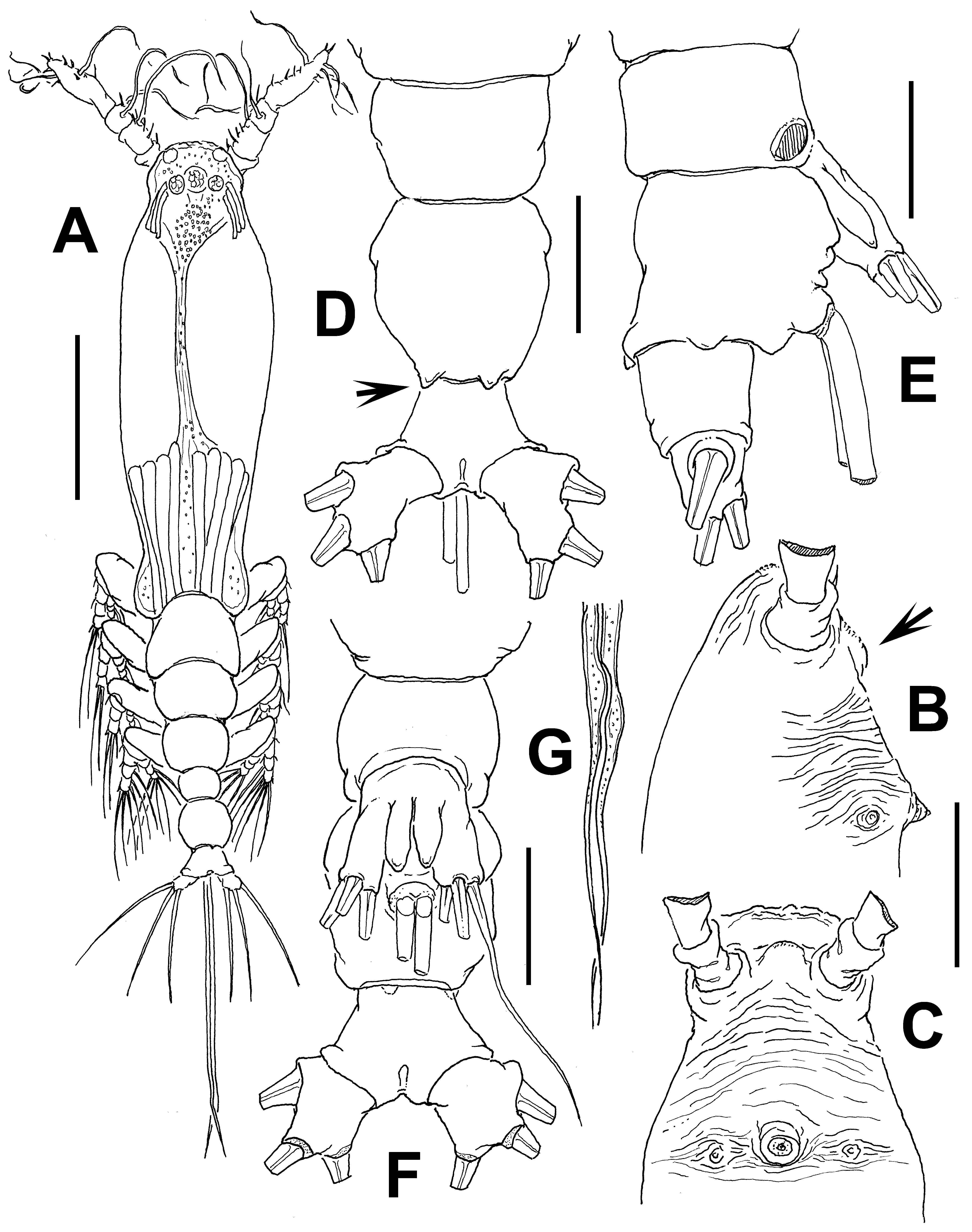
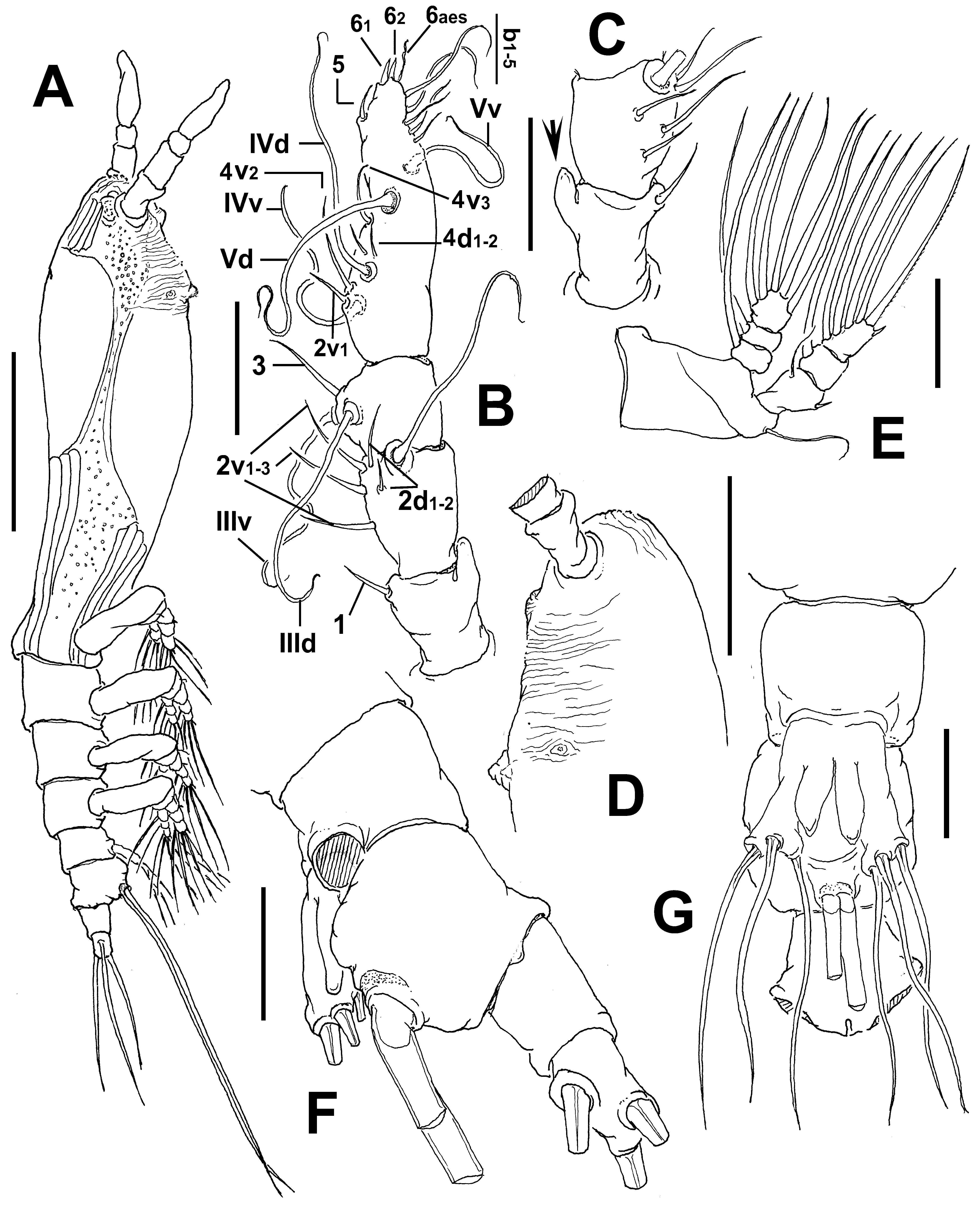
( Figs 20 View FIGURE 20 , 21 View FIGURE 21 )
Material examined. Holotype: adult female from Warneet inlet, Western Port Bay, Victoria, Australia ( 38°27.432’ S, 145°17.951’ E), ethanol-preserved; dissected parts mounted on two slides in glycerine, sealed with Entellan®. Date of collection: 20th March 1985. Paratype adult female from same locality and date, partially dissected, mounted on two slides in glycerine, sealed with Entellan®. Slides deposited in the collection of MTQ, Australia (cat. MTQ W34382, MTQ W34383, respectively).
Description of adult female. Body moderately robust; body length of holotype female 0.93 mm. Cephalothorax approximately 0.55 mm long, representing nearly 61% of total body length. Midventral oral papilla protuberant, located at 25% of cephalothorax length. Pair of relatively small ocelli present, pigment cups weakly developed, separated by one eye diameter, lightly pigmented; ventral cup larger than lateral cups ( Fig. 20 View FIGURE 20 A). Cephalic region not laterally protuberant. Frontal area ornamented with pattern of shallow longitudinal striations ( Figs 20 View FIGURE 20 B, C) no frontal sensilla were observed. Dorsal surface of cephalothorax smooth, ventral surface ornamented with transverse striae between antennule bases and oral area. Additional ornamentation of ventral surface including: 1) low medial protuberance on ventral surface posterior to antennule bases, ornamented with striae (arrowed in Fig. 20 View FIGURE 20 B); 2) pair of symmetrical, nipple-like processes on anterior ventral surface located at both sides of oral papilla.
Urosome consisting of fifth pedigerous somite, genital double-somite and anal somite, together representing 14% of total body length. Relative lengths of urosomites (fifth pedigerous, genital double and free anal somites) as: 31: 45.9: 23.1 = 100, respectively ( Fig. 20 View FIGURE 20 D, E). Lateral margins of fifth pedigerous somite moderately expanded. Genital double-somite longest of urosome, with smooth dorsal surface, anteroventral surface corrugated anterior to insertion of ovigerous spines ( Fig. 20 View FIGURE 20 E). Anterior half of double-somite expanded, with moderately developed lateral processes and paired subtriangular protuberances on middle posterodorsal margin (arrowed in Fig. 20 View FIGURE 20 D). Ovigerous spines paired, separated at base, relatively short, 50% of total body length ( Fig. 20 View FIGURE 20 A). Spines slender, straight at their base and along shaft, with weak distal expansions and tapering apically ( Fig. 20 View FIGURE 20 G), both spines equally long; spines 0.48 mm long ( Fig. 20 View FIGURE 20 A). Anal somite without medial constriction. Caudal ramus subquadrate, about as long as wide, armed with three caudal setae ( Fig. 20 View FIGURE 20 D, E, G).
Antennule length 0.21 mm, representing about 22% of total body length and 35% of cephalothorax length, 4- segmented, segments 3 and 4 separated. First segment with thumb-like protuberance on outer margin (arrowed in Fig. 21 View FIGURE 21 C). Relative length of distal antennulary segment 46.3%. In terms of pattern described by Grygier & Ohtsuka (1995) for female monstrilloid antennulary armature, short, spiniform element 1 present on first segment; elements on second segment: 2d1-2, 2v 1-3, and IId. Third segment with slender element 3, setae IIId and IIIv of normal aspect. Segment 4 bearing elements 4d1,2, 4v 1-2, element 4v 2 being longest of group. Setae IVd, IVv, Vd, Vv, and element 4aes present. Element 5 short, slender. Subterminal elements b1-5 present, unbranched, elements 61-2 and 6 aes present ( Fig. 21 View FIGURE 21 B).
Incorporated first pedigerous somite and succeeding three free pedigerous somites each bearing a pair of biramous legs. Pedigerous somites 2–4, together accounting for 23% of total body length. Legs 1–4 slightly increasing in size posteriorly. Intercoxal sclerites of legs 1–4 subrectangular, surface and posterior margin smooth. Bases of legs articulating with large, rectangular coxae along oblique line; with hair-like lateral seta; on leg 3, this seta about 3.5 times longer, thicker than those on other legs ( Fig. 21 View FIGURE 21 E). Endopods and exopods of legs 1–4 triarticulated. Ramal setae all biserially plumose except spiniform outer seta on exopodal segments 1 and 3, and inner seta of first exopodal segment, these latter being short, slender. Outermost distal spines on third exopodal segment of legs 1–4 short, 0.2 times as long as segment. Outermost apical exopodal setae of legs 1–4 with inner margin naked, outer margin spinulose.
Armature formula of legs 1–4:
Fifth legs medially conjoined, bilobate, outer (exopodal) lobe cylindrical, distally blunt. Outer lobe armed with three equally long apical setae ( Fig. 21 View FIGURE 21 G). Inner lobe digitiform, shorter than outer lobe, reaching about ¾ the length of exopodal lobe, unarmed ( Fig. 21 View FIGURE 21 F).
Male: unknown.
Type locality. Warneet inlet, Western Port Bay, Victoria, Australia ( 38°27.432’S, 145°17.951’E).
Etymology. The species name, an adjective derived from the Latin dentatum (= with teeth) with the addition of the prefix bi - (=two), refers to the two tooth-like processes on the anterior half and along the posterior margin of the female genital double-somite.
Diagnosis. Cymbasoma with relatively robust cephalothorax, representing 61% of total body length, third antennulary segment representing more than 46% of antennule length, with low medial process on ventral surface of cephalic region. Cephalothorax with smooth dorsal surface. First antennulary segment with outer thumb-like process. Genital double-somite with lateral processes on proximal half and pair of tooth-like processes on posterior margin. Somite with corrugated anteroventral surface. Anal somite without medial constriction. Fifth leg with elongate, thumb-like outer lobe with three subdistal setae with equally long setae, inner lobe digitiform, smaller than outer lobe, unarmed.
Remarks. This species has affinities with other Cymbasoma species with a fifth leg bearing an outer lobe armed with three subequally long setae and an elongate, digitiform inner lobe arising basally. These include C. rigidum from which the new species differs in the length of the innermost seta of the fifth leg outer lobe, which is clearly the shortest and thinnest in species of this group (see Bourne1890, non Sars, 1921). The new species also resembles C. germanicum ( Timm, 1893) ( Suárez-Morales 2006) , C. gracile ( cf. Gurney 1927), and C. davisi Suárez-Morales & Pilz, 2008 ( Suárez-Morales & Pilz 2008) . The fifth leg pattern is most similar to that of C. gracile ( cf. Gurney 1927) but C. bidentatum sp. nov. differs in the body shape and proportions. In C. gracile the cephalothorax is long, slender, representing 67% of the total body length, whereas it is 61% in the new species which is clearly more robust. Also, in C. gracile the antennule has segments 3–4 fused ( Gurney 1927: fig. C) and they are clearly separated in the new species. The fifth leg of C. bidentatum sp. nov. is similar to that of C. germanicum ( cf. Suárez-Morales 2006: fig. 4b), but these species differ in the shape of the genital somite, with an expanded, rounded proximal half and a constricted posterior half in C. germanicum ( cf. Suárez-Morales 2006: fig. 4d) and a uniformly globose shape in the new species. The new species has a low ventral wrinkled process on the cephalic region (arrowed in Fig. 20 View FIGURE 20 B) which is absent in C. germanicum , with a smooth surface in the same area ( Suárez-Morales 2006: fig. 1b). In C. davisi the fifth leg inner lobe is remarkably narrow, slender and arises medially from the inner margin of the outer lobe and the innermost seta is shorter than the two outer setae (Suárez- Morales & Pilz 2008: fig. 3A), thus diverging from the pattern observed in the new species. Also, the genital double-somite is different in these species; in C. davisi the anterior half is expanded, with straight margins and the posterior half is narrower but has also straight margins ( Suárez-Morales & Pilz 2008: fig. 3A, B) vs. a rounded, globose genital double-somite in the new species. Overall, the most distinctive characters of the new species include a thumb-like protuberance on the distal outer margin of the first antennulary segment (arrowed in Fig. 21 View FIGURE 21 C) but most importantly, the presence of two anterior lateral processes and two spiniform processes on the posterior margin of the genital double-somite and a corrugate ventral surface of the same somite, visible in lateral view. This combination of characters is absent in C. gracile ( cf. Gurney 1927), C. germanicum ( cf. Suárez-Morales 2006), C. davisi ( cf. Suárez-Morales & Pilz 2008), and other species of the genus. It can easily be distinguished from the Australian C. annulocolle and C. markhasevae in the lack of a fringe of striae covering part of the cephalothorax.
No known copyright restrictions apply. See Agosti, D., Egloff, W., 2009. Taxonomic information exchange and copyright: the Plazi approach. BMC Research Notes 2009, 2:53 for further explanation.
|
Kingdom |
|
|
Phylum |
|
|
Class |
|
|
Order |
|
|
Family |
|
|
Genus |