
Cymbasoma agoense Sekiguchi, 1982
|
publication ID |
https://doi.org/10.11646/zootaxa.4102.1.1 |
|
publication LSID |
lsid:zoobank.org:pub:9A7BA798-AA7C-4CAA-B42C-1E260CA573E4 |
|
DOI |
https://doi.org/10.5281/zenodo.6091341 |
|
persistent identifier |
https://treatment.plazi.org/id/03C4CA6D-D540-FFC2-FF12-505E92592E5F |
|
treatment provided by |
Plazi |
|
scientific name |
Cymbasoma agoense Sekiguchi, 1982 |
| status |
|
Cymbasoma agoense Sekiguchi, 1982
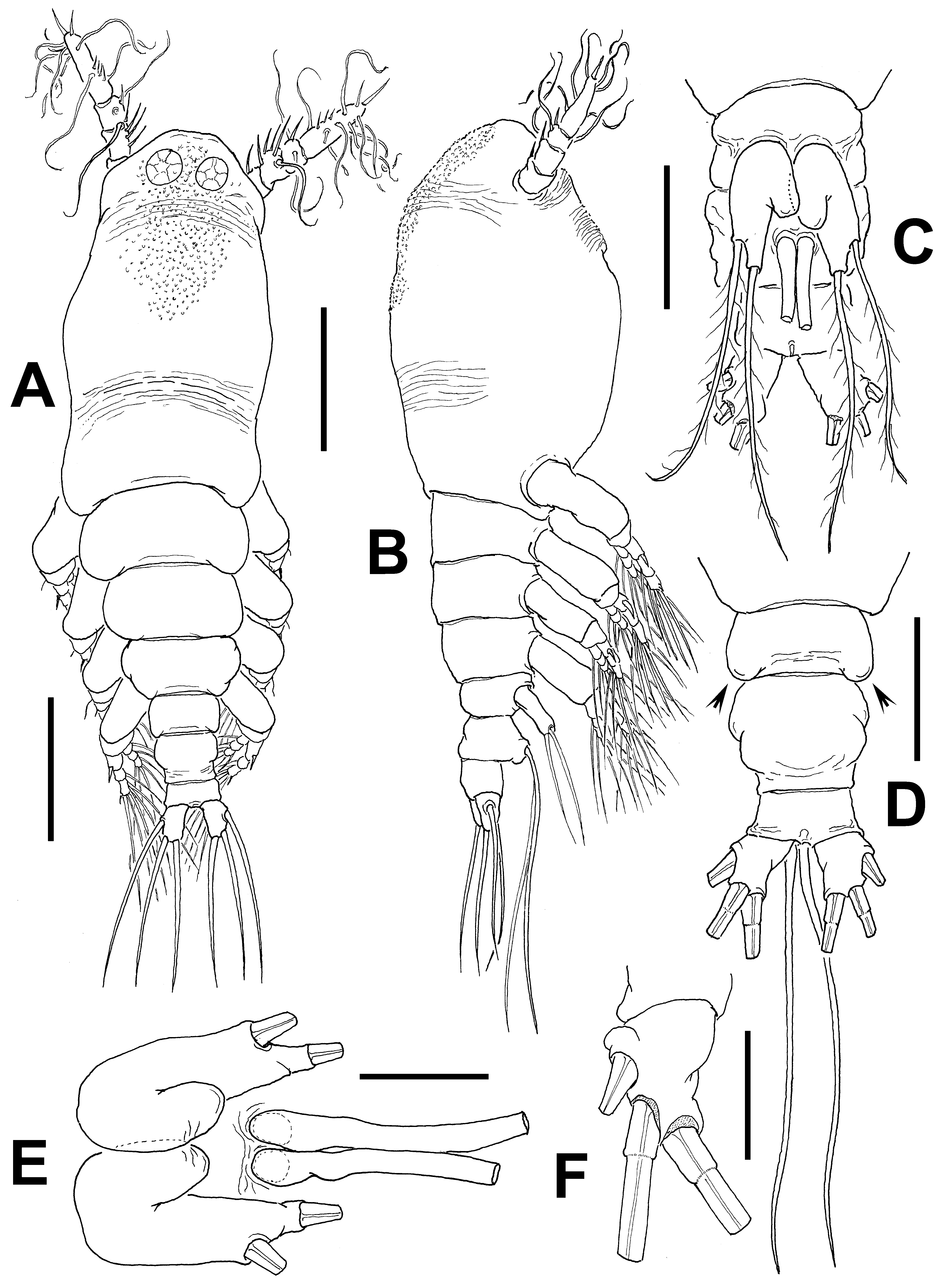
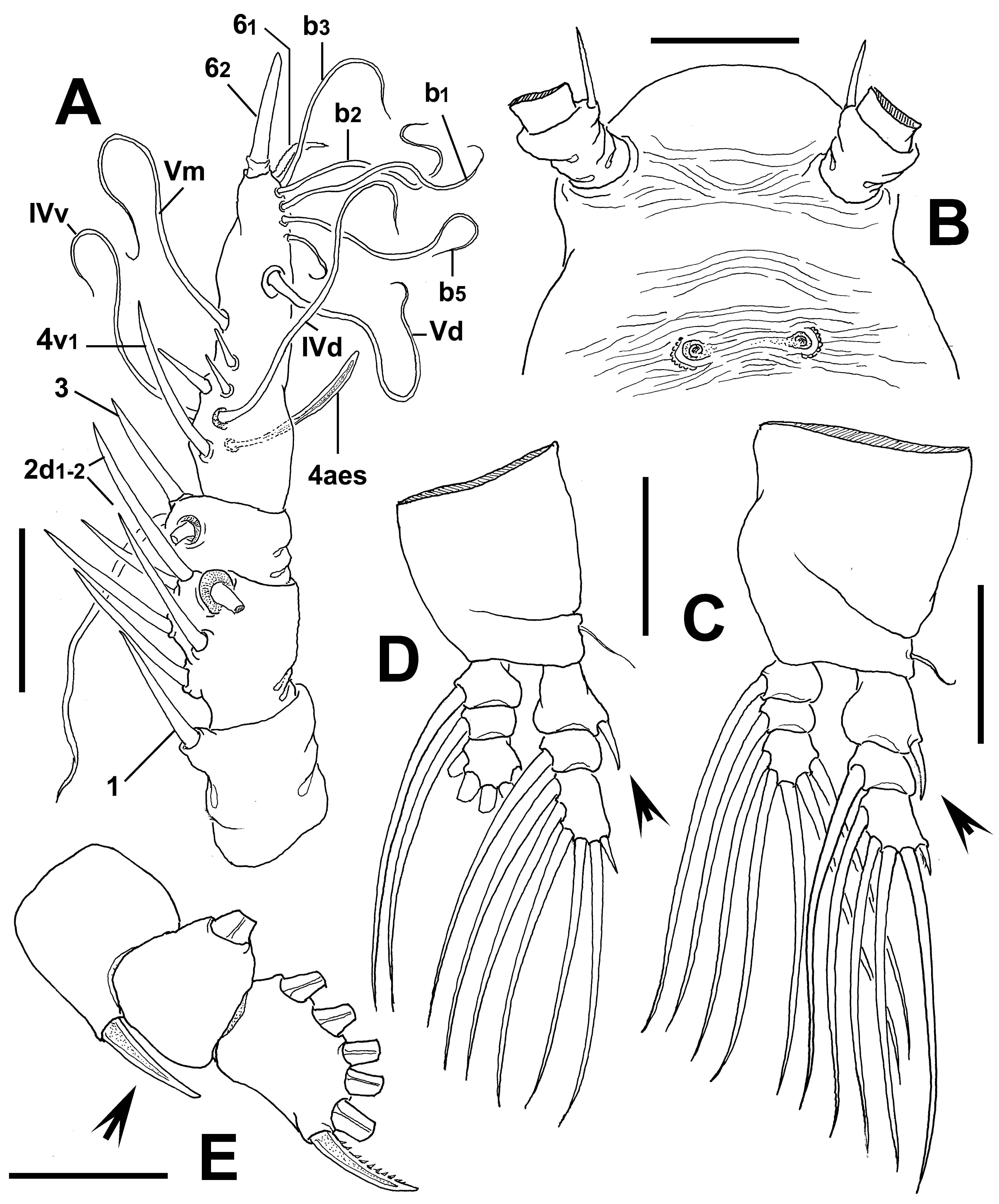
( Figs 56 View FIGURE 56 , 57 View FIGURE 57 )
Material examined. Adult female from Davies Reef, Queensland, Australia ( 19°7.340’ S, 146°53.024’ E), partially dissected, ethanol-preserved; dissected parts mounted on slide in glycerine, sealed with Entellan®. Date of collection: 12th December 1989. Slide deposited in the collection of MTQ, Australia (cat. MTQ W34400).
Description of adult female. Body noticeably robust, relatively wide in dorsal view, globose in lateral view; body length of holotype female 0.97 mm. Cephalothorax approximately 0.52 mm long, representing 53% of total body length. Midventral oral papilla not protuberant, located at 30% of cephalothorax length. Pair of relatively large ocelli present, pigment cups medially conjoined, weakly pigmented; ventral cup as large as lateral cups ( Fig. 56 View FIGURE 56 A). Cephalic area wide, with moderately produced "forehead”. Cephalic frontal area wide. Dorsal surface of cephalothorax smooth except for large field of minute wart-like cuticular processes overlying ocelli area and stretching to medial dorsal surface; wrinkled areas posterior to eye region and in distal 1/3 of cephalothorax ( Fig. 56 View FIGURE 56 A, B). Ventral surface with field of transverse cuticular wrinkles between antennulary bases and preoral area. Ventral surface also with pair of nipple-like processes connected medially by transverse striae ( Fig. 57 View FIGURE 57 B).
Urosome consisting of fifth pedigerous somite, genital double-somite and anal somite, together representing 16% of total body length. Relative lengths of urosomites (fifth pedigerous, genital double- and free anal somites) 30.9: 47.3: 21.8 = 100, respectively ( Figs. 56 View FIGURE 56 D). Lateral margins of fifth pedigerous somite straight, with few transverse wrinkles on dorsal surfaces and postero-lateral corners produced into rounded protuberances (arrowed in Fig. 56 View FIGURE 56 D). Genital double-somite longest of urosome, with dorsal surface ornamented with few wrinkles ( Fig. 56 View FIGURE 56 D); anterior half swollen, posterior half with straight margins. Ovigerous spines paired, basally separated, slender, straight at their bases ( Fig. 56 View FIGURE 56 C), relatively short ( 0.41 mm), 43% of total body length. Anal somite short, without medial constriction. Caudal ramus subquadrate, about as long as wide, armed with three caudal setae, distal two proximally thickened ( Fig. 56 View FIGURE 56 D, F).
Antennule length 0.21 mm, representing about 22% of total body length and 41% of cephalothorax length; 4- segmented. Relative length of distal antennulary segment 48.7%. Remarkably long, spiniform element 1 present on first segment; elements on second segment 2d1-2, 2v 1-3, all these being noticeably strong and long, plus setiform element IId. Third segment with element 3 strong, stout, spiniform; elements IIId and IIIv setiform, of normal aspect. Segment 4 bearing elements 4d1,2, 4v 1-2, element 4v 3 not observed, element 4v 1 extremely long; setae IVd, IVv, Vd, Vm, and 4aes present. Element 5 absent. Subterminal elements b1-5 present, unbranched, apical surface with extremely long, stout element 62 ( Fig. 57 View FIGURE 57 A).
Incorporated first pedigerous somite and succeeding three free pedigerous somites each bearing a pair of biramous legs. Pedigerous somites 2–4, together accounting for 31% of total body length. Bases of legs 1–4 with hair-like lateral seta ( Fig. 57 View FIGURE 57 C, D); on leg 3, this seta about three times longer, thicker than those on other legs. Endopods and exopods of legs 1–4 triarticulated. Ramal setae all biserially plumose except spiniform outer seta on exopodal segments 1 and 3, and inner seta of first exopodal segment, these latter being short, slender. Outer distal spines on first exopodal segment of legs 1–4 noticeably long, reaching beyond distal margin of second exopodal segment (arrowed in Fig. 57 View FIGURE 57 C, D); outermost distal spine of third exopodal segment 0.6 times as long as segment, with inner margin spinulose ( Fig. 57 View FIGURE 57 E). Outermost apical exopodal setae of legs 1–4 with inner and outer margins naked.
Armature formula of legs 1–4: Fifth legs medially separate, bilobate, outer lobe subrectangular, tapering distally, armed with two setae, one apical, one subapical ( Fig. 56 View FIGURE 56 C). Inner lobe represented by large, wide thumb-shaped protuberance arising from proximal margin of exopodal lobe, unarmed ( Fig. 56 View FIGURE 56 E).
Male: unknown.
Type locality. Ago Bay, Japan ( Sekiguchi 1982).
New locality. Davies Reef, Queensland, Australia.
Diagnosis. Cymbasoma with body noticeably robust, short, relatively wide in dorsal view, globose in lateral view. Dorsal surface of cephalothorax with field of minute wart-like cuticular processes overlying ocelli area and stretching to medial dorsal surface. Antennules representing about 22% of total body length and 41% of cephalothorax length; second antennulary segment with noticeably long, strong spiniform elements 2d1-2 and 2v 1- 3. Fifth pedigerous somite with postero-lateral corners produced into rounded protuberances. Genital doublesomite subquadrate, with anterior half swollen in dorsal view. Legs 1–4 with enlarged spines on exopodal segments 1 and 3. Fifth leg with outer lobe bearing two setae; inner lobe large, wide, arising from proximal half of exopodal lobe, unarmed. Terminal caudal setae proximally thickened. Ovigerous spines representing 43% of total body length.
Remarks. This species clearly belongs to the C. agoense species-group, now comprising C. agoense , and the Australian C. dakini , C. lourdesae , and C. tharawalorum , all of which display two setae on the fifth leg outer lobe. We attribute our specimen to C. agoense , the first described species of Cymbasoma with this peculiar character ( Sekiguchi 1982) and provide additional data not included in the original description, like the details of the antennulary armature; this is also relevant because it is the most representative species of the agoense species group. This species can readily be distinguished from other members of this group but also from most other females of Cymbasoma by the characteristic globose shape of the body, particularly when observed in lateral view. Within Cymbasoma this characteristic has so far been observed only in C. agoense , previously known exclusively from Japan. A similar strongly globose cephalothorax has been observed only among species of Monstrilla like M. hamatapex and M. obesa Isaac, 1975 . The Australian specimen was identified as C. agoense because it shares with the type material (original description by Sekiguchi1982) not only the body shape but also the distinctive fifth leg. In particular, the wide exopodal lobe armed with two setae (one apical and one subapical) differs from the usual pattern observed in the other members of the C. agoense group, all of which have both setae terminally inserted. The wide, robust inner lobe is another character that is present in both the Japanese ( Sekiguchi 1982: fig. 6F) and the Australian specimens. In addition, both have an almost identical antennulary structure and armature, including long, spiniform elements 2v 1-3, 2d 1-2 and 4v, and the presence of only four elements of the 4v-d group on the fourth segment ( Sekiguchi 1982: fig. 6C). It should be noted that most elements are relatively longer in our specimen ( Fig. 57 View FIGURE 57 A). Element 1 on the first segment is slightly shorter in C. agoense from Japan than in our specimen, which has a very strong and long spine. Element 5 is present as a stout, spiniform element in C. agoense from Japan ( Sekiguchi 1982: fig. 6C) while it is absent (probably broken off) in our specimen. The strong spiniform apical elements 61-2 are long and slender in Japanese C. agoense vs. a stronger, basally wide element 62 occupying the entire apex of the segment in the Australian specimen. The body is more robust and wider in our specimen than in C. agoense from Japan. Finally, the pedigerous somites 2–4 are 2.5, 2.1, 2.2 times wider than long, respectively, in the Australian specimen, whereas according to the corresponding figures of these somites the width: length ratios are 1.1, 1.4, 1.3, respectively, in C. agoense ( Sekiguchi 1982: fig. 6A); thus the body is relatively longer and more slender in the Japanese C. agoense .
Aside from the conspicuous differences in the body shape and the structure of the female fifth leg, the Australian C. agoense can be distinguished from the other species of the agoense group by its dorsal field of papilla-like cuticular ornamentations on the cephalic region (see Fig. 56 View FIGURE 56 A), which differs from the reduced set of wrinkles in C. lourdesae , the fringe of spinules around the body of C. tharawalorum and the fringe of wrinkles in C. dakini . Also, the antennulary armature has some interesting features in C. agoense from Australia, i.e. element 1 is remarkably long and stout, and apical element 62 is remarkably long and strong, its base covering most of the apical surface of the antennule, and is accompanied by a shorter apical element 61 ( Fig. 57 View FIGURE 57 A). Sekiguchi (1982: fig. 6C) depicted elements 61-2 as two subequally long, relatively slender apical elements in the Japanese specimen ( vs. a relatively smaller element 61 in the Australian specimen). A large apical element has been observed only on the antennules of C. striatus ( Suárez-Morales 2000b: fig. 3) but the two species can be separated by the structure of the fifth leg, having a single lobe (endopodal lobe absent) in C. striatus vs. a bilobate condition in C. agoense .
Cymbasoma quintanarooense has a discontinuous fringe covering only part of the dorsal surface of the cephalothorax (Suárez- Morales & Escamilla 2001), but has also a unilobate fifth leg. Cymbasoma nicolettae shares the discontinuous striation fringe covering part of the ventral surface of the cephalothorax ( Suárez-Morales 2002) but differs from the new species in the number of exopodal setae (3) on the fifth leg and the shape of the inner lobes which are mammiliform in C. nicolettae and robust in C. agoense . The antennulary segments 3–4 are separated in C. agoense and completely fused in C. nicolettae ( Suárez-Morales 2002) .
No known copyright restrictions apply. See Agosti, D., Egloff, W., 2009. Taxonomic information exchange and copyright: the Plazi approach. BMC Research Notes 2009, 2:53 for further explanation.
|
Kingdom |
|
|
Phylum |
|
|
Class |
|
|
Order |
|
|
Family |
|
|
Genus |
Cymbasoma agoense Sekiguchi, 1982
| Suárez-Morales, Eduardo & Mckinnon, David 2016 |
C. nicolettae ( Suárez-Morales 2002 )
| Suarez-Morales 2002 |