
Cymbasoma solanderi, Suárez-Morales, Eduardo & Mckinnon, David, 2016
|
publication ID |
https://doi.org/10.11646/zootaxa.4102.1.1 |
|
publication LSID |
lsid:zoobank.org:pub:9A7BA798-AA7C-4CAA-B42C-1E260CA573E4 |
|
DOI |
https://doi.org/10.5281/zenodo.6091335 |
|
persistent identifier |
https://treatment.plazi.org/id/03C4CA6D-D544-FFFE-FF12-51E195832ECE |
|
treatment provided by |
Plazi |
|
scientific name |
Cymbasoma solanderi |
| status |
sp. nov. |
Cymbasoma solanderi sp. nov.
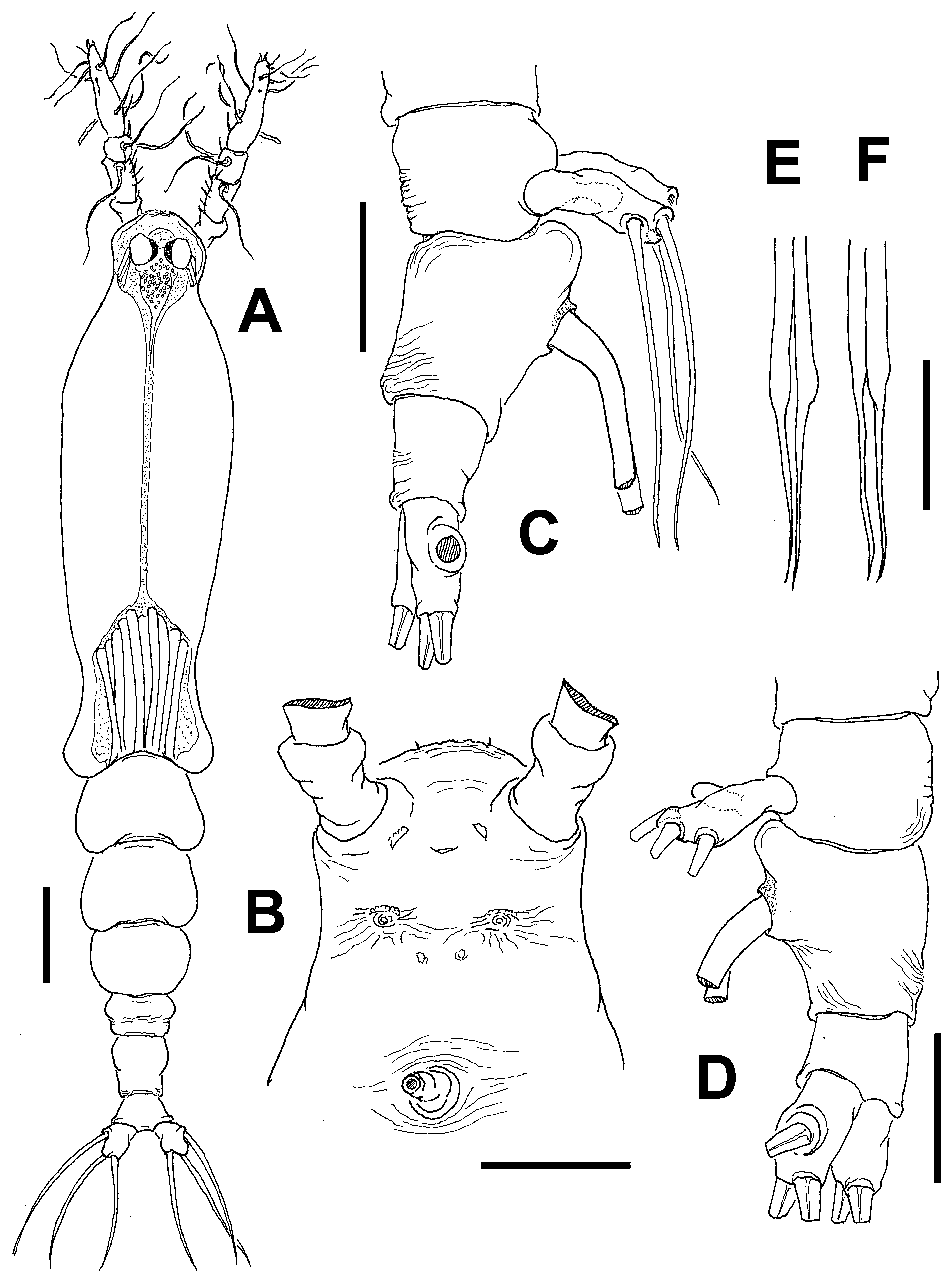
( Figs 54 View FIGURE 54 , 55 View FIGURE 55 )
Material examined. Holotype: adult female from Scott Reef, Western Australia ( 14°02.500’ S, 121°52.800’ E), partially dissected, ethanol-preserved; dissected parts mounted on 2 slides in glycerine, sealed with Entellan®. Date of collection: 6th June 2009. Slides deposited in the collection of the Museum of Western Australia (cat. WAM C61309).
Description of adult female. Body elongate, slender ( Fig. 54 View FIGURE 54 A, B); body length of holotype female 1.77 mm. Cephalothorax approximately 1.16 mm long, representing 66% of total body length. Midventral oral papilla protuberant, located at 18% of cephalothorax length, papilla flanked by nipple-like processes ( Fig. 54 View FIGURE 54 C). Pair of medium- sized ocelli present, pigment cups moderately developed, medially separated, weakly pigmented; ventral cup about as large as lateral cups ( Fig. 54 View FIGURE 54 C, D). Cephalic area narrower than post oral region, with straight lateral margins and moderately produced "forehead”. Frontal area with slight medial depression flanked by pair of sensilla and field of transverse wrinkles ( Fig. 54 View FIGURE 54 D). Dorsal surface with field of faint transverse wrinkles overlying area posterior to ocelli and two small sensilla in medial position (arrowed in Fig. 54 View FIGURE 54 D). Ventral surface with additional cuticular elements: 1) pair of symmetrical, crescent-shaped cuticular processes on anterior ventral surface between bases of antennules, with no adjacent striae (arrows in Fig. 54 View FIGURE 54 C); 2) nipple-like processes with adjacent transverse and concentric wrinkles, processes located at each side of oral papilla; 3) perioral transverse wrinkles.
Urosome consisting of fifth pedigerous somite, genital double-somite and anal somite, together representing 14% of total body length. Relative lengths of urosomites (fifth pedigerous, genital double- and free anal somite) 31.7:42.8: 25.5 = 100, respectively ( Fig. 54 View FIGURE 54 E). Fifth pedigerous somite with slightly expanded lateral margins, dorsal and ventral surfaces smooth. Genital double-somite longest of urosome, lateral surface with few longitudinal wrinkles, dorsal and ventral surfaces smooth ( Figs 54 View FIGURE 54 E, 55B). Anterior half of genital double-somite moderately swollen in dorsal view, with weak antero-ventral protuberance ( Fig. 55 View FIGURE 55 B). Ovigerous spines paired, basally separated, slender, straight at their bases, distally acute; spines about 40% of total body length ( 0.76 mm long) ( Fig. 54 View FIGURE 54 A, B). Anal somite relatively narrow, elongate, with few wrinkles on lateral surface, lacking medial constriction; dorsal and ventral surfaces smooth ( Fig. 54 View FIGURE 54 E). Caudal ramus subrectangular, about 1.6 times as long as wide, armed with three subequally long caudal setae.
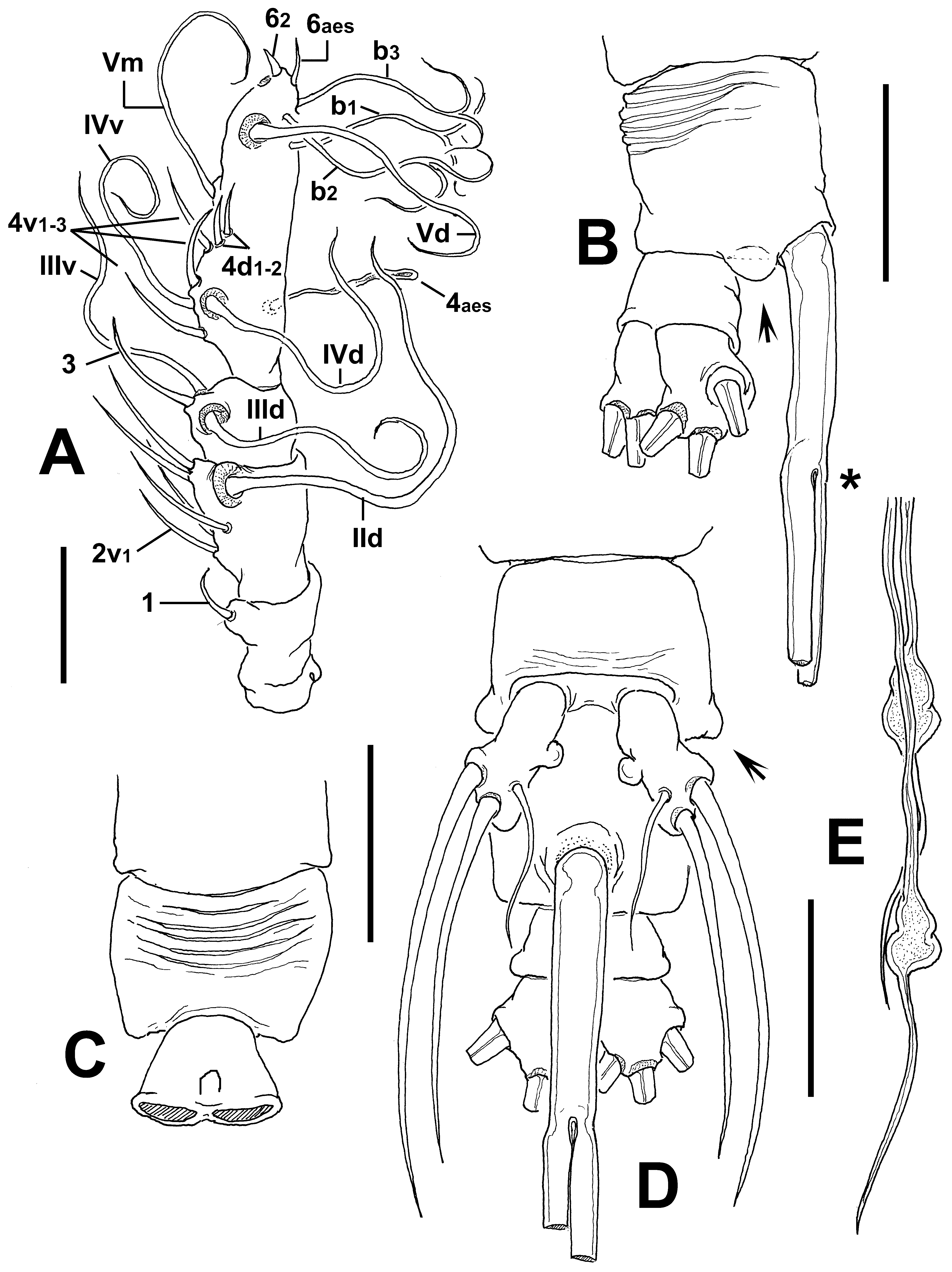
Antennule length 0.26 mm, relatively short, representing about 15% of total body length and 23% of cephalothorax length; 4-segmented, segments 3 and 4 separated. Relative length of distal antennulary segment 49%. Short spiniform element 1 present on first segment; elements on second segment noticeably short, reduced elements 2d1-2 and 2v 1-3, plus normal element IId. Third segment with remarkably short spiniform element 3, elements IIId and IIIv setiform, of normal aspect. Segment 4 bearing elements 4d1,2, 4v 1-2; setae IVd, IVv, Vd, Vv, Vm present; 4aes not observed. Element 5 short, spiniform. Subterminal elements b1-3 and b5 present, unbranched; apical elements 61-2 and 6aes not present in specimens, but sockets were observed ( Fig. 55 View FIGURE 55 A).
Incorporated first pedigerous somite and succeeding three free pedigerous somites each bearing a pair of biramous legs. Pedigerous somites 2–4, together accounting for 21% of total body length. Intercoxal sclerites of legs 1–4 subrectangular, narrow, surface with pattern of finely spinulose subquadrate patches, posterior margin curved, smooth. Bases of legs 1–4 with hair-like lateral seta ( Figs. 55 View FIGURE 55 D–F); on leg 3, this seta about 2.2 times longer than those on other legs ( Fig. 55 View FIGURE 55 F). Endopods and exopods of legs 1–4 triarticulated. Ramal setae all biserially plumose except spiniform outer seta on exopodal segments 1 and 3, and inner seta of first exopodal segment, these latter being slender, sparsely setulated ( Fig. 55 View FIGURE 55 D–F). Outermost distal spines on third exopodal segment of legs 1–4 being 0.3 times as long as segment. Outermost apical exopodal setae of legs 1–4 with inner margin setulated at proximal 1/5 only, naked otherwise; outer margin spinulose.
Armature formula of legs 1–4:
Fifth legs medially conjoined, bilobate; outer (exopodal) lobe short, its distal end barely reaching insertion of ovigerous spines ( Fig. 55 View FIGURE 55 B, C); subrectangular, distally expanded, armed with two distal and one subdistal inner setae, latter seta slightly narrower but as long as other two ( Fig. 55 View FIGURE 55 C). Inner lobe relatively large, strongly globose, arising from proximal half of exopodal lobe, unarmed ( Fig. 55 View FIGURE 55 C).
Male: unknown.
Type locality. Scott Reef, Western Australia ( 14°2.500’ S, 121°52.800’ E).
Etymology. This species is named for the RV Solander, which has conducted marine research on behalf of the Australian Institute of Marine Science in NW Australia since 2007 and, and was in turn named for Dr. Daniel Solander, naturalist on James Cook’s 1768 voyage to Australia on the Endeavour.
Diagnosis. Cymbasoma with frontal area bearing weak medial depression flanked by sensilla and with transverse wrinkles. Antennules representing about 15% of total body length and 23% of cephalothorax length; third and fourth antennulary segments separate, second segment with noticeably short, reduced elements 2d1-2 and 2v 1-3. Genital double-somite with anterior half moderately swollen in dorsal view, with few longitudinal wrinkles on lateral margin. Antero-ventral process weak. Anal somite relatively narrow, elongate, slightly shorter than genital double-somite, with few wrinkles on lateral surface. Fifth leg with short, robust outer lobe armed with two distal and one subdistal setae, innermost seta being narrower but about as long as other two setae; inner lobe relatively large, globose, arising from proximal half of exopodal lobe, unarmed. Ovigerous spines representing 40% of total body length.
Remarks. The female of C. solanderi sp. nov. shares with the Australian C. constrictum and C. lentilum but also with C. striifrons ( cf. Chang 2012) a remarkably long anal somite, which is almost as long as the genital double-somite, and a short fifth leg inner lobe. It also shares with C. lenticula a protuberant anteroventral process on the genital double-somite. The new species C. solanderi differs from these other two species in having a strongly globose fifth leg inner lobe vs. a digitiform, narrower lobe in both C. constrictum and C. lenticula and a poorly developed lobe in C. striifrons ( Chang 2012: fig. 2C). A wide, globose fifth leg inner lobe is known in other species of Cymbasoma including C. sinopense ( cf. Üstün et al. 2014), C. morii (Martin Thompson 1973; Grygier 1994), C. chelemense ( cf. Suárez-Morales & Escamilla 1997) and C. californiense (Suárez-Morales & Palomares- García 1999). These four species are members of the C. longispinosum -group and clearly differ from C. solanderi in several other characters including the shape and ornamentation of the genital double-somite, the proximal fusion of the ovigerous spines and the relative length of the cephalothorax. Cymbasoma solanderi does not belong to the C. longispinosum species-group. The length of the fifth leg is another character that can be helpful in recognizing this species. In the new species the outer lobe does not reach the point of insertion of the ovigerous spines while in most Australian species it reaches this level or beyond it. Only in C. bali ( Fig. 28 View FIGURE 28 D; Desai & Krishnaswamy 1962: fig. 5) and in C. jinigudira the outer lobe is as short as in the new species; it can readily be distinguished from C. bali by the shape and size of the inner lobe of the fifth leg. It is reduced to a small protuberance in C. bali ( Desai & Krishnaswamy 1962: fig. 10), thus diverging from the relatively large, strongly globose lobe displayed by the new species. Also, the antennulary elements of the 2v-d group ( sensu Grygier & Ohtsuka 1995) are long and normally developed in C. bali ( Desai & Krishnaswamy 1962: fig. 6) as well as in most species of the genus, however, are remarkably short in the new species. In addition, it differs from C. jinigudira in the size and armature of the fifth leg. The inner lobe is clearly smaller in C. jinigudira and it has also a short, narrow innermost exopodal seta ( Fig. 49 View FIGURE 49 D), thus diverging from the pattern observed in the new species.
| WAM |
Western Australian Museum |
No known copyright restrictions apply. See Agosti, D., Egloff, W., 2009. Taxonomic information exchange and copyright: the Plazi approach. BMC Research Notes 2009, 2:53 for further explanation.
|
Kingdom |
|
|
Phylum |
|
|
Class |
|
|
Order |
|
|
Family |
|
|
Genus |