
Cymbasoma buckleyi, Suárez-Morales, Eduardo & Mckinnon, David, 2016
|
publication ID |
https://doi.org/10.11646/zootaxa.4102.1.1 |
|
publication LSID |
lsid:zoobank.org:pub:9A7BA798-AA7C-4CAA-B42C-1E260CA573E4 |
|
DOI |
https://doi.org/10.5281/zenodo.6091313 |
|
persistent identifier |
https://treatment.plazi.org/id/03C4CA6D-D55A-FFE8-FF12-540D97022DF7 |
|
treatment provided by |
Plazi |
|
scientific name |
Cymbasoma buckleyi |
| status |
sp. nov. |
Cymbasoma buckleyi sp. nov.
( Figs 42 View FIGURE 42 , 43 View FIGURE 43 )
Material examined. Holotype: adult female from Warneet, Western Port Bay, Victoria, Australia ( 38°13.200’ S, 145°18.758’ E), partially dissected, ethanol-preserved; dissected parts mounted on 2 slides in glycerine, sealed with Entellan®. Date of collection: 12th June 1984. Vial and slides deposited in the collection of MTQ, Australia (cat. MTQ W34396).
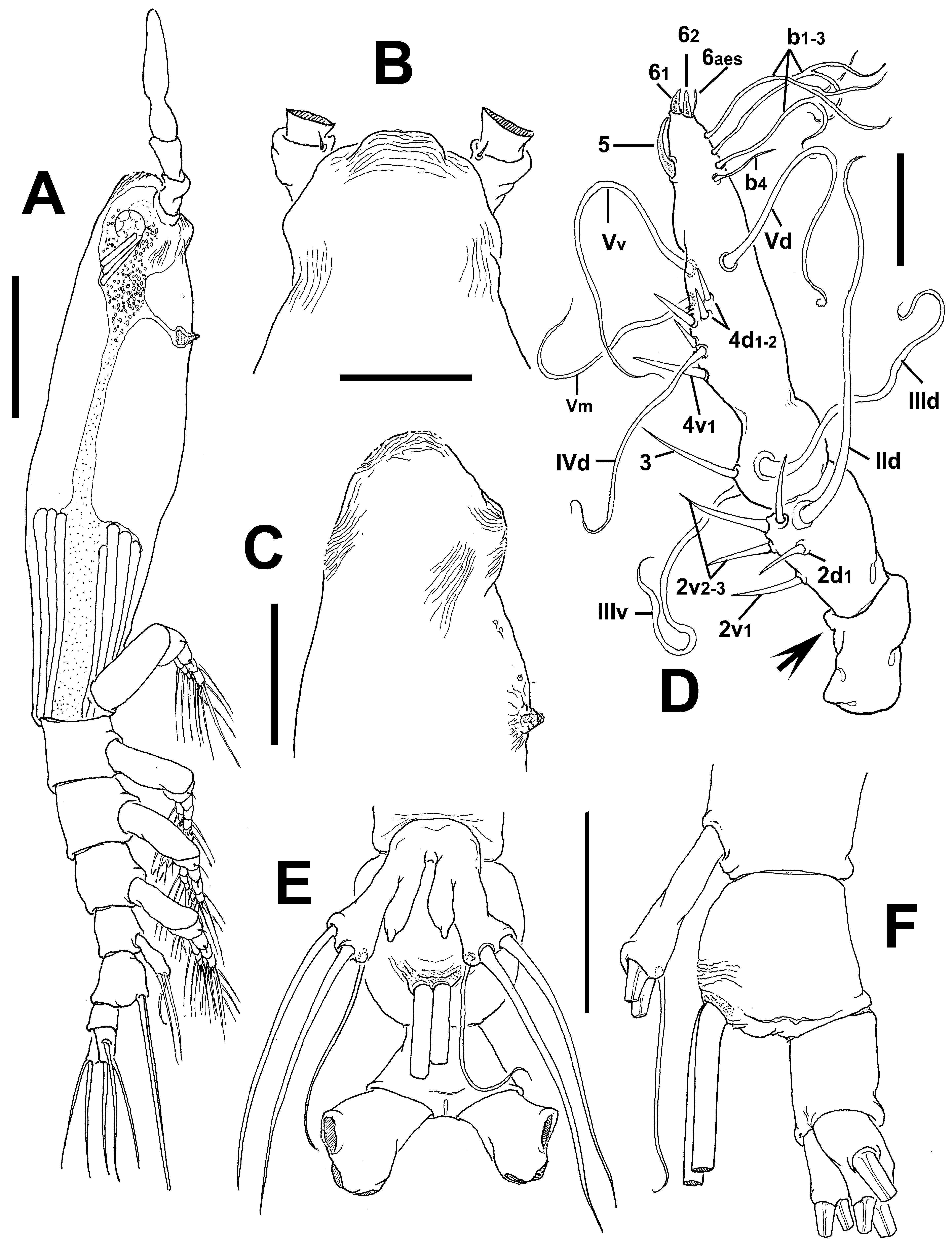
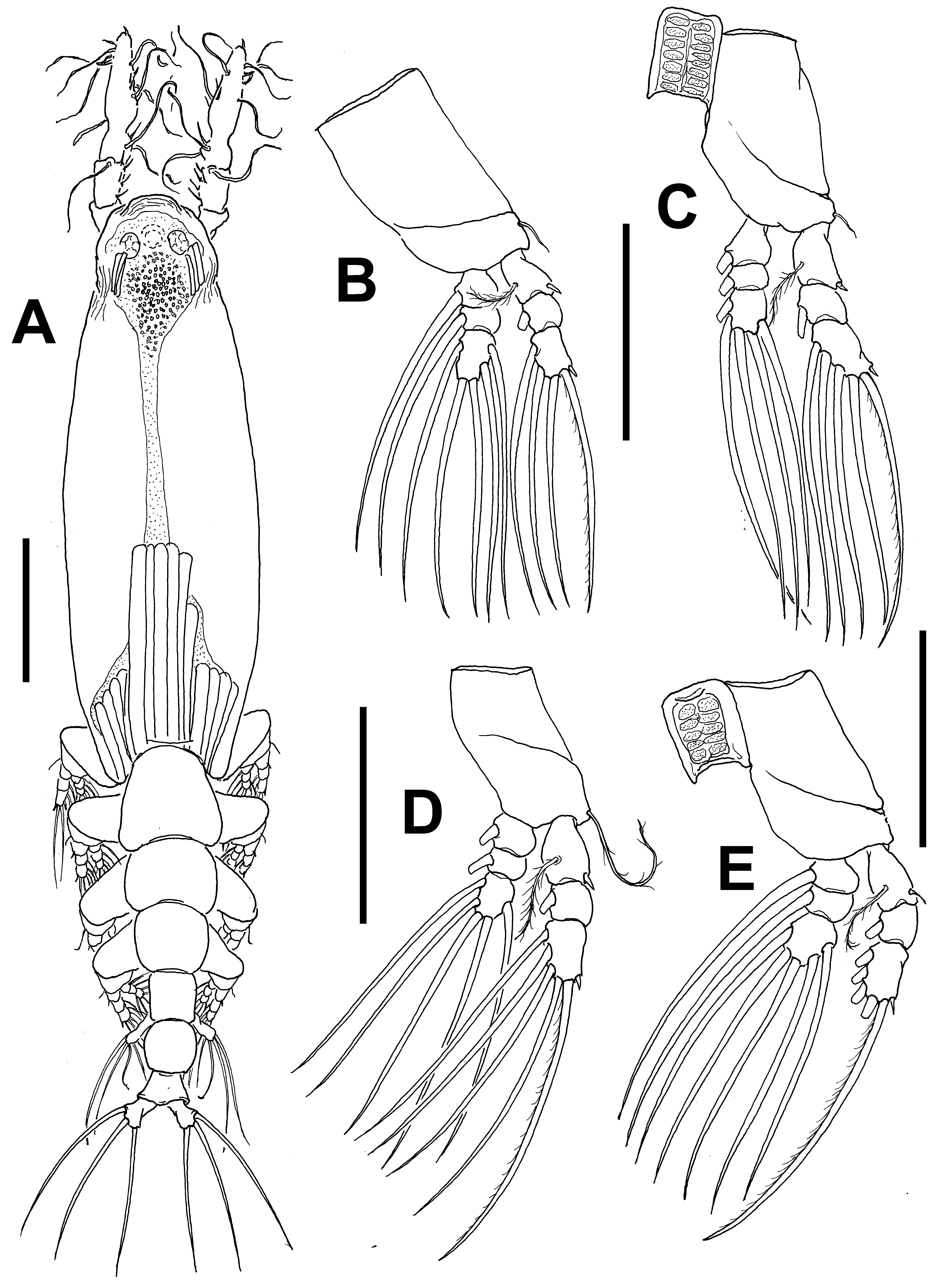
Description of adult female. Body relatively robust, with cephalothoracic margins expanded in dorsal view ( Figs 42 View FIGURE 42 A, 43A); body length of holotype female 1.24 mm. Cephalothorax approximately 0.73 mm long, representing 60% of total body length. Midventral oral papilla weakly protuberant, located at 28% of cephalothorax length. Pair of relatively small ocelli present, pigment cups moderately developed, separated by 1.2 times an eye diameter, weakly pigmented; ventral cup about as large as lateral cups ( Fig. 43 View FIGURE 43 A). Cephalic area relatively wide, with slightly produced "forehead” ornamented with transverse wrinkles ( Fig. 42 View FIGURE 42 B, C), frontal sensilla absent ( Fig. 42 View FIGURE 42 B). Dorsal surface of cephalothorax smooth except for shallow longitudinal wrinkles posterior to ocellar area ( Fig. 42 View FIGURE 42 B, C). Ventral surface ornamented with 1) medial rounded protuberance between bases of antennules, ornamented with transverse wrinkles; 2) two pairs of weakly developed papilla-like processes; 3) perioral transverse wrinkles.
Urosome consisting of fifth pedigerous somite, genital double-somite and anal somite, together representing 15% of total body length. Relative lengths of urosomites (fifth pedigerous, genital double- and free anal somites) 36.6: 37: 26.4 = 100, respectively ( Fig. 42 View FIGURE 42 E, F). Fifth pedigerous somite with straight lateral margins, dorsal and ventral surfaces smooth. Genital double-somite longest of urosome, ventrally produced, forming evenly rounded surface instead of usual anteroventral process ( Fig. 42 View FIGURE 42 F). Somite with lateral margins expanded into globose shape; with ventral surface ornamented with few transverse wrinkles at insertion of ovigerous spines ( Figs 42 View FIGURE 42 E, 43A). Ovigerous spines paired, basally separated, slender, straight at their bases ( Fig. 42 View FIGURE 42 A, E), relatively short, 20% of total body length ( 0.25 mm). Anal somite shortest of urosome not constricted; dorsal and ventral surfaces smooth ( Fig. 42 View FIGURE 42 E, F). Caudal ramus subrectangular, about 1.3 times as long as wide, armed with three caudal setae, ramus about as long as anal somite.
Antennule length 0.26 mm, representing about 21% of total body length and 35% of cephalothorax length; 4- segmented, segments 3–4 almost completely fused. Relative length of distal antennulary segment 46%. In terms of pattern described by Grygier & Ohtsuka (1995) for female monstrilloid antennulary armature, element 1 absent on both antennules (arrowed position in Fig. 42 View FIGURE 42 D); elements on second segment: 2d1-2, 2v 1-3, and IId. Third segment with element 3 being long, slender, spiniform, reaching proximal 1/3 of succeeding second segment; elements IIId and IIIv setiform, of normal aspect. Segment 4 bearing elements 4d1,2, 4v 1-3, element 4v 1 longest of group; setae IVd, Vd, Vv, Vm present. Element 5 spiniform, short, curved. Subterminal elements b1–4 present, former three distally branched, elements 61 and 62 present in specimens ( Fig. 42 View FIGURE 42 D).
Incorporated first pedigerous somite and succeeding three free pedigerous somites each bearing a pair of biramous legs. Pedigerous somites 2–4, together accounting for 24.5% of total body length. Intercoxal sclerites of legs 1–4 subrectangular; surface with patches of minute spinules; posterior margin smooth, slightly curved. Bases of legs 1–4 articulating with large, rectangular coxa along oblique line; with hair-like lateral seta ( Fig. 43 View FIGURE 43 B–E); on leg 3, this seta sparsely setulose, about four times longer, thicker than those on other legs ( Fig. 43 View FIGURE 43 D). Endopods and exopods of legs 1–4 triarticulated. Ramal setae all biserially plumose except spiniform outer seta on exopodal segments 1 and 3, and inner seta of first exopodal segment, these latter being noticeably long, reaching proximal half of third exopodal segment; setae slender, slightly setulose. Outermost distal spines on third exopodal segment of legs 1–4 short, 0.25 times as long as segment. Outermost apical exopodal setae of legs 1–4 with inner margin setulose, outer margin naked.
Armature formula of legs 1–4:
Fifth legs medially conjoined, bilobate, outer (exopodal) lobe elongate, cylindrical, distally truncate. Outer lobe armed with three apical setae, innermost seta noticeably shorter and narrower than other two ( Fig. 42 View FIGURE 42 E). Inner lobe mammiliform, unarmed, shorter than outer lobe, arising proximally from exopodal lobe and reaching about ¾ its length ( Fig. 42 View FIGURE 42 E).
Male: unknown.
Type locality. Warneet, Western Port Bay, Victoria, Australia ( 38°13.200’ S, 145°18.758’ E).
Etymology. The species was named after the escaped English convict William Buckley who prior to European settlement lived with aboriginal tribes of the area where the species was collected.
Diagnosis. Cymbasoma with third and fourth antennulary segments partially fused, fourth antennulary segment representing more than 45% of antennule length. Cephalic area with frontal, dorsal and ventral fields of cuticular wrinkles. Medial cephalic protuberance ornamented with transverse wrinkles on ventral surface. Antennules with element 1 absent. Legs 1–4 with noticeably long inner seta on first exopodal segment. Genital double-somite without anteroventral process but ventrally produced into widely rounded surface in lateral view; globose in dorsal view. Fifth leg with outer lobe armed with three setae, innermost being shorter and thinner than the other; inner lobe distinctively mammiliform, unarmed. Ovigerous spines short, representing 20% of total body length.
Remarks. Together with C. nicolettae from the Mediterranean ( Suárez-Morales 2002), this is the only known species of Cymbasoma with a mammiliform fifth leg inner lobe. Both species also share a corrugated frontal area, antennules with fused segments 3–4, a globose genital double- somite and a short and thin innermost seta on the fifth leg outer lobe ( Suárez-Morales 2002). They differ in several characters, namely in the new species the body is wider, more robust than in C. nicolettae ( Suárez-Morales 2002: figs. 1, 2); in C. nicolettae the frontal area of the cephalic region is flat and lacks a ventral protuberance between the antennulary bases and the oral papilla (Suárez- Morales 2002: fig. 4) vs. a produced forehead and a large rounded, wrinkled ventral cephalic process in the new species. The relative antennule length is different in these species; it is remarkably long and represents 28.4% of the total body length in C. nicolettae ( Suárez-Morales 2002: fig. 1) vs. 22% in the new species. In addition, the innermost seta of the fifth leg exopodal lobe is relatively shorter in C. nicolettae being as long as the lobe (Suárez- Morales 2002: fig. 8) whereas it is about 1.5 times longer in the new species.
This species is also related to other Australian Cymbasoma with a fifth leg bearing an elongate inner lobe that arises basally from the outer lobe but can readily be distinguished from them by the mammiliform fifth leg inner lobe. In addition, it differs by the protuberant, rounded shape of the genital double-somite (lateral view), thus diverging from those species with an anteroventral process, including C. bali , C. markhasevae , C. apicale , or C. lenticula .
No known copyright restrictions apply. See Agosti, D., Egloff, W., 2009. Taxonomic information exchange and copyright: the Plazi approach. BMC Research Notes 2009, 2:53 for further explanation.
|
Kingdom |
|
|
Phylum |
|
|
Class |
|
|
Order |
|
|
Family |
|
|
Genus |