
Cymbasoma strzeleckii, Suárez-Morales, Eduardo & Mckinnon, David, 2016
|
publication ID |
https://doi.org/10.11646/zootaxa.4102.1.1 |
|
publication LSID |
lsid:zoobank.org:pub:9A7BA798-AA7C-4CAA-B42C-1E260CA573E4 |
|
DOI |
https://doi.org/10.5281/zenodo.6091355 |
|
persistent identifier |
https://treatment.plazi.org/id/03C4CA6D-D56B-FFD6-FF12-51B697922AD7 |
|
treatment provided by |
Plazi |
|
scientific name |
Cymbasoma strzeleckii |
| status |
sp. nov. |
Cymbasoma strzeleckii sp. nov.
( Figs 68 View FIGURE 68 , 69 View FIGURE 69 )
Material examined. Holotype: adult male from Rhyll, Western Port Bay, Victoria, Australia ( 38°27.792’ S, 145°18.496’ E), partially dissected, ethanol-preserved; dissected parts mounted on slide in glycerine, sealed with Entellan®. Date of collection: 10th March 1984. Slide deposited in the collection of MTQ, Australia (cat. MTQ W34405).
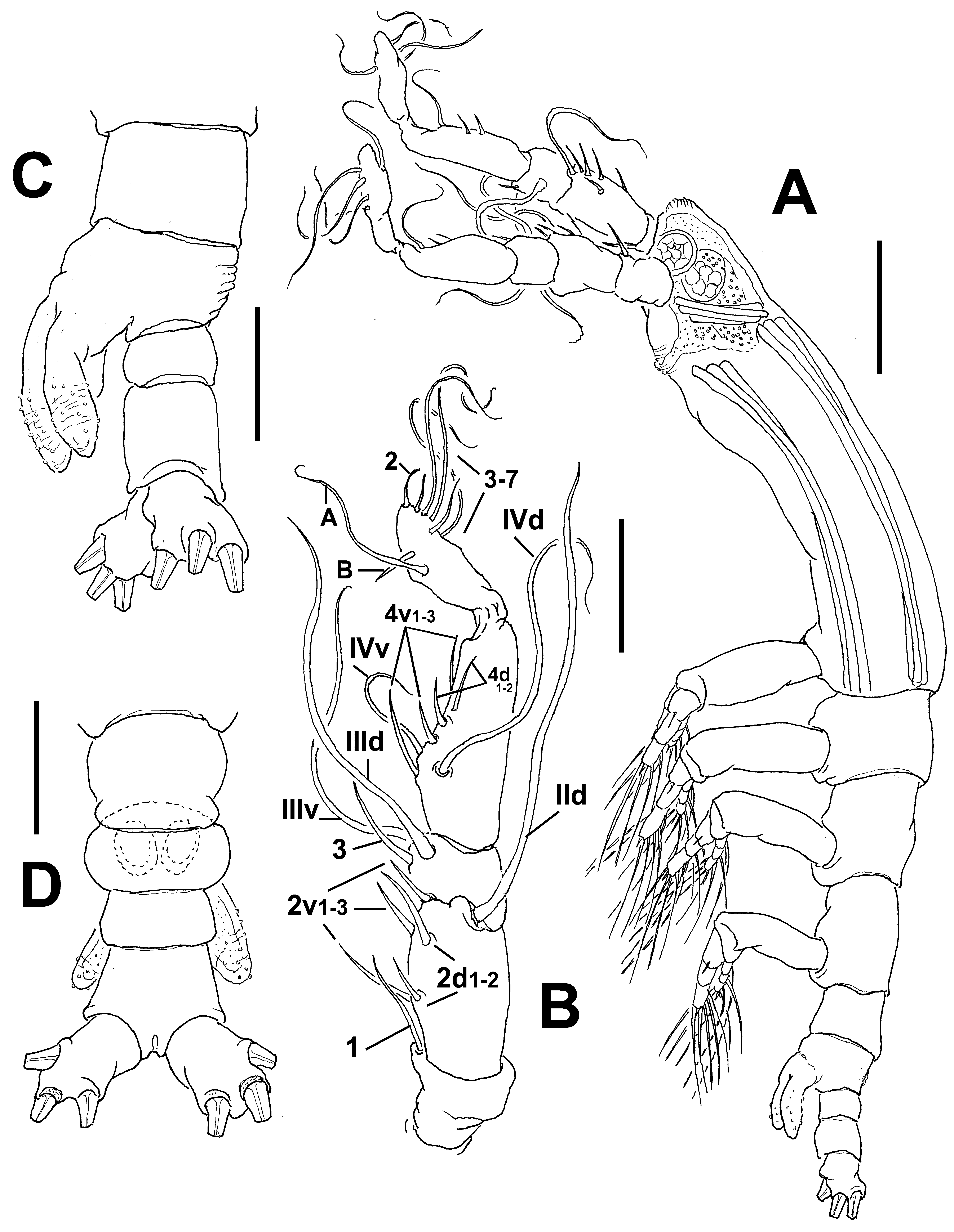
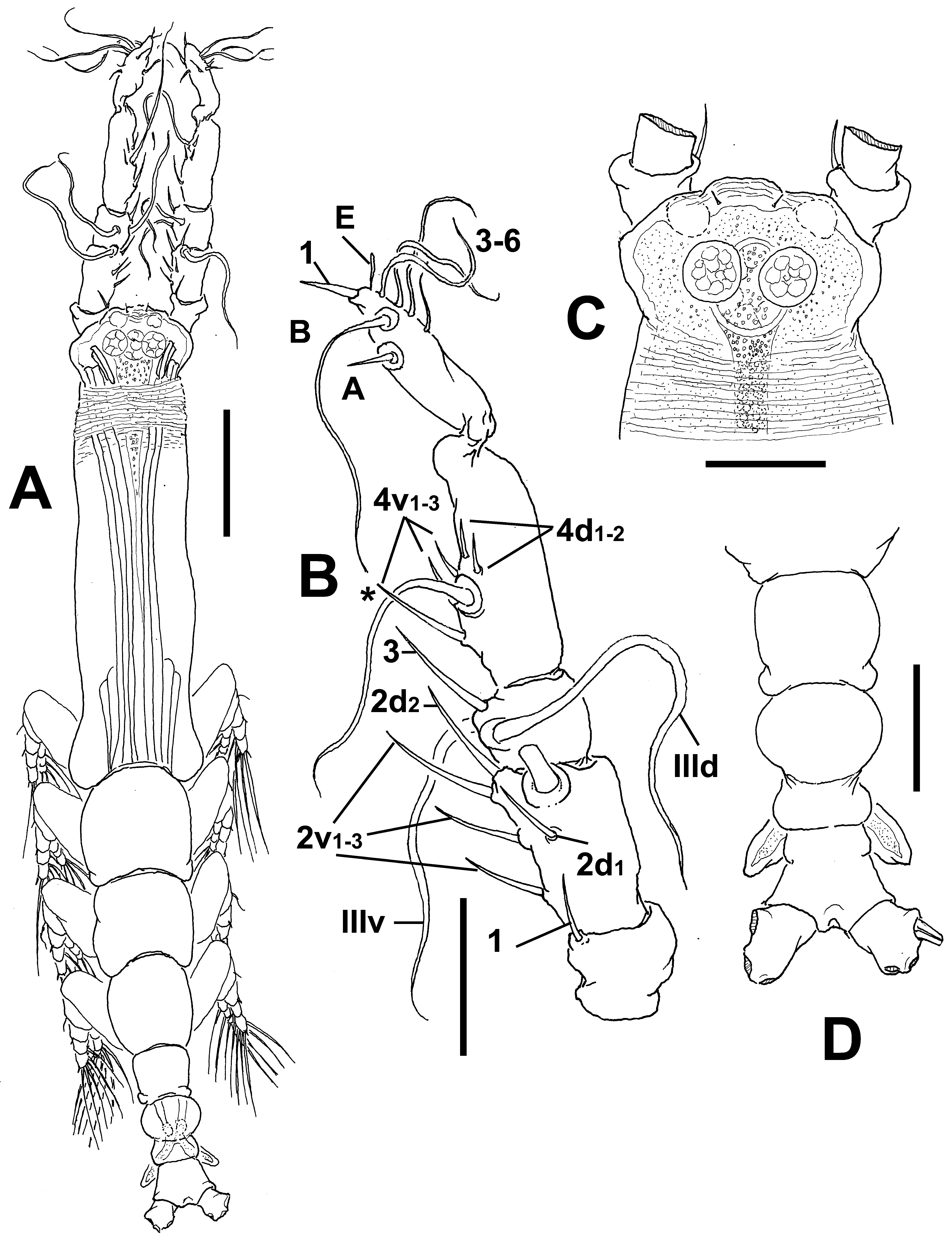
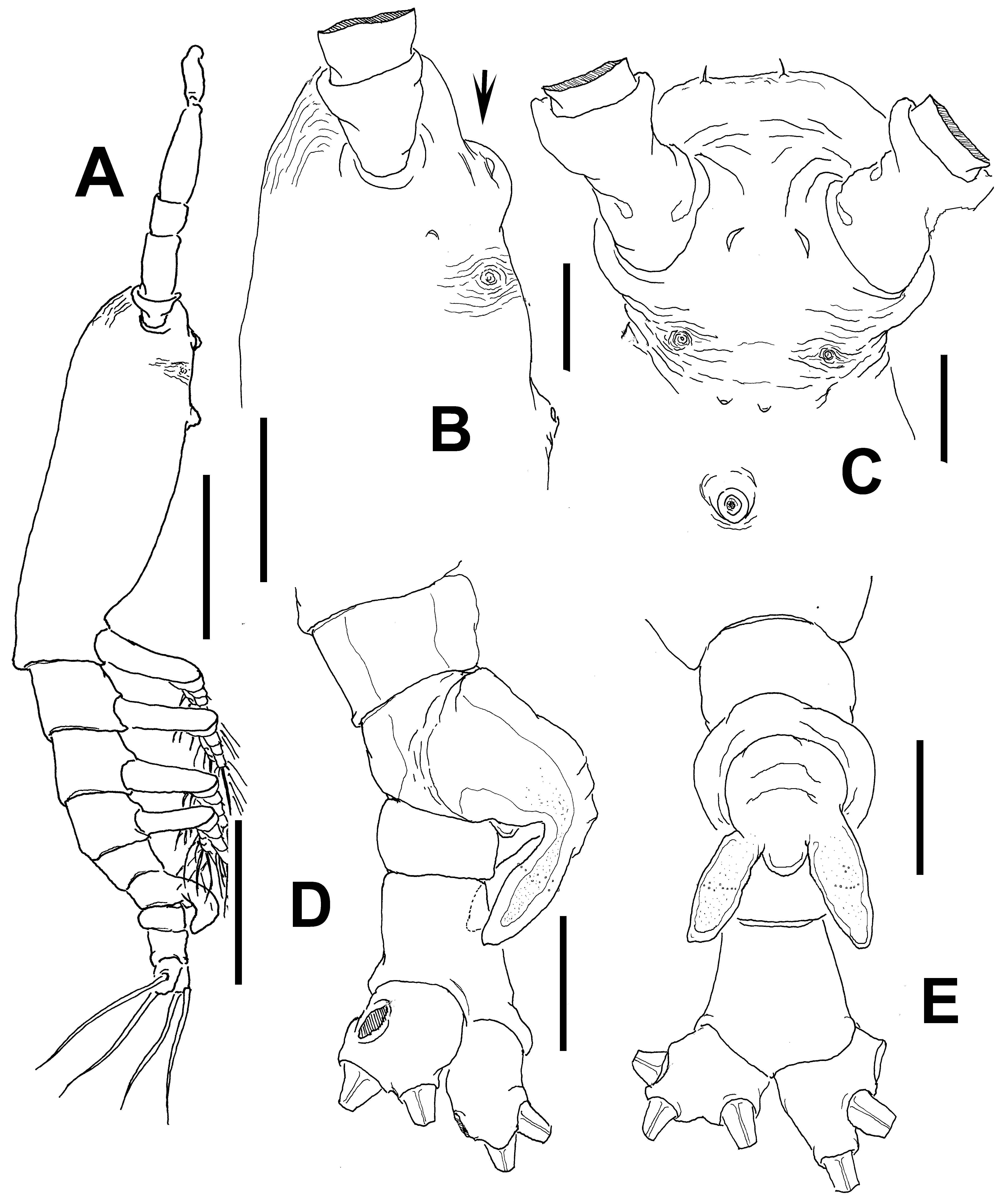
Description of adult male. Total body length 0.85 mm. Cephalothorax 0.42 mm long, representing 49% of total body length ( Fig. 68 View FIGURE 68 A). Midventral oral papilla well developed, located at 31% of cephalothorax length ( Fig. 68 View FIGURE 68 A, B). Cephalic region slightly protuberant bilaterally in dorsal and ventral views ( Fig. 68 View FIGURE 68 C). Pair of dorsal ocelli present, weakly developed; pigment cups medium-sized. Ocelli faintly pigmented. Pair of frontal sensilla between antennulary bases. Forehead area moderately produced anteriorly ( Fig. 68 View FIGURE 68 C); ventral process arising between the antennulary bases (arrowed in Fig. 68 View FIGURE 68 B). Other cuticular ventral processes including pair of crescent-shaped minute processes and nipple-like processes, the latter with adjacent wrinkles.
Urosome consisting of fifth pedigerous somite, genital somite (carrying genital complex), preanal somite, and anal somite. Fifth pedigerous somite with smooth ventral surface. Genital somite slightly shorter than fifth pedigerous somite. Genital complex of type II, represented by pair of robust, moderately divergent genital lappets ( Fig. 68 View FIGURE 68 E), these being slightly asymmetrical, reaching to halfway down the long anal somite ( Fig. 68 View FIGURE 68 D). Rounded, protuberant medial process present at common basal joint of lappets, lappets smooth. Anal somite about twice as long as preanal somite in dorsal and lateral views, comprising 30% of urosome length; no suture visible on ventral or dorsal surfaces ( Fig. 68 View FIGURE 68 D, E). Caudal rami small, subquadrate, approximately 1.1 times as long as wide, about as long as anal somite. Each ramus with three setae.
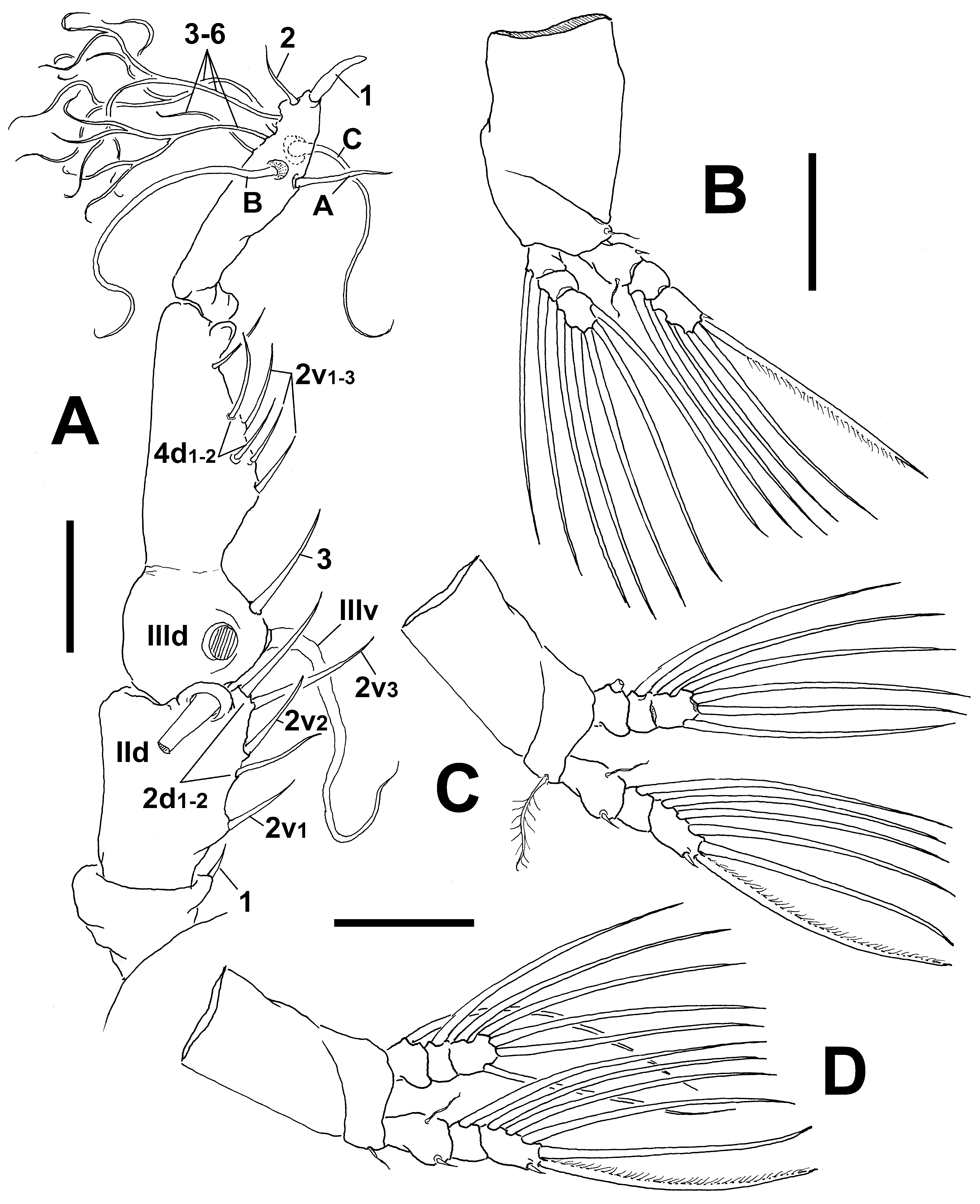
Antennulary length 0.31 mm. Antennules relatively long, representing 44% of total body length, and 80% of cephalothorax length; 5-segmented, all segments separated, with segment 5 located distal to geniculation ( Fig. 69 View FIGURE 69 A). Length ratio of antennulary segments, from first to fifth 10.4: 26.1: 11.3: 34.2: 18 (= 100). Setal element 1 on first segment remarkably short and slender, spiniform. Antennulary elements 2v 1-3, 2d1,2, and IId present on second segment. Setal elements IIId, IIIv, and 3 present on third segment; element 3 slender, curved. Fourth segment with elements 4d 1-2, 4v 1–3. Fifth segment with 5 “b”-group setae, elements b1-3 long, distally branched. According to Huys et al. (2007) setal nomenclature of the distal segment, elements A–C and 1–6 present; element 1 being remarkably long, spiniform.
Pedigerous somites 2–4, together accounting for 28% of total body length in dorsal view. Coxae of each pair unarmed, joined by intercoxal sclerite which is slightly longer than wide. Bases of legs 1–4 separated from coxae posteriorly by oblique articulation; with hair-like lateral seta ( Fig. 69 View FIGURE 69 B-D); on leg 3, this seta about 3.5 times longer, than in other legs ( Fig. 69 View FIGURE 69 C). Exopods of legs 1–4 longer than endopods. Ramal setae all biserially plumose except spiniform outer seta on exopodal segments 1 and 3. Flexible, slender, inner seta present on first exopodal segment of legs 1–4. Outer spine on distal exopodal segment of legs 1–4 about 0.3 times as long as segment. Also, outermost apical exopodal setae of legs 1–4 with inner margin smooth, outer margin sparsely spinulose.
Armature formula of legs as follows:
basis endopodexopod
leg 1 0-1 0-1; 0-1; 1,2, 2 I- 1; 0-1; I,2,2 legs 2–4 0-1 0-1; 0-1; 1,2, 2 I- 1; 0-1; I,2,3 Female: unknown.
Type locality. Rhyll, Western Port Bay, Victoria, Australia ( 38°26.792’ S, 145°18.496’ E).
Etymology. This species is named in honour of Sir Pawel Edmund Strzelecki , a Polish explorer who reached Western Port Bay in 1840.
Diagnosis. Cymbasoma with conspicuous medial ventral protuberance on cephalic area; protuberance smooth. Oral papilla well developed. Antennule representing 44% of total body length and 80% of cephalothorax length. Fifth pedigerous somite with smooth ventral surface. Genital complex of type II, with thumb-like, slightly asymmetrical lappets, inner margins and distal tips of lappets smooth. Basal joint between lappets produced into rounded protuberance. Three caudal setae. Element 1 on first antennulary segment short. Element 1 on distal antennulary segment long, spiniform.
Remarks. Cymbasoma strzeleckii sp. nov. has a unique combination of characters including a frontal smooth protuberance, a genital somite with smooth dorsal surface, a genital complex with moderately divergent, smooth thumb-like lappets and a strong medial rounded protuberance at the insertion of the lappets. There are six other species with a relatively strong, rounded medial protuberance of the male genital complex and lappets resembling those of the new species, namely C. tropicum ( cf. Martin Thompson & Easterson 1983), C. quadridens ( cf. Suárez- Morales & Pilz 2008), C. gracile ( cf. Gurney 1927), C. bullatum ( cf. Suárez-Morales 2007), and the Australian C. galerus and C. annulocolle . Cymbasoma strzeleckii differs from these species in the presence of a smooth ventral cephalic protuberance. A similar cephalic ventral protuberant process is present in C. rochai ( Suárez-Morales & Dias 2001) , but in both C. rochai ( cf. Suárez-Morales & Dias 2001) and C. tropicum ( cf. Martin Thompson & Easterson 1983) four caudal setae are present thus differing from the of new species, with three caudal setae. In C. quadridens , C. annulocolle and C. galerus the lappets are ornamented with spinules or papillae, whereas in the new species these structures are smooth. As for C. gracile , the available comparative data are limited ( Gurney 1927), but it shares with C. strzeleckii the smooth lappets and the presence of three caudal setae; however, in C. gracile the anal somite has a constriction and a suture ( Gurney 1927: fig. D), thus differing from the non-constricted anal somite of the new species. Additionally, these species can also be distinguished using details of the antennulary armature. Element 1 ( sensu Grygier & Ohtsuka, 1995) on the first segment is very small in the new species; although being similarly spiniform it is clearly longer in C. tropicum ( Martin Thompson & Easterson 1983: fig. 2g), C. quadridens ( Suárez-Morales & Pilz 2008: fig. 5C), C. annulocolle ( Fig. 15 View FIGURE 15 B), and C. galerus ( Fig. 9 View FIGURE 9 B). The element is very long, setiform and setulated in C. rochai ( Suárez-Morales & Dias 2001: fig. 26) and C. bullatum ( Suárez-Morales 2007: fig. 2A,B); the condition in C. gracile is unknown since the element was not illustrated ( Gurney 1927: fig. E).
No known copyright restrictions apply. See Agosti, D., Egloff, W., 2009. Taxonomic information exchange and copyright: the Plazi approach. BMC Research Notes 2009, 2:53 for further explanation.
|
Kingdom |
|
|
Phylum |
|
|
Class |
|
|
Order |
|
|
Family |
|
|
Genus |