
Cymbasoma paraconstrictum, Suárez-Morales, Eduardo & Mckinnon, David, 2016
|
publication ID |
https://doi.org/10.11646/zootaxa.4102.1.1 |
|
publication LSID |
lsid:zoobank.org:pub:9A7BA798-AA7C-4CAA-B42C-1E260CA573E4 |
|
DOI |
https://doi.org/10.5281/zenodo.6091353 |
|
persistent identifier |
https://treatment.plazi.org/id/03C4CA6D-D56F-FFD5-FF12-54CD952A2C66 |
|
treatment provided by |
Plazi |
|
scientific name |
Cymbasoma paraconstrictum |
| status |
sp. nov. |
Cymbasoma paraconstrictum sp. nov.
( Figs 66 View FIGURE 66 , 67 View FIGURE 67 )
Material examined. Holotype: Adult female from Garden Island, Western Australia ( 32°10.48’ S, 115°40.72’ E), partially dissected, ethanol-preserved; dissected parts mounted on slide in glycerine, sealed with Entellan®. Date of collection: 19th September 2012. Slides deposited in the collection of the Museum of Western Australia (cat. WAM C61310).
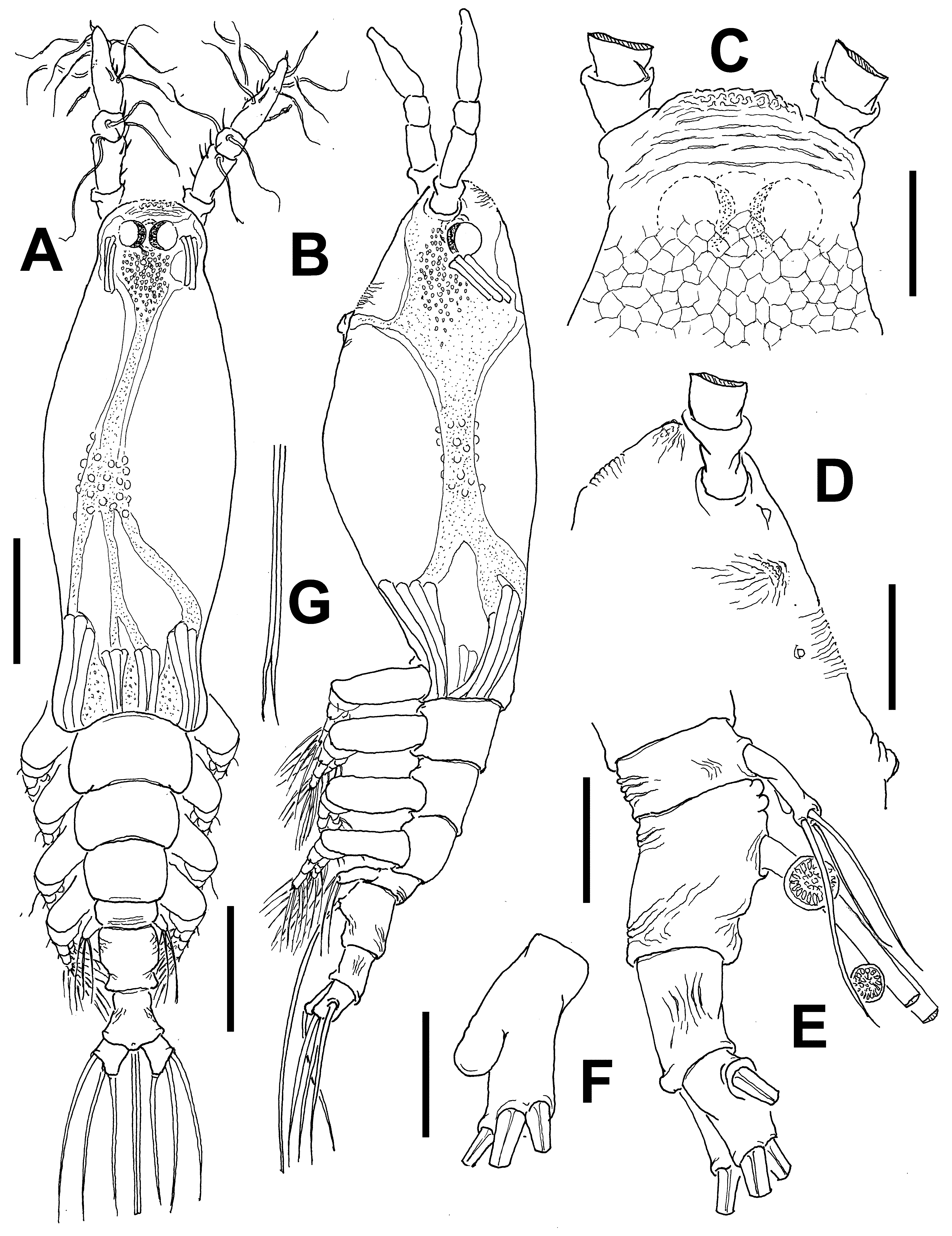
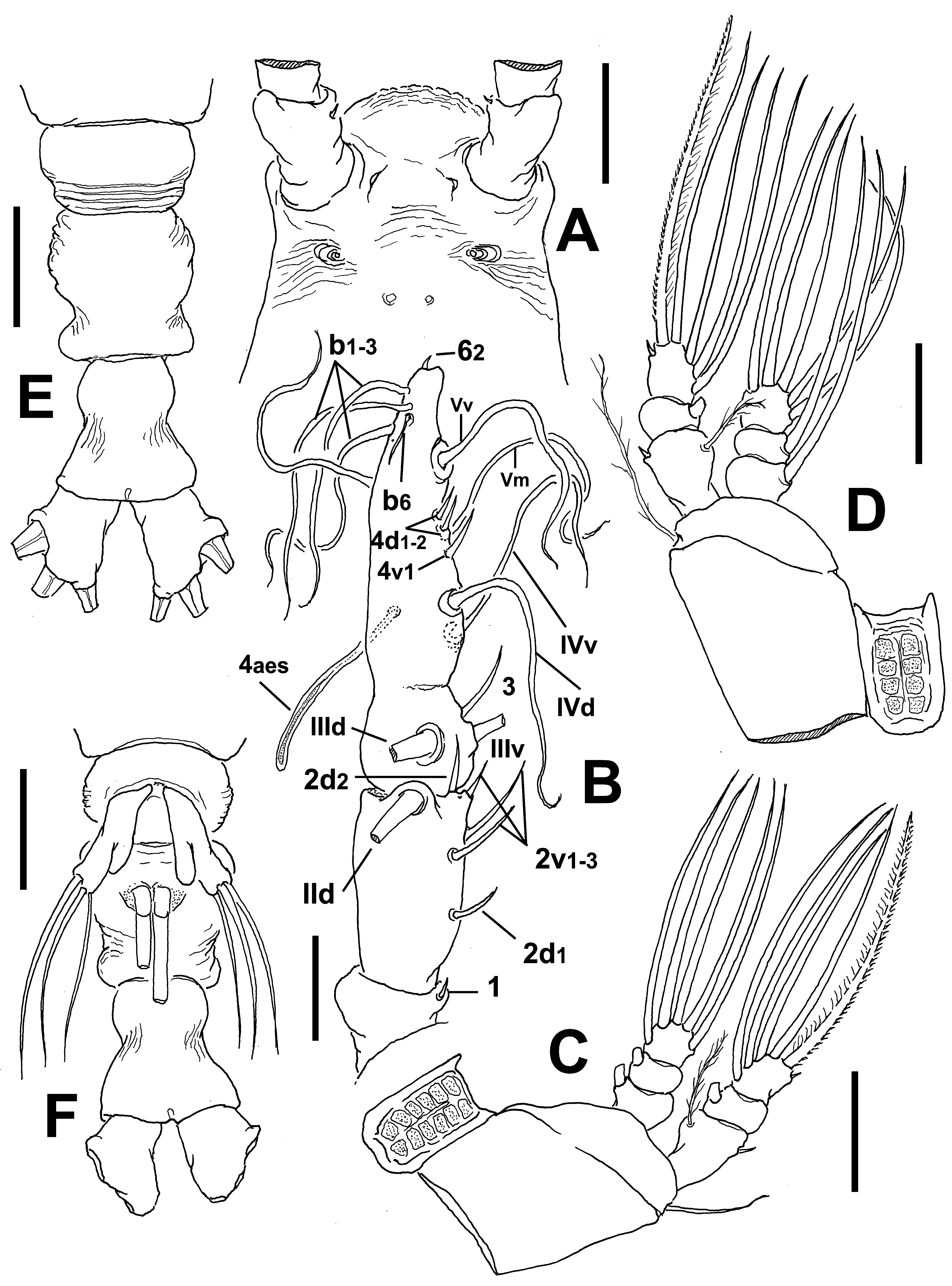
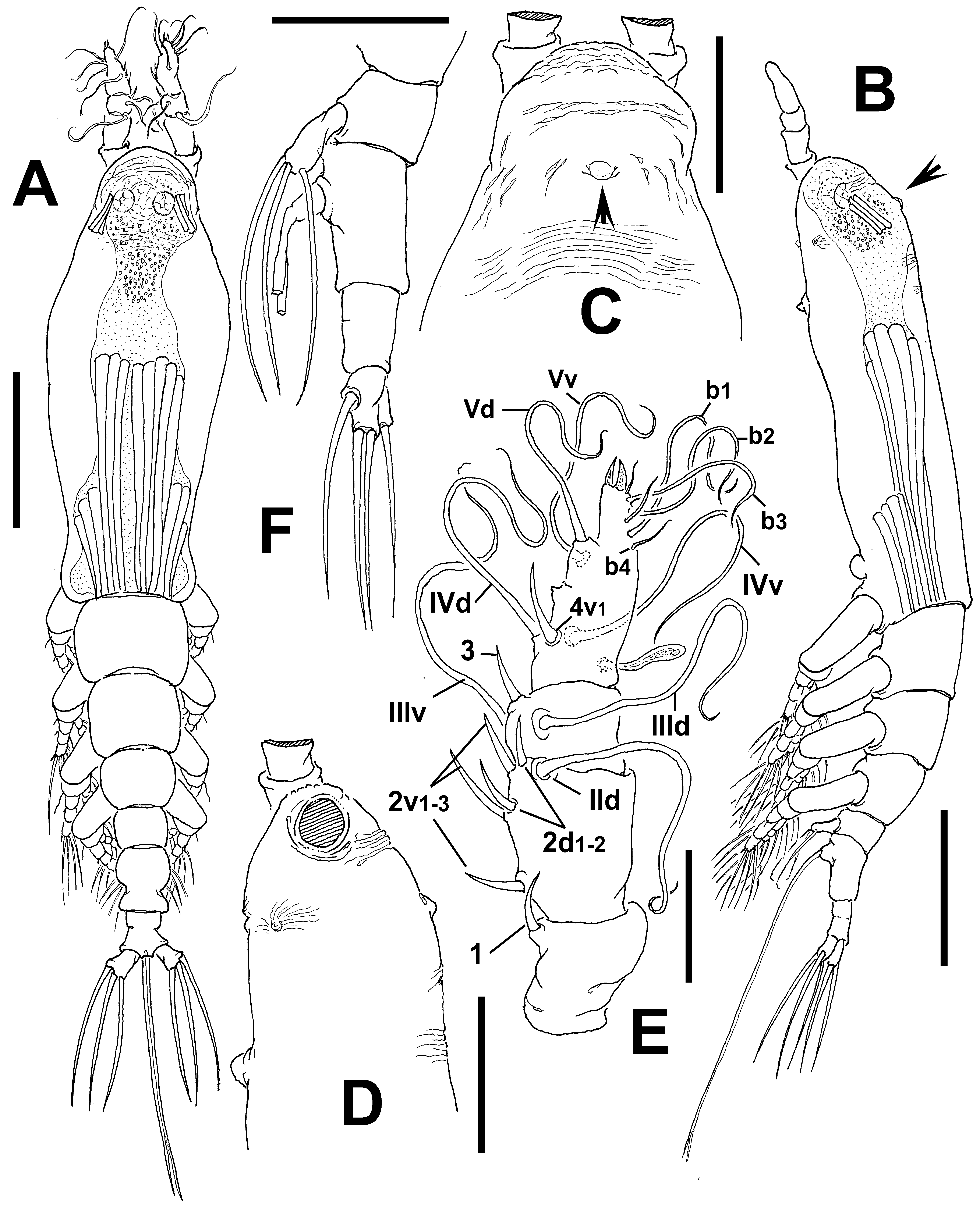
Description of adult female. Body moderately robust, particularly in dorsal position, widest at proximal 1/3 of cephalothorax ( Fig. 66 View FIGURE 66 A); body relatively slender in lateral view ( Fig. 66 View FIGURE 66 B). Total length of holotype female 1.1 mm. Cephalothorax approximately 0.59 mm long, representing 55% of total body length. Midventral oral papilla moderately protuberant, located at 33% of cephalothorax length. Pair of relatively small ocelli present, pigment cups medially separated by about one eye diameter, weakly pigmented; ventral cup larger than lateral cups. Frontal area smooth except for a few shallow wrinkles; frontal sensilla absent ( Fig. 66 View FIGURE 66 C). Anterior part of cephalothorax with dorsal field of transverse and oblique wrinkles and low, small medial dorsal protuberance ( Fig. 66 View FIGURE 66 C (arrow), 66D). Additional ornamentation of ventral surface including pair of nipple-like processes on ventral surface located anterior to oral papilla, processes with adjacent field of transverse and curved wrinkles ( Fig. 67 View FIGURE 67 C).
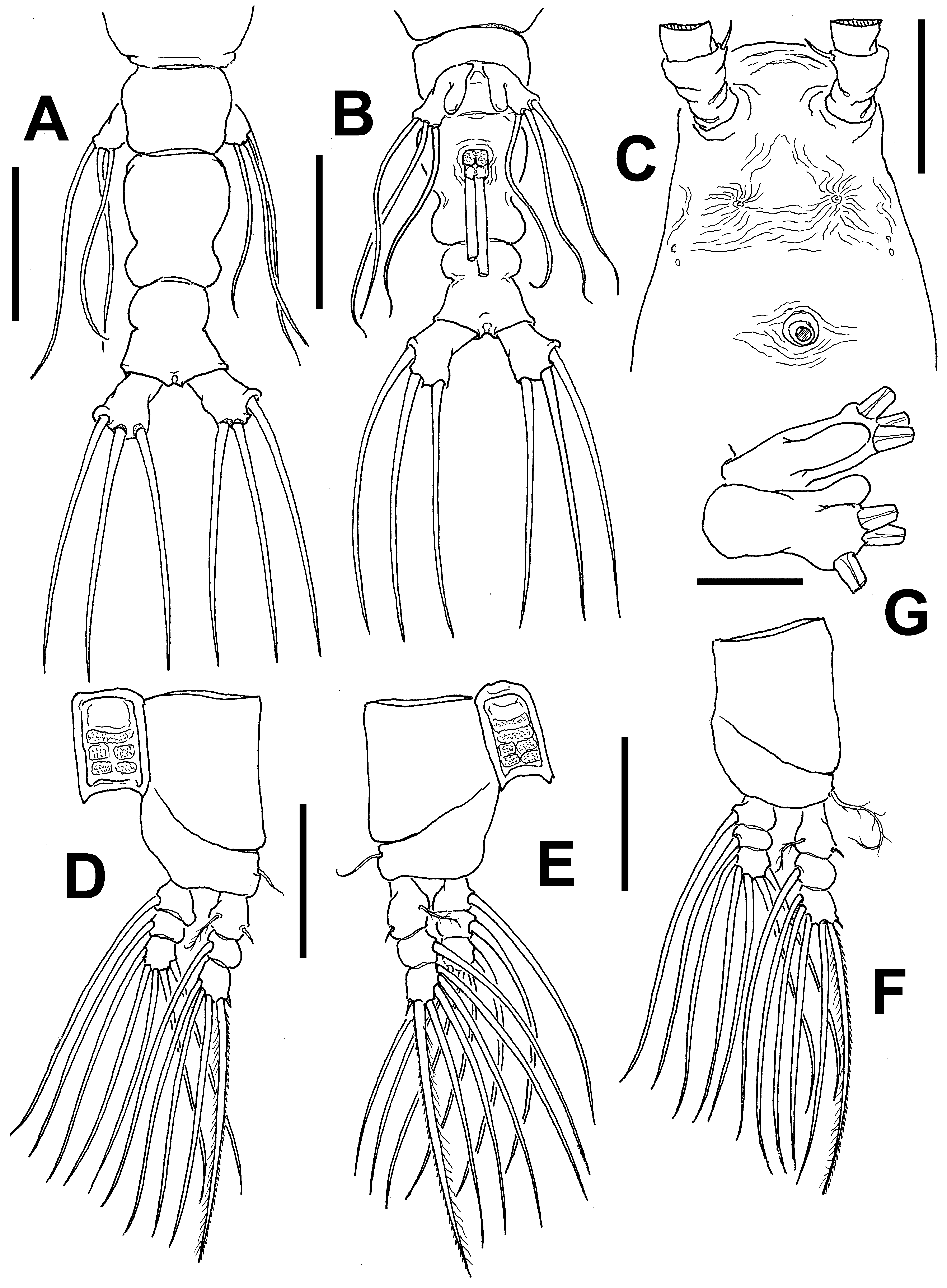
Urosome consisting of fifth pedigerous somite, genital double-somite, and anal somite, together representing 19% of total body length. Relative lengths of urosomites (fifth pedigerous, genital double- and free anal somites) 26.5: 41.2: 32.3 = 100, respectively ( Figs. 66 View FIGURE 66 F). Fifth pedigerous somite shortest of urosome, with smooth dorsal and ventral surfaces. Genital double-somite longest of urosome; anterior half expanded, smooth; with constriction; posterior half expanded forming rounded lateral protuberances ( Fig. 67 View FIGURE 67 A, B). Ovigerous spines paired, separated at base, relatively short, 45% of total body length ( 0.44 mm). Spines slender, straight at their bases and along shaft, distally acute, subequally long ( Fig. 66 View FIGURE 66 A, B). Anal somite constricted medially, anterior half with rounded margins; posterior half with divergent margins; dorsal and ventral surfaces smooth ( Fig. 67 View FIGURE 67 A, B). Caudal rami divergent, subrectangular, about 1.3 times as long as wide, armed with three subequally long caudal setae.
Antennule short, thick; length 0.16 mm, representing about 17% of total body length and 32% of cephalothorax length; 4-segmented. Relative length of remarkably short distal antennulary segment 34.7%. Short, curved spiniform element 1 present on first segment; elements on second segment: 2d1-2, 2v 1-3, and IId. Third segment with short, spiniform element 3; IIId and IIIv of normal aspect. Segment 4 bearing long, spiniform element 4v 1, other elements of group not observed. Setae IVd, IVv, Vd, Vv, and 4aes present. Element 5 absent. Subterminal elements b1-3 present, long, unbranched. Apical elements 61-2 and 6aes present in specimen ( Fig. 66 View FIGURE 66 E).
Incorporated first pedigerous somite and succeeding three free pedigerous somites each bearing a pair of biramous legs. Pedigerous somites 2–4, together accounting for 26% of total body length. Intercoxal sclerites of legs 1–4 subrectangular, surface and posterior margin smooth. Bases of legs 1–4 articulating with large, rectangular coxa along oblique line; with hair-like lateral seta ( Fig. 67 View FIGURE 67 D–F); on leg 3, this seta sparsely setulated, about three times longer, thicker than those on other legs ( Fig. 67 View FIGURE 67 F). Endopods and exopods of legs 1–4 triarticulated. Ramal setae all biserially plumose except spiniform outer seta on exopodal segments 1 and 3, and inner seta of first exopodal segment, these latter being short, slender. Outermost distal spines on third exopodal segment of legs 1–4 short, 0.25 times as long as segment. Outermost apical setae on third exopodal segment of legs 1–4 with inner margin sparsely setulose, outer margin spinulose.
Armature formula of legs 1–4:
Fifth legs medially conjoined, bilobate. Outer (exopodal) lobe cylindrical, distally truncate, barely reaching beyond posterior margin of fifth pedigerous somite; armed with three apical subequally long setae ( Fig. 67 View FIGURE 67 A, B). Inner lobe arising from middle inner margin of outer lobe, thumb-like, almost reaching midlength of exopodal lobe ( Fig. 67 View FIGURE 67 G).
Male: unknown.
Type locality. Garden Island, Western Australia ( 32°10.48’ S, 115°40.72’ E).
Etymology. The specific name, using the prefix “para” (= near) denotes the resemblance of this species to another Australian species ( C. constrictum ), both species have constricted anal and genital double-somites.
Diagnosis. Cymbasoma with relatively robust cephalothorax, widest at level of oral papilla. Dorsal protuberance and field of transverse wrinkles on cephalic area. Antennule short, representing about 17 % of total body length and 32% of cephalothorax length. Third antennulary segment remarkably short, representing only 34.7% of antennule length. Oral papilla moderately protuberant. Legs 1–4 with first and second endopodal segments with inner margin not protuberant; outermost distal spines on third exopodal segment of legs 1–4 short, modified, mammiliform. Urosomites smooth, genital double-somite longest, constricted. Anal somite strongly constricted medially, anterior half with rounded margins. Fifth leg with three distal subequally long setae on short outer lobe. Inner lobe thumb-like, unarmed. Ovigerous spines representing 45% of total body length.
Remarks. This species most closely resembles the Australian C. constrictum in the body proportions, the general structure and armature of the fifth leg, its constricted genital double- somite, and in its long, medially constricted anal somite. The shape of the cephalothorax differs in both species; in C. constrictum the ventral margin is clearly globose in lateral view ( Fig. 36 View FIGURE 36 B) and it is straight in the new species ( Fig. 66 View FIGURE 66 B). The new species has a dorsal cephalic protuberance (arrowed in Fig. 66 View FIGURE 66 B, C) which is absent in C. constrictum . In addition, the eyes are large and strongly pigmented (medially) in C. constrictum ( Fig. 36 View FIGURE 36 A), thus differing from the relatively smaller, unpigmented eyes in the new species ( Fig. 66 View FIGURE 66 A). The antennule structure is also different in these two species, segments 3–4 being fused in C. constrictum ( Fig. 37 View FIGURE 37 B) and clearly separated in the new species. In addition, the last antennulary segment is remarkably short in C. constrictum whereas it is robust in the new species (34.7% of total antennule length ( Fig. 66 View FIGURE 66 E) vs. 45% in C. constrictum ). Cymbasoma constrictum can readily be distinguished by the elongate anal somite which is clearly longer than the genital double-somite. Only in C. striifrons ( Chang 2012) and in the new species ( Fig. 67 View FIGURE 67 A) the anal somite is almost as long as the genital double-somite. In addition, the fifth pedigerous somite of C. constrictum has a set of strong transverse striae across the somite ( Fig. 37 View FIGURE 37 E) which is absent from the otherwise smooth dorsal surface in the new species. Also, in C. constrictum the constrictions of both the genital double- and anal somites are ornamented with wrinkles ( Fig. 37 View FIGURE 37 E, F), whereas they are smooth in the new species. A strongly constricted genital double-somite combined with a weakly constricted anal somite is known in C. germanicum ( Suárez-Morales 2006: figs. 4b, d), but the anal somite is clearly shorter than the genital double-somite. In addition, the fifth leg inner lobe is digitiform and relatively longer in C. germanicum , reaching the distal end of the outer lobe ( Suárez-Morales 2006: fig. 4b). In the new species the inner lobe is short, barely reaching beyond half of the inner margin of the outer lobe.
| WAM |
Western Australian Museum |
No known copyright restrictions apply. See Agosti, D., Egloff, W., 2009. Taxonomic information exchange and copyright: the Plazi approach. BMC Research Notes 2009, 2:53 for further explanation.
|
Kingdom |
|
|
Phylum |
|
|
Class |
|
|
Order |
|
|
Family |
|
|
Genus |