
Cymbasoma tharawalorum, Suárez-Morales, Eduardo & Mckinnon, David, 2016
|
publication ID |
https://doi.org/ 10.11646/zootaxa.4102.1.1 |
|
publication LSID |
lsid:zoobank.org:pub:9A7BA798-AA7C-4CAA-B42C-1E260CA573E4 |
|
DOI |
https://doi.org/10.5281/zenodo.6091351 |
|
persistent identifier |
https://treatment.plazi.org/id/03C4CA6D-D572-FFD1-FF12-50B0977129BB |
|
treatment provided by |
Plazi |
|
scientific name |
Cymbasoma tharawalorum |
| status |
sp. nov. |
Cymbasoma tharawalorum sp. nov.
( Figs 64 View FIGURE 64 , 65 View FIGURE 65 )
Material examined. Holotype: adult female from Port Hacking, New South Wales, Australia (34°4.670’ S, 151°9.100’ E), partially dissected, ethanol-preserved; dissected parts mounted on 2 slides in glycerine, sealed with Entellan®. Date of collection: 30th October 1986. Slides deposited in the collection of MTQ, Australia (cat. MTQ W34404).
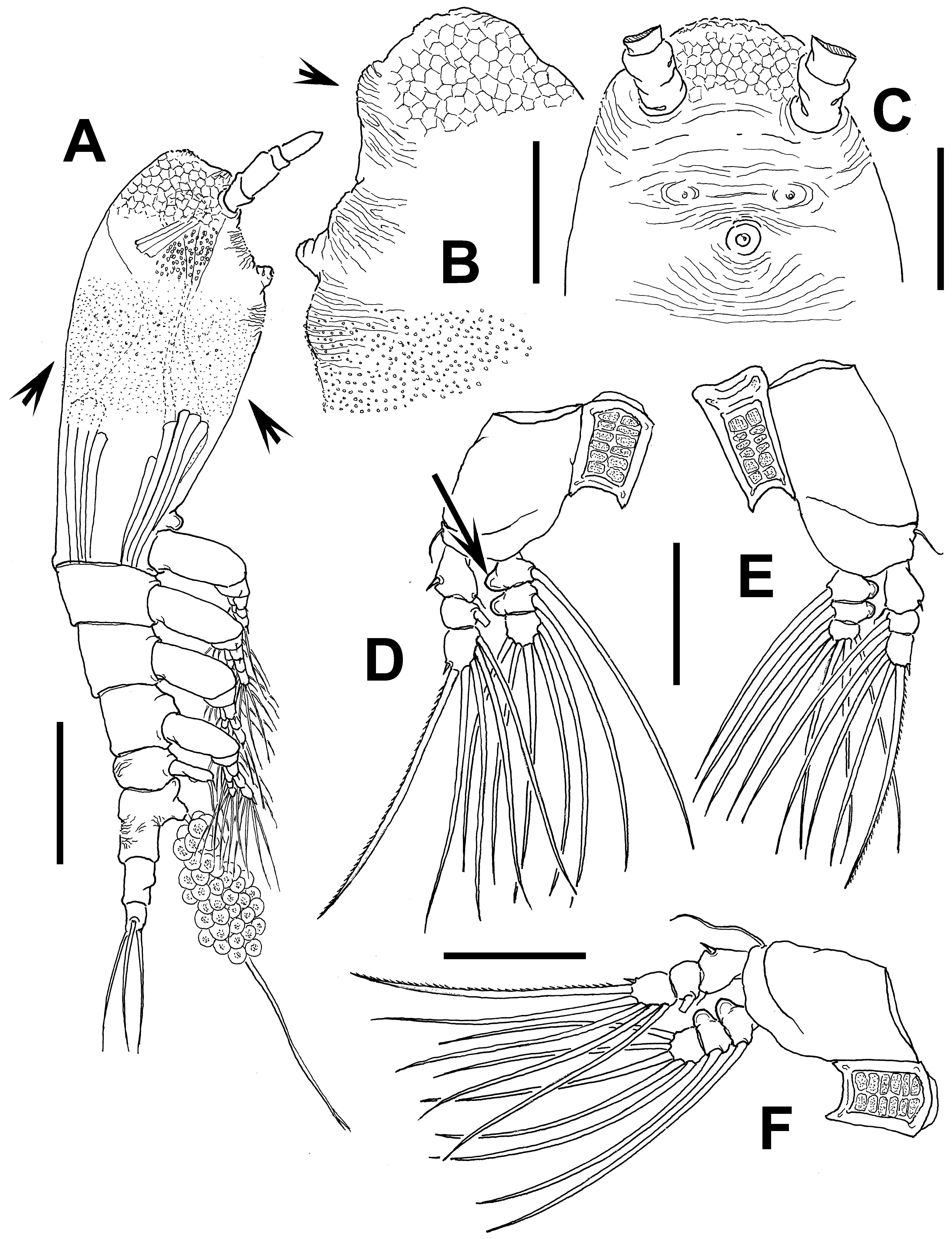
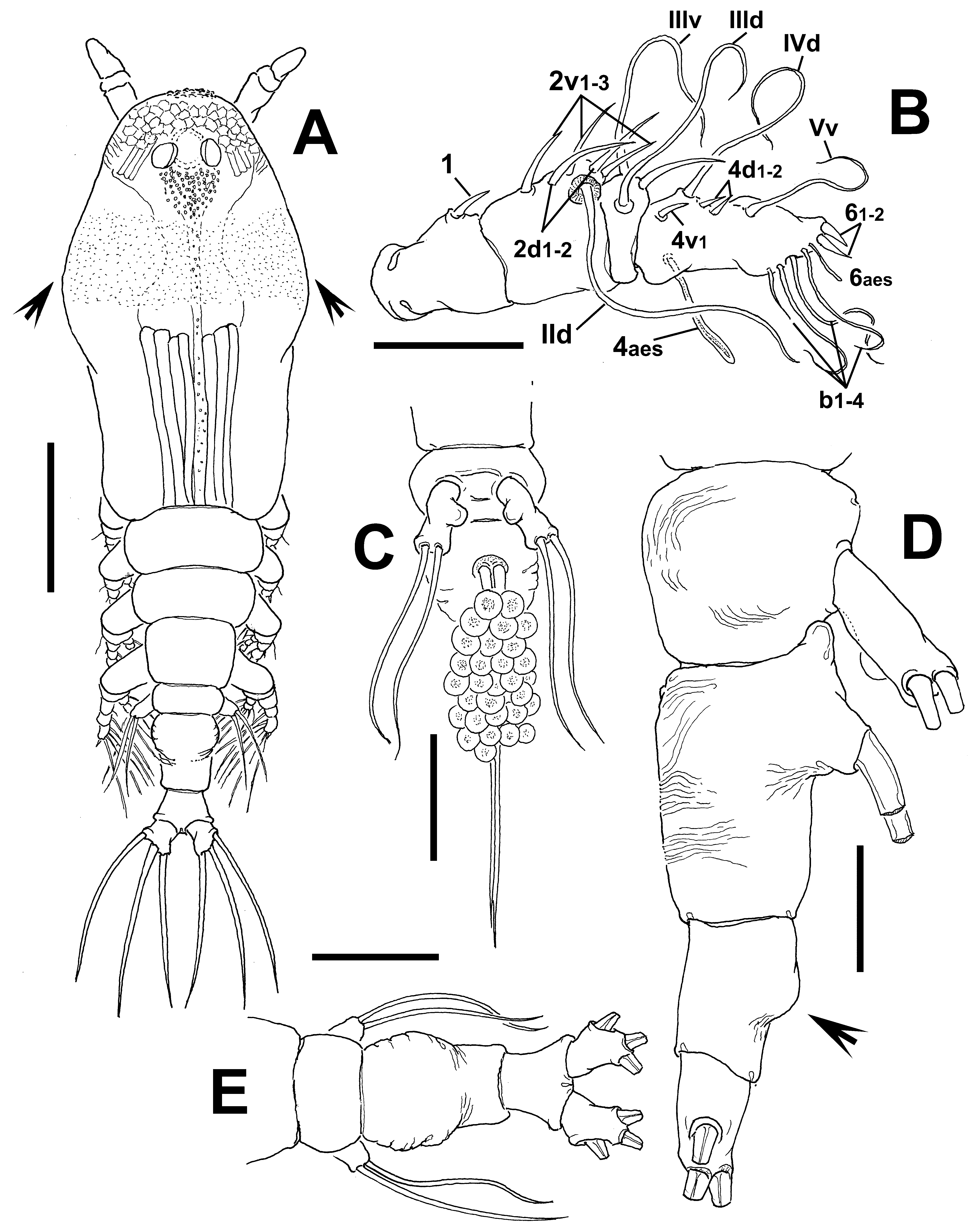
Description of adult female. Body robust, relatively short ( Figs 64 View FIGURE 64 A, 65A); cephalothorax with expanded anterior half, widest at midlength. Body length of holotype female 1.07 mm. Cephalothorax 0.56 mm long, representing approximately 57% of total body length. Midventral oral papilla protuberant, located at 26% of cephalothorax length. Pair of relatively small ocelli present, pigment cups moderately developed, medially separated, weakly pigmented on reduced inner area; ventral cup larger than lateral cups ( Fig. 65 View FIGURE 65 A). Cephalic area with “forehead” with low crest ornamented with shallow transverse striations ( Fig. 64 View FIGURE 64 B, C), frontal sensilla absent. Field of shallow reticulation covering anteriormost surface of cephalic area ( Fig. 64 View FIGURE 64 B, C) and fringe of minute papilla-like cuticular processes partially covering anterior half of cephalothorax ( Figs 64 View FIGURE 64 A, 65A). Ventral rounded low protuberance ornamented with transverse striae arising between antennulary bases (arrowed in Fig. 64 View FIGURE 64 B). Ventral surface with additional features including: 1) transverse striations in perioral area; 2) pair of symmetrical nipple-like processes on anterior ventral surface at each side of oral area ( Fig. 64 View FIGURE 64 C).
Urosome consisting of fifth pedigerous somite, genital double-somite and anal somite, together measuring 0.23 mm and representing 23% of total body length. Relative lengths of urosomites (fifth pedigerous, genital double- and free somites) 33.5: 42.8: 23.7 = 100, respectively ( Fig. 65 View FIGURE 65 D, E). Fifth pedigerous somite with few wrinkles on dorsal and lateral surfaces. Genital double-somite with wrinkles on dorsal, lateral, and ventral surfaces ( Fig. 65 View FIGURE 65 D, E); somite with strong ventral process on anterior margin ( Fig. 65 View FIGURE 65 D). Anal somite with ventral protuberance (arrowed in Fig. 65 View FIGURE 65 D). Caudal ramus subrectangular, 1.6 times longer than wide, armed with three subequally long, sparsely setulated caudal setae. Ovigerous spines paired, relatively short, 45% of total body length (0.44 mm) ( Fig. 64 View FIGURE 64 A). Spines basally separated, slender, straight at their bases and along shaft, without distal expansions and tapering apically, one spine slightly shorter; spines 0.44 mm long.
Antennule relatively short, length 0.17 mm, representing about 16.5 % of total body length and 30% of cephalothorax length; 4-segmented, segments 3 and 4 compressed. Relative length of short distal antennulary segment 42.5%. In terms of pattern described by Grygier & Ohtsuka (1995) for female monstrilloid antennulary armature: spiniform element 1 present on first segment; elements on second segment: 2d1-2, 2v 1-3, and IId. Third segment with long spiniform, curved element 3, and setiform elements IIId, and IIIv. Segment 4 bearing elements 4d1,2, 4v 1; elements 4v 2-3 not observed; setae IVd, Vv and 4aes present. Element 5 absent. Subterminal elements b1–4 present, unbranched, apical elements 61-2 and 6aes present in specimen ( Fig. 65 View FIGURE 65 B).
Incorporated first pedigerous somite and succeeding three free pedigerous somites each bearing a pair of biramous legs. Pedigerous somites 2–4, together accounting for 24 % of total length in dorsal view in each of the specimens examined. Legs 1–4 slightly decreasing in size posteriorly. Intercoxal sclerites of legs 1–4 subrectangular, widest at base, slightly tapering distally, surface with finely spinulose patches ( Fig. 64 View FIGURE 64 D–F). Bases of legs 1–4 with hair-like lateral seta; on leg 3, this seta about 2.5 times longer and slightly thicker than those on the other legs ( Fig. 64 View FIGURE 64 F). Endopods and exopods of legs 1–4 triarticulated; first and second endopodal segments of legs 1–4 with outer margin protuberant (arrowed in Fig. 64 View FIGURE 64 D). Ramal setae all biserially plumose except minute spiniform outer seta on exopodal segments 1 and 3, and inner seta of first exopodal segment, these latter being remarkably short, slender ( Fig. 64 View FIGURE 64 D, E). Spine on distal exopodal segment of right leg 4 noticeably short, 0.25 times as long as segment. Also, outermost apical exopodal setae of legs 1–4 with inner margin naked, outer margin spinulose.
Armature formula of legs 1–4:
Fifth legs medially separated, bilobate, outer (exopodal) lobe elongate, distally truncate, reaching about 1/3 length of genital double-somite, armed with two subequally long setae on distal position. Inner lobe represented by small, rounded process inserted at middle inner margin of main lobe.
Male: unknown.
Type locality. Port Hacking, New South Wales, Australia (34°4.670’ S, 151°9.100’ E).
Etymology. The species is named in recognition of the Tharawal people, traditional owners of the Port Hacking area.
Diagnosis. Cymbasoma with expanded, robust cephalothorax with reticulation on cephalic area and fringe of minute papilla-like processes, covering part of anterior half of cephalothorax. Antennule short, representing about 16.5 % of total body length and 30% of cephalothorax length. Third antennulary segment representing more than 40% of antennule length. Oral papilla protuberant. Legs 1–4 with first and second endopodal segments with inner margin protuberant. Fifth pedigerous and genital double-somite with wrinkles. Fifth leg with two distal setae on elongate outer lobe, inner lobe small, rounded, unarmed. Anal somite with ventral protuberance. Ovigerous spines representing 45% of total body length.
Remarks. Cymbasoma tharawalorum sp. nov. is assignable to the agoense group and is easily recognizable by a combination of characters including a short, globose inner lobe inserted medially along the inner margin of the outer (exopodal) lobe of the fifth leg. The other species of the group have either an elongate, digitiform inner lobe ( C. dakini , C. lourdesae ) or a strong, thumb-like lobe arising proximally ( C. agoense ). The cephalic cuticular ornamentation is also distinctive; it has a reticulation field on the anterior part of the frontal surface and a wide fringe of papilla-like elements covering 1/3 of the cephalothorax ( Figs. 65 View FIGURE 65 A). The antennule has some additional distinctive characters including relatively short third and fourth segments; the latter representing less than 40% of the total length of the antennule, thus diverging from the other species in which the last segment is longer (more than 50% of the antennule length). Cymbasoma tharawalorum differs from C. agoense in the shape and structure of the fifth leg inner lobe; both setae on the outer lobe are distal in the new species whereas one is distal and one is subdistal in C. agoense ( Sekiguchi 1982: fig. 6F). They differ also in the body proportions, with pedigers 2–3 being short in C. tharawalorum , but clearly longer in C. agoense . In addition, the fifth pedigerous somite is almost as long as the subquadrate genital double-somite ( Sekiguchi 1982: fig. 6A). In the new species the fifth pediger is about 2.5 times shorter than the genital double-somite, which is rounded at its anterior half and has straight margins in its posterior half.
No known copyright restrictions apply. See Agosti, D., Egloff, W., 2009. Taxonomic information exchange and copyright: the Plazi approach. BMC Research Notes 2009, 2:53 for further explanation.