
Cymbasoma tranteri, Suárez-Morales, Eduardo & Mckinnon, David, 2016
|
publication ID |
https://doi.org/ 10.11646/zootaxa.4102.1.1 |
|
publication LSID |
lsid:zoobank.org:pub:9A7BA798-AA7C-4CAA-B42C-1E260CA573E4 |
|
DOI |
https://doi.org/10.5281/zenodo.6091349 |
|
persistent identifier |
https://treatment.plazi.org/id/03C4CA6D-D576-FFCC-FF12-54EC95FE2D64 |
|
treatment provided by |
Plazi |
|
scientific name |
Cymbasoma tranteri |
| status |
sp. nov. |
Cymbasoma tranteri sp. nov.
( Figs 62 View FIGURE 62 , 63 View FIGURE 63 )
Material examined. Holotype: Adult female from Port Hacking, New South Wales, Australia (34°4.670’ S, 151°9.100’ E), partially dissected, ethanol-preserved; dissected parts mounted on slide in glycerine, sealed with Entellan®. Date of collection: 30th October 1986. Slide desposited in the collection of MTQ, Australia (cat. MTQ W34403).
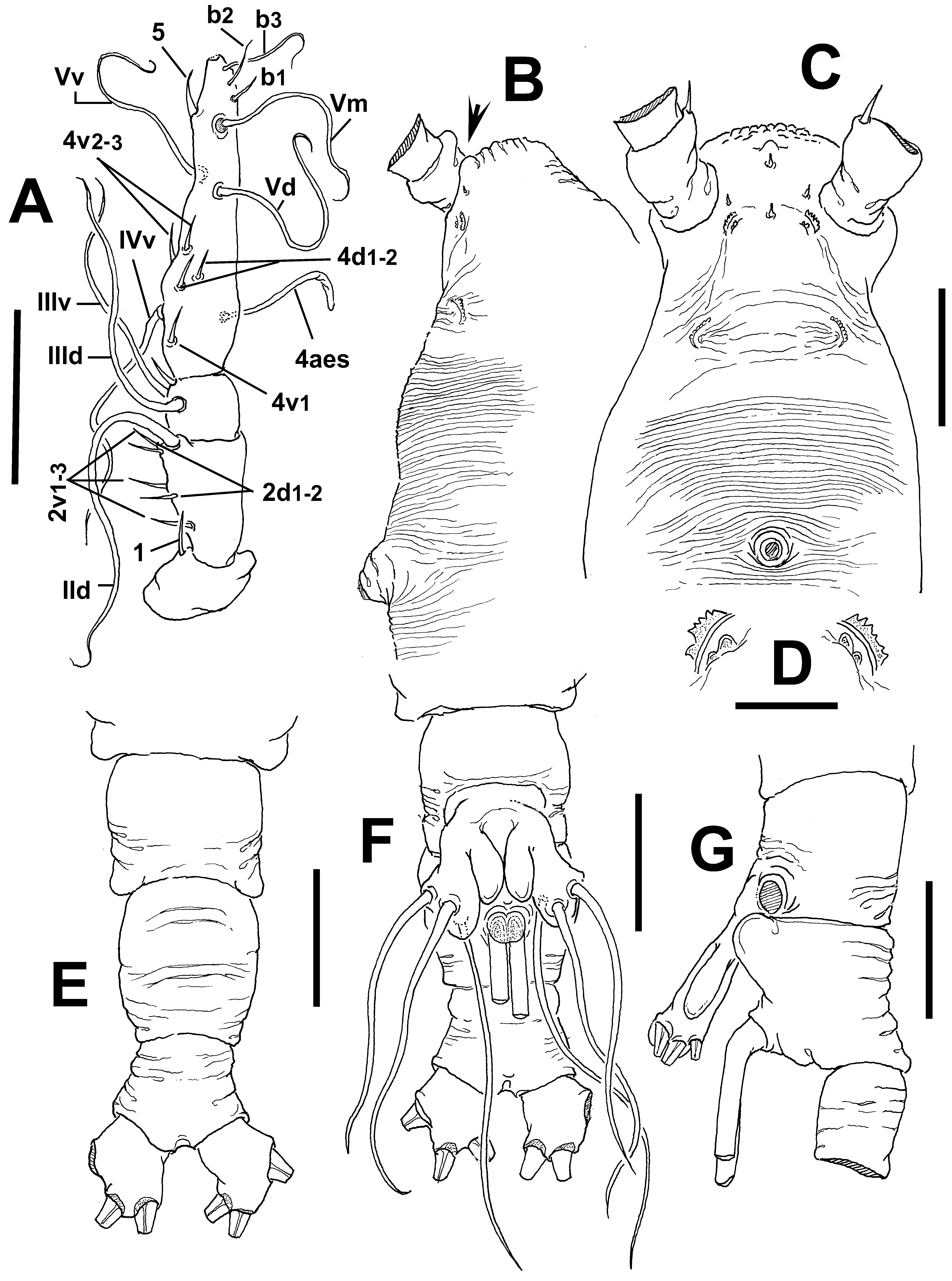
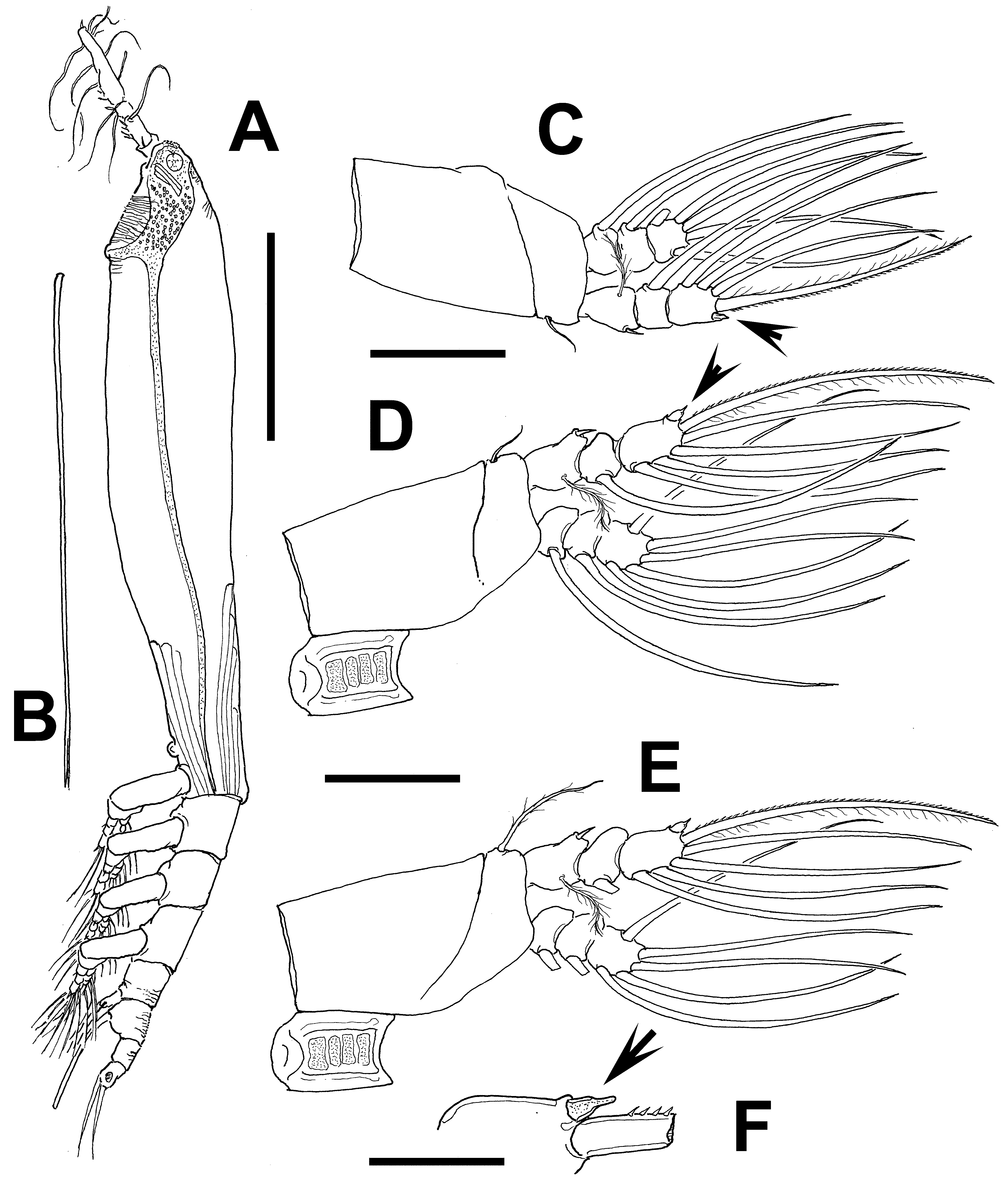
Description of adult female. Body elongate, slender ( Fig. 63 View FIGURE 63 A); total length of holotype female 2.23 mm. Cephalothorax approximately 1.48 mm long, representing 68% of total body length. Midventral oral papilla moderately protuberant, located at 15% of cephalothorax length. Pair of relatively large ocelli present, pigment cups well developed, medially conjoined, separated by less than half an eye diameter, weakly pigmented; ventral cup as large as lateral cups. Frontal area ornamented with deep transverse striations ( Fig. 62 View FIGURE 62 B, C) and medial rounded protuberance (arrowed in Fig. 62 View FIGURE 62 B); frontal sensilla absent. Anterior part of cephalothorax with field of transverse wrinkles limited to the ventral surface between the nipple-like processes and post-oral area. Additional ornamentation of ventral surface including: 1) four sensillum-like elements, one in medial position posterior to small rounded protuberance, the other three adjacent to nipple like processes; 2) two pairs of small, cuticular crescent-shaped processes between antennulary bases, with few adjacent striae ( Fig. 62 View FIGURE 62 D); 3) pair of symmetrical nipple-like processes on anterior ventral surface located anterior to oral papilla, processes connected medially by transverse striae.
Urosome consisting of fifth pedigerous somite, genital double-somite and anal somite, together representing 12% of total body length. Relative lengths of urosomites (fifth pedigerous, genital double- and free anal somite) 32.6: 40: 27.4 = 100, respectively ( Fig. 62 View FIGURE 62 E–G). Fifth pedigerous somite shorter than genital double-somite, with straight lateral margins, posterolateral corners forming rounded protuberances; somite with wrinkles on dorsal and ventral surfaces ( Fig. 62 View FIGURE 62 E). Genital somite longest of urosome, medial margin of somite moderately expanded, with transverse wrinkles on dorsal surface ( Fig. 62 View FIGURE 62 E). Antero-ventral surface of somite with strong, rounded protuberance ( Fig. 62 View FIGURE 62 G). Ovigerous spines paired, separated at base, relatively long (1.14 mm), 52% of total body length ( Fig. 63 View FIGURE 63 B). Spines slender, straight at their bases and along shaft, distally acute, equally long. Anal somite with weak medial constriction and with striae on dorsal and ventral surfaces. Caudal rami not divergent, subrectangular, about 1.3 times as long as wide, armed with three caudal setae.
Antennule length 0.36 mm, representing about 16.2% of total body length and 23.4% of cephalothorax length; 4-segmented. Relative length of distal antennulary segment 57%. Short, spiniform element 1 present on first segment; elements on second segment: 2d1-2, 2v 1-3, and IId. Third segment with short spiniform element 3; IIId and IIIv of normal aspect. Segment 4 bearing short, spiniform elements 4d1,2, 4v 1-3; setae IVv, Vd, Vv, Vm, and 4aes present. Element 5 spiniform, curved. Subterminal elements b1-3 present, short, unbranched. Apical elements 61 and 62 not present in specimens, but sockets were observed ( Fig. 62 View FIGURE 62 A).
Incorporated first pedigerous somite and succeeding three free pedigerous somites each bearing a pair of biramous legs. Pedigerous somites 2–4, together accounting for 17% of total body length. Intercoxal sclerites of legs 1–4 subrectangular, surface and posterior margin smooth. Bases of legs 1–4 with hair-like lateral seta ( Fig. 63 View FIGURE 63 C–E); on leg 3, this seta about three times longer, thicker than those on other legs ( Fig. 63 View FIGURE 63 E). Endopods and exopods of legs 1–4 triarticulated. Ramal setae all biserially plumose except spiniform outer seta on exopodal segments 1 and 3, and inner seta of first exopodal segment, these latter being short, slender. Outermost distal spines on third exopodal segment of legs 1–4 short, 0.25 times as long as segment; spines modified, mammiliform (arrowed in Fig. 63 View FIGURE 63 C, D, F). Outermost apical setae on third exopodal segment of legs 1–4 with inner margin sparsely setulose, outer margin spinulose.
Armature formula of legs 1–4:
Fifth legs medially conjoined, bilobate, outer (exopodal) lobe cylindrical, with distal rounded process. Inner lobe arising basally, thumb-like, reaching midlength of exopodal lobe. Outer lobe armed with three subapical setae, innermost seta longest ( Fig. 62 View FIGURE 62 F).
Male: unknown.
Type locality. Port Hacking, New South Wales, Australia (34°4.670’ S, 151°9.100’ E).
Etymology. This species is named for David Tranter, in recognition of his contributions to the study of Australian marine zooplankton.
Diagnosis. Cymbasoma with slender body and ventral field of wrinkles on cephalic area. Ventral surface of cephalic area with four sensillum-like elements, one in medial position posterior to small rounded protuberance, the other three adjacent to nipple-like processes. Antennule short, representing about 16.2 % of total body length and 23% of cephalothorax length. Third antennulary segment remarkably long, representing 57% of antennule length. Oral papilla moderately protuberant. Legs 1–4 with first and second endopodal segments with inner margin not protuberant; outermost distal spines on third exopodal segment of legs 1–4 short, modified, mammiliform. Fifth pedigerous and genital double-somite with wrinkles on dorsal surface and produced posterolateral corners; genital double-somite longest of urosome. Fifth leg with three subdistal setae on elongate outer lobe, innermost seta longest. Inner lobe thumb-like, unarmed. Anal somite not medially constricted. Ovigerous spines representing 52% of total body length.
Remarks. This new species, C. tranteri , can be distinguished from its Australian congeners that have a fifth leg inner lobe being shorter than the outer lobe by details of the armature of the outer lobe. In C. tranteri the exopodal lobe has three subdistal setae of which the innermost is longest whereas in the other species the outer lobe bears two or three distal setae, the innermost being the shortest. Cymbasoma tranteri also has a remarkably long fourth antennulary segment, representing up to 57% of the total length of the antennule, a character not found in any other known congener. For instance, the length of this segment among the species of the longispinosum -group ranges between 46 and 50%. Two other species, C. reticulatum and C. gracilis , also have a long terminal segment, the relative length of which is 52% (cf. Giesbrecht 1893; Gurney 1927). Another distinctive apomorphy of C. tranteri is the peculiar mammiliform shape of the outermost spine of the third exopodal segment of legs 1–4 ( Fig. 63 View FIGURE 63 C-F), a character not previously observed in any other known Cymbasoma .
No known copyright restrictions apply. See Agosti, D., Egloff, W., 2009. Taxonomic information exchange and copyright: the Plazi approach. BMC Research Notes 2009, 2:53 for further explanation.