
Cymbasoma leighrandalli, Suárez-Morales, Eduardo & Mckinnon, David, 2016
|
publication ID |
https://doi.org/10.11646/zootaxa.4102.1.1 |
|
publication LSID |
lsid:zoobank.org:pub:9A7BA798-AA7C-4CAA-B42C-1E260CA573E4 |
|
DOI |
https://doi.org/10.5281/zenodo.6091345 |
|
persistent identifier |
https://treatment.plazi.org/id/03C4CA6D-D57C-FFC7-FF12-53AE931B2A02 |
|
treatment provided by |
Plazi |
|
scientific name |
Cymbasoma leighrandalli |
| status |
sp. nov. |
Cymbasoma leighrandalli sp. nov.
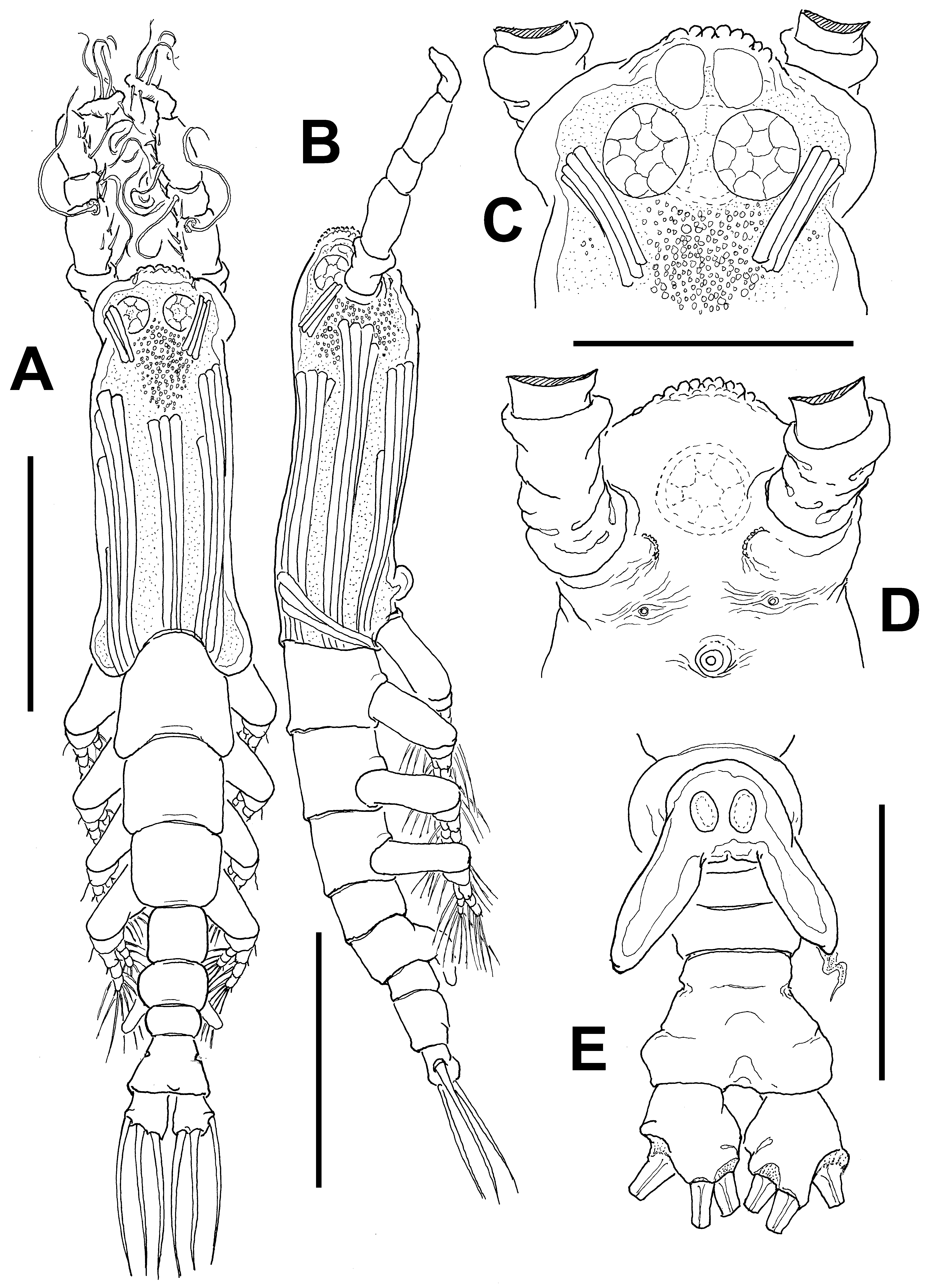
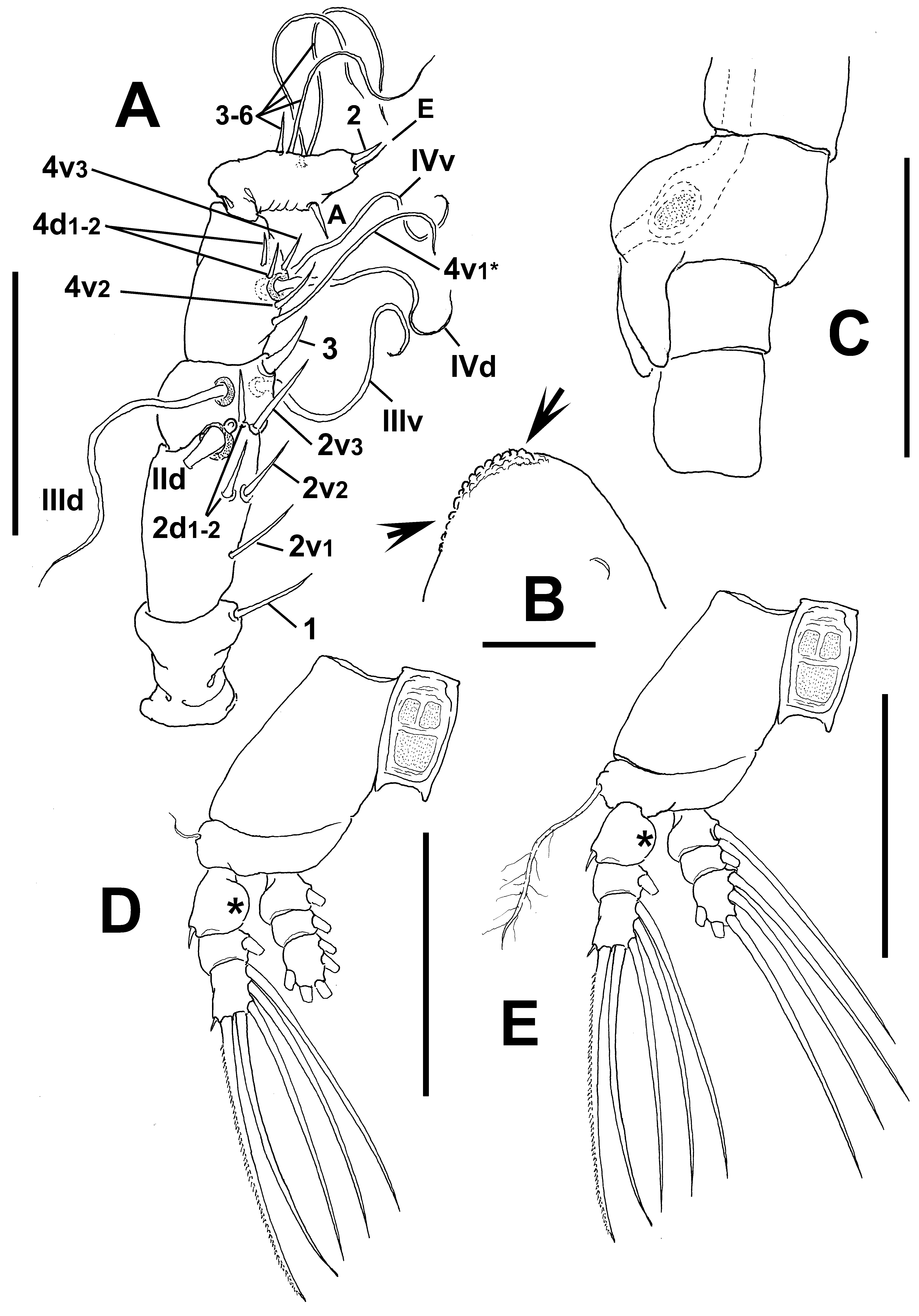
( Figs 58 View FIGURE 58 , 59 View FIGURE 59 )
Material examined. Holotype: adult male from Ross Creek, Townsville, Queensland, Australia ( 19°16.500’ S, 146°48.500’ E), partially dissected, ethanol-preserved; dissected parts mounted on slide in glycerine, sealed with Entellan®. Date of collection: 15th May 1983. Slide desposited in the collection of MTQ, Australia (cat. MTQ W34401).
Description of adult male. Total body length 0.66 mm. Cephalothorax 0.28 mm long, representing 44% of total body length ( Fig. 58 View FIGURE 58 A, B). Midventral oral papilla weakly developed, located at 22% of cephalothorax length ( Fig. 58 View FIGURE 58 B). Cephalic region moderately protuberant bilaterally in dorsal view. Pair of large dorsal ocelli present, moderately developed; pigment cups relatively large. Ocelli separated by the length of less than half an eye diameter, faintly pigmented. Ventral ocellus as large as lateral cups ( Fig. 58 View FIGURE 58 C). No frontal sensilla between antennulary bases. Forehead produced forming lumpy medial crest flanked by few wrinkles ( Figs 58 View FIGURE 58 C, D, arrowed in 59B). Ventral surface of head between antennulary bases and oral papilla smooth. Dorsal surface of cephalic area smooth. Ventral surface with additional cuticular elements: 1) pair of symmetrical, crescent-shaped cuticular processes on anterior ventral surface between bases of antennules, with few adjacent striae ( Fig. 58 View FIGURE 58 D); 2) nipplelike processes with adjacent transverse wrinkles; 3) few perioral transverse wrinkles.
Urosome consisting of fifth pedigerous somite, genital somite (carrying genital complex), preanal somite, and anal somite. Genital somite longer than fifth pedigerous somite. Genital complex of type II, represented by pair of moderately divergent, slightly asymmetrical genital lappets ( Fig. 58 View FIGURE 58 E), right lappet slightly longer than left lappet. Lappets posteriorly directed in lateral view, relatively short, reaching posterior margin of preanal somite ( Fig. 59 View FIGURE 59 C). Common basal joint of lappets flat, with small medial notch ( Fig. 58 View FIGURE 58 E). Lappet surface smooth. Anal somite noticeably long, about 2.6 times as long as preanal somite in dorsal view, comprising 33% of urosome length; constriction at proximal 1/3 of somite visible in ventral aspect, cuticular hyaline frill absent. Caudal rami subquadrate, approximately 1.1 times as long as wide, about 0.7 times as long as anal somite. Each ramus with three setae.
Antennulary length 0.19 mm. Antennules representing 30% of total body length, and 70% of cephalothorax length; 5-segmented, all segments separated, with segment 5 located distal to geniculation ( Fig 59 View FIGURE 59 A). Length ratio of antennulary segments, from first to fifth 15:25.3:12.3: 21.8: 25.6 (= 100). Setal element 1 on first segment relatively long, setiform, reaching proximal 1/3 of second segment. Antennulary elements 2v 1-3, 2d1,2 stiff, spiniform; element IId present on second segment. Elements IIId, IIIv, and strong, spiniform element 3 present on third segment, the latter reaching 1/3 of fourth segment. Fourth segment with elements 4d1,2, 4v 1– 3 present; element 4v 1 setiform (asterisk in Fig. 59 View FIGURE 59 A). Setae IVd, IVv present. Fifth segment with 4 “b”-group setae, elements b1-3 long, unbranched; element 61 present in distal position. According to Huys et al. (2007) setal nomenclature of the distal segment, elements A, E and 2–6 present.
Incorporated first pedigerous somite and succeeding three pedigerous somites each bearing well-developed biramous legs. Pedigerous somites 2–4, together accounting for 31% of total body length in dorsal view. Coxae of each pair unarmed, joined by intercoxal sclerite ornamented with spinulose patches. Exopods of legs 1–4 longer than endopods. Bases of legs 1–4 with hair-like lateral basipodal seta ( Fig. 59 View FIGURE 59 D, E); on leg 3, this seta about 4.5 times longer, sparsely setulated from distal half and slightly thicker than those on the other legs ( Fig. 59 View FIGURE 59 E). Endopods and exopods of legs 1–4 triarticulated. Ramal setae all biserially plumose except spiniform outer seta on exopodal segments 1 and 3. Flexible, slender, sparsely setulated inner seta present on first exopodal segment of legs 1–4. Also, outermost apical exopodal setae of legs 1–4 with outer margin sparsely spinulose, inner margin naked. Inner seta on first exopodal segment of legs 1–4 absent (asterisks in Fig. 59 View FIGURE 59 D, E).
Armature formula of legs as follows: Female: unknown.
Type locality. Ross Creek, Townsville, Queensland, Australia ( 19°16.5’ S, 146°48.5’ E).
Etymology. This species is dedicated to Leigh Randall McKinnon, son of the second author (DM).
Diagnosis. Cymbasoma with conspicuous medial protuberance on cephalic area with lumpy surface. First antennulary segment with long, stout spiniform element 1; second segment with stout spiniform elements 2d1,2 and 2v 2-3. Element 2v 3 setiform, long. Antennule representing 30% of total body length and 70% of cephalothorax length. Genital complex of type II, slightly asymmetrical, tapering distally, with rounded tips, basal joint between lappets flat, with medial notch. Anal somite 2.6 times as long as preanal somite, with proximal constriction. Three caudal setae.
Remarks. The male of C. leighrandalli sp. nov. differs from the other Australian Cymbasoma by having smooth genital lappets and a flat margin with a notch between the lappets. The other species have a rounded medial process, basal spines or rows of large or small spinules along the inner margin, as in C. quadridens ( cf. Suárez- Morales & Pilz 2008), and the Australian C. colefaxi , C. annulocolle , and C. pseudoquadridens . A flat medial margin between lappets is present in other congeners like C. rigidum , C. longispinosum , C. thompsonii ( cf. Sars 1921), C. chelemense ( cf. Suárez-Morales & Escamilla 1997), and C. quintanarooense ( cf. Suárez-Morales 2000). Among this group of species, only C. rigidum has a constricted anal somite ( Sars 1921). These two species share other characters including similar body proportions, a long anal somite being about twice as long as the preanal somite, and a similar structure and length of both genital lappets. Comparison with Sars’s (1921) redescription of C. rigidum reveal differences in several other important characters such as the oral papilla which is weakly developed and located at 22% of the cephalothorax length in the new species whereas it is normally developed and situated more posteriorly, at 33% of the cephalothorax length in C. rigidum . In C. leighrandalli the distal antennulary segment is short, robust and has a short apical element 2 ( sensu Huys et al. 2007), whereas the same segment is slender and has a remarkably long apical element 2 C. rigidum (cf. Sars 1921). Cymbasoma rigidum ( cf. Sars 1921) lacks a corrugate frontal process, a character present in the new species ( Fig. 58 View FIGURE 58 C). The constriction of the anal somite is medial in C. rigidum ( cf. Sars 1921) whereas it is located on the proximal 1/3 of the somite in C. leighrandalli ( Fig. 58 View FIGURE 58 E). Also, four caudal setae are present in C. rigidum and three in the new species.
No known copyright restrictions apply. See Agosti, D., Egloff, W., 2009. Taxonomic information exchange and copyright: the Plazi approach. BMC Research Notes 2009, 2:53 for further explanation.
|
Kingdom |
|
|
Phylum |
|
|
Class |
|
|
Order |
|
|
Family |
|
|
Genus |