
Triplonychus tibialatus, Rosa, Simone Policena, 2011
|
publication ID |
https://doi.org/ 10.5281/zenodo.277308 |
|
DOI |
https://doi.org/10.5281/zenodo.6188953 |
|
persistent identifier |
https://treatment.plazi.org/id/03C58797-BE08-2923-FF57-F90CFAF1F3A3 |
|
treatment provided by |
Plazi |
|
scientific name |
Triplonychus tibialatus |
| status |
sp. nov. |
Triplonychus tibialatus sp. nov.
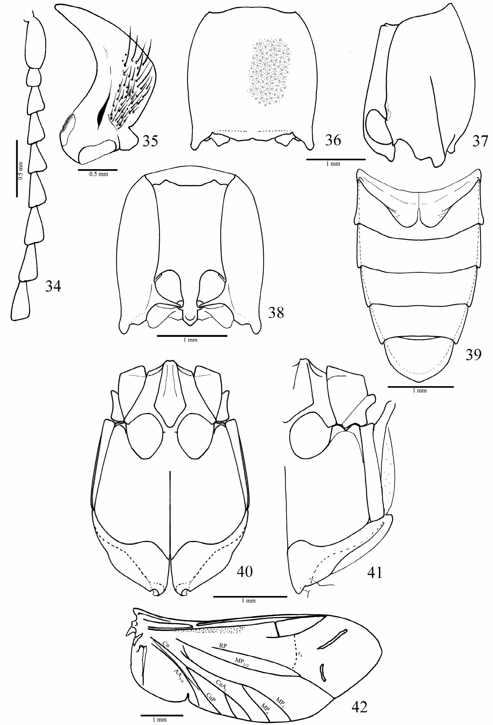
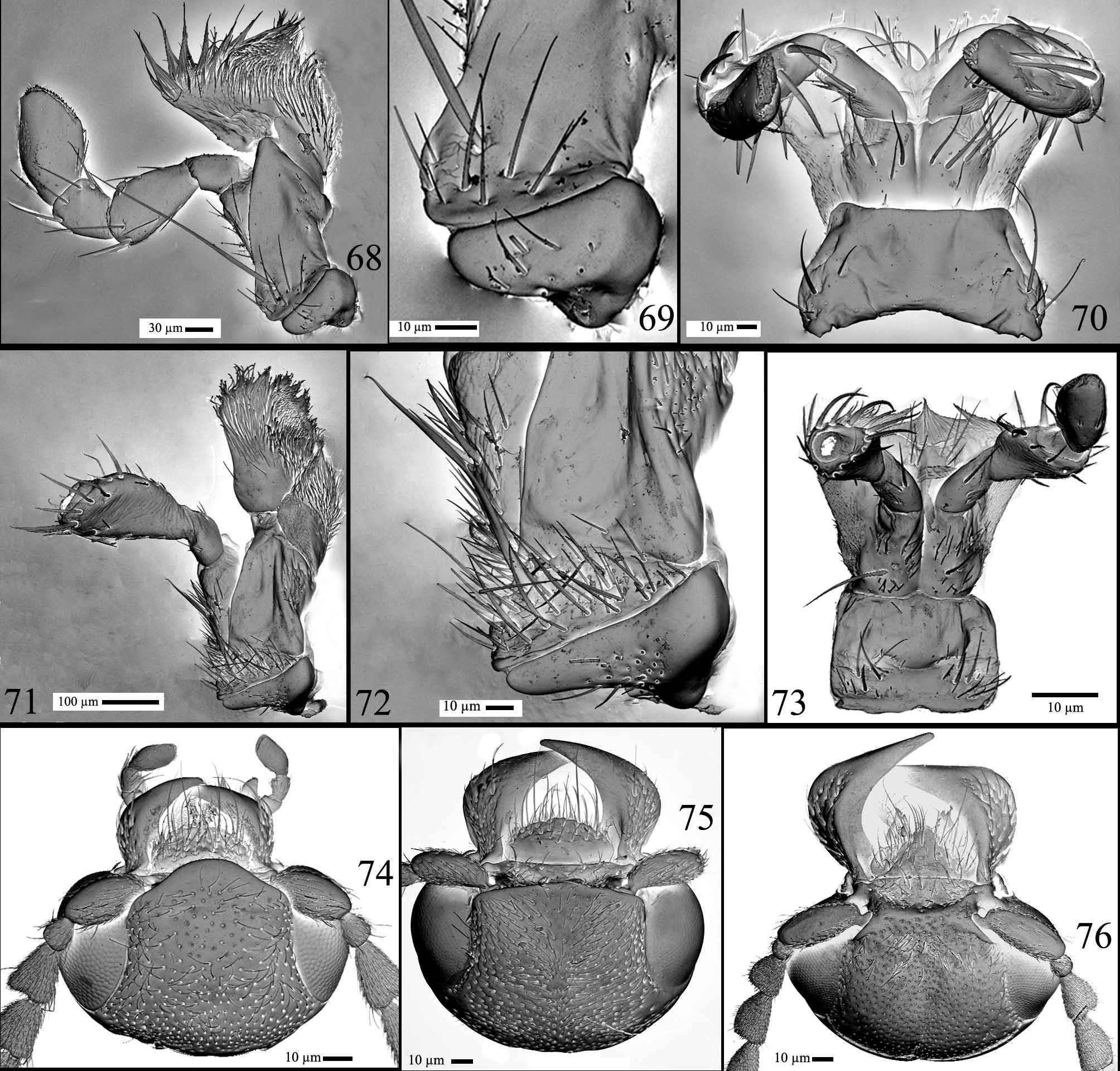
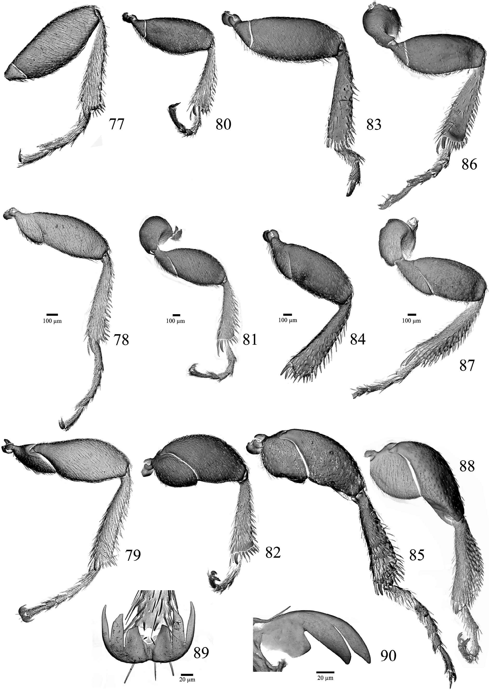
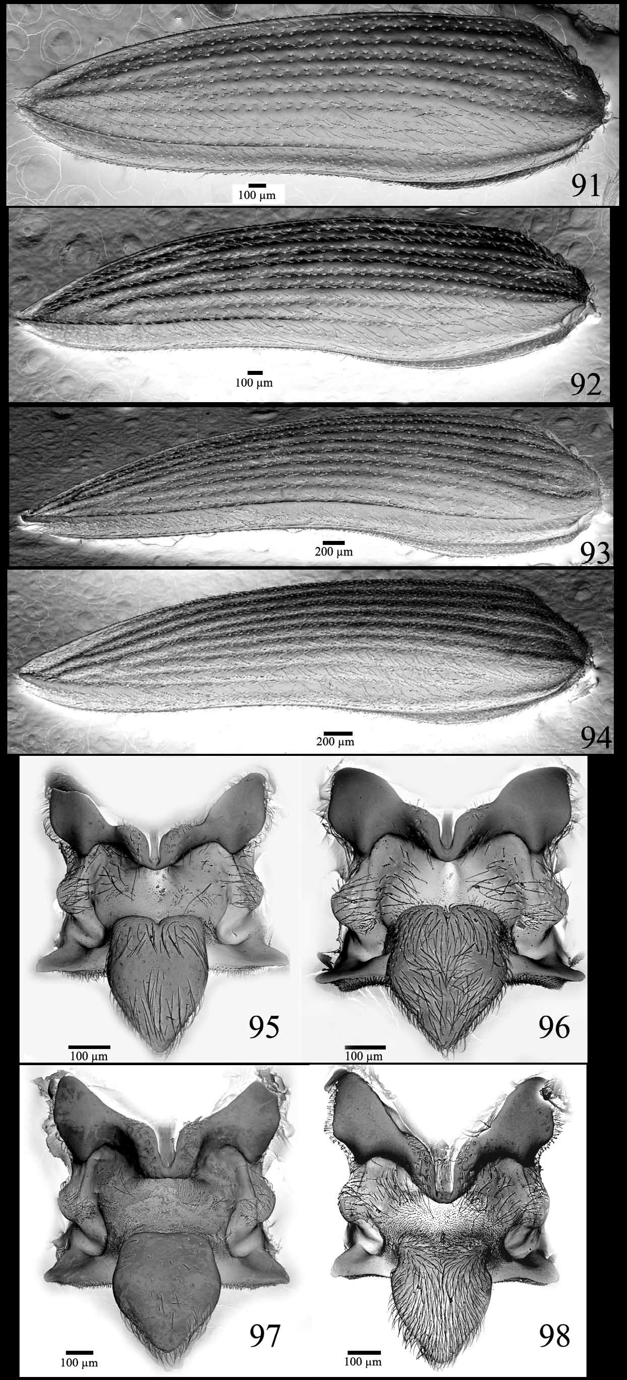
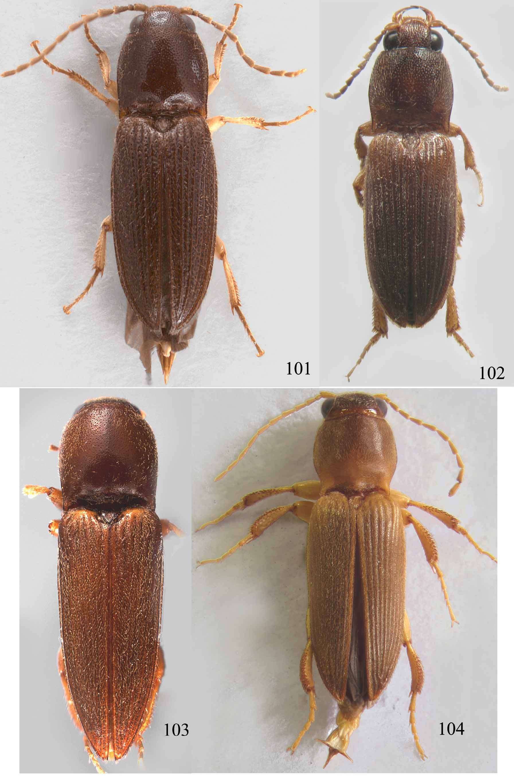
( Figs. 34–48 View FIGURES 34 – 42 View FIGURES 43 – 48 , 71–73, 75 View FIGURES 68 – 76 , 83–85 View FIGURES 77 – 90 , 93, 97 View FIGURES 91 – 98 , 103 View FIGURES 101 – 104 )
Etymology. From Latin, tibia = tibia; latus = broad, alluding to the broadened tibiae of this species.
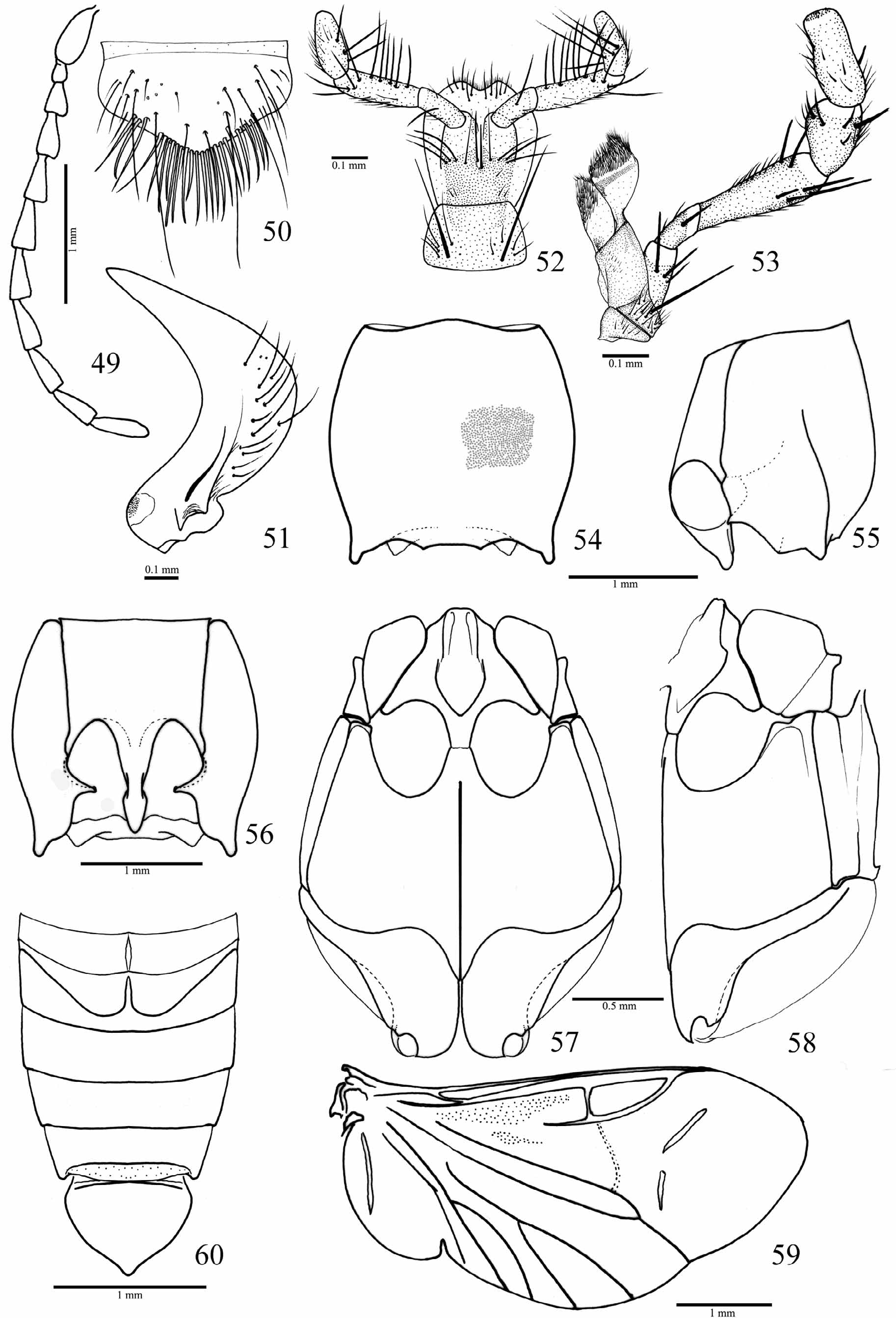
Diagnosis. Anterior margin of frons carinate, nearly straight, not produced ( Fig. 75 View FIGURES 68 – 76 ); mandibles large, unidentate ( Fig. 35 View FIGURES 34 – 42 ); pronotum 1.0–1.1 times longer than wide with punctation double ( Fig. 36 View FIGURES 34 – 42 ); prosternum with a pair of lateral spiniform projections on anterior margin ( Fig. 38 View FIGURES 34 – 42 ), prosternal process curved dorsad ( Fig. 37 View FIGURES 34 – 42 ); metacoxal plate short ( Fig. 41 View FIGURES 34 – 42 ); ninth elytral interstice entirely carinate ( Fig. 93 View FIGURES 91 – 98 ); metatrochanter and metafemur convex; meso- and metatibiae broadened with dorsal margin curved ventrad, outer surface densely covered with stout spiniform setae ( Figs. 84–85 View FIGURES 77 – 90 ).
Description. Male ( Fig. 103 View FIGURES 101 – 104 ). Body convex, densely covered with fine and short setae; setae yellowish and decumbent. Integument bright; head and pronotum evenly light yellowish-brown or light brown to brown, elytra lighter with basal margin yellow. Light brown specimens with pronotal and elytral punctures slightly darker than the surrounding area. Total length: 7.0–8.0 mm; elytra 2.6–2.7 times longer than prothorax; elytral base as wide as prothorax.
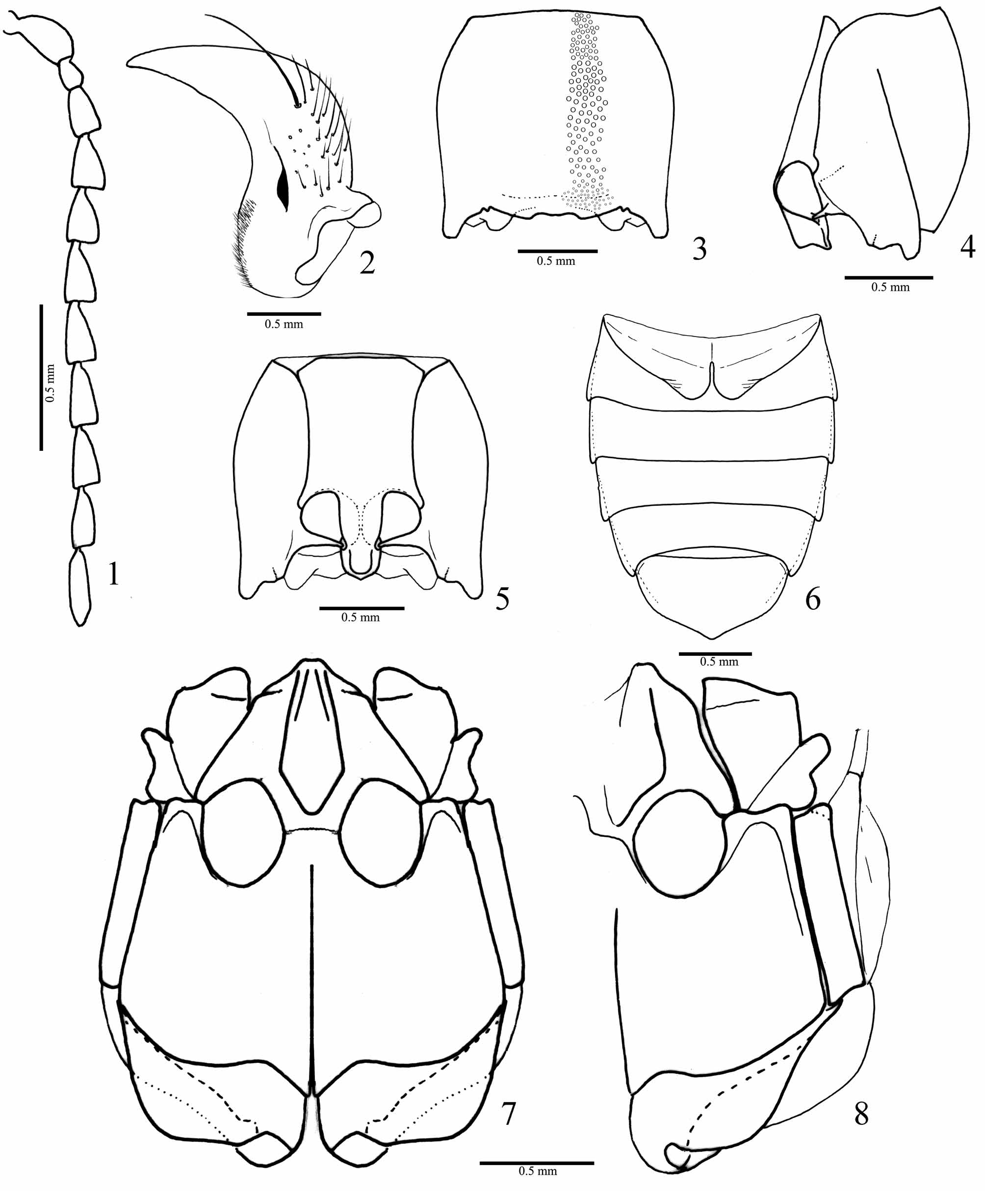
Head ( Fig. 75 View FIGURES 68 – 76 ). Frons usually concave, anterior margin carinate and nearly straight, frontoclypeal region steeply declivous to base of labrum and about 2 times wider than long. Punctures smaller than the larger punctures of pronotum, umbilicate 0.5–1.0 diameter apart. Antenna ( Fig. 34 View FIGURES 34 – 42 ) surpassing the hind angles of pronotum at antennomere 11; antennomere 3 1.7 times longer than 2, subequal to the 4. Dorsal index of eye prominence 0.4. Labrum semielliptical, transversely convex with distal half strongly punctate with long setae, basal half smooth. Mandibles ( Fig. 35 View FIGURES 34 – 42 ) large (distal third superimpose each other), lateroventral margin slightly expanded laterad then abruptly curved to apex, unidentate; mesobasal margin with a small translucent area covered with microsetae, dorsal surface with a longitudinal short and narrow carina. Maxillae ( Figs 71, 72 View FIGURES 68 – 76 ) with basistipe partly fused to medistipe, with a long stout seta and several fine, short setae; galea oval, densely setose. Labium ( Fig. 73 View FIGURES 68 – 76 ) with mentum rectangular or trapezoidal, densely setose on posterior angles; prementum slightly emarginate at anteromedian margin, lateroanterior angles weakly produced; second labial palpomere with stout setae apically; apical maxillary and labial palpomeres securiform.
Thorax. Prothorax ( Figs. 36–38 View FIGURES 34 – 42 ) 1.0–1.1 times longer than wide, lateral margins rounded, carinate on posterior 1/2 to 3/5. Pronotum convex evenly punctate with punctation double, larger punctures 1–2 diameters apart. Pronotal hind angles acute, parallel to slightly divergent; posterior margin between hind angle and median line with a very small notch. Hypomera ( Fig. 38 View FIGURES 34 – 42 ) with punctation double; notosternal suture curved. Prosternum ( Fig. 38 View FIGURES 34 – 42 ) about 1.4 times longer than wide, with a pair of spiniform short projections on anterior margin; median longitudinal area smooth, remaining area with punctation double, larger punctures smaller than the pronotal large punctures, 1– 3 diameters apart, denser on lateral borders. Prosternal process 2.2 times longer than coxal diameter with dorsal surface posteriorly to coxae flattened laterally and strongly curved dorsad up to apex or steeply sloping dorsad near the apex. Procoxal cavities closed. Scutellar shield ( Fig. 97 View FIGURES 91 – 98 ) cordiform, elevated about 45o above the level of mesoscutum. Mesoventrite ( Fig. 40 View FIGURES 34 – 42 ) with posterior region inclined 45o above the level of anterior region; mesoventral cavity widened up to anterior border of mesocoxae than abruptly convergent. Mesocoxal cavity oval, closed. Mesepisternum with anterior inner angle acute. Meso-metaventral suture absent or weakly impressed. Metaventrite ( Figs. 40, 41 View FIGURES 34 – 42 ) with punctuation double; metacoxal plate short with posterior margin rounded, decreasing in length gradually outwards. Elytra ( Fig. 93 View FIGURES 91 – 98 ) tapering from base to apex; elytral striae with 2–4 irregular rows of small and large punctures; interstices 2–8 convex on apical region; flattened on anterior region; interstice 9 entirely carinate. Legs ( Fig. 83–85 View FIGURES 77 – 90 ). Pro- and mesofemur convex, metafemur strongly convex. Tibiae widened apicad, meso- and metatibiae with dorsal margin curved ventrad; tibiae with spiniform setae on dorsal and apical margin, metatibiae with stout spiniform setae on outer surface shorter and wider than the dorsal ones; tibial spurs less than 0.5 times as long as tarsomere 1; tarsomeres densely covered with fine and spiniform setae.
Abdomen ( Fig. 39 View FIGURES 34 – 42 ) tapered from base to apex, with punctation double, denser on lateral margin. First ventrite 3.7 times longer on lateral border than at middle, hind angles of ventrites 1–4 acute, last ventrite subpentagonal. Tergite 8 ( Fig. 43 View FIGURES 43 – 48 ) evenly sclerotized, densely covered with microsetae, with several short setae on lateroposterior and posterior margins, anterior sclerotized border V-shaped emarginated. Sternite 8 ( Fig. 44 View FIGURES 43 – 48 ), pentagonal, with two rounded membranous areas medially and short setae lateroposteriorly; apex inverted U-shaped notched. Sternite 9 ( Fig. 45 View FIGURES 43 – 48 ) tapering to apex, median anterior margin membranous, anterior median half translucent, apex with short and elongate setae. Tergite 9 ( Fig. 46 View FIGURES 43 – 48 ) with anterior margin nearly straight, posterior lobes acute, tergite 10 ( Fig. 46 View FIGURES 43 – 48 ) semioval.
Aedeagus ( Figs. 47, 48 View FIGURES 43 – 48 ). Parameres with apex abruptly flattened laterally with two setae, articulated to penis by a transparent membrane. Penis with basal struts about 0.5 times as long as its total length, median region between basal struts with a short process, lateral sides posterior to basal struts rounded then parallel up to apex; ventral sclerite absent.
Type material. Holotype. [BR-MS-Três Lagoas, International Paper, Horto Rio Verde, Black light FIT, Eucalyptus grandis stand, Flechtmann, C. A. H. col., 08/XII/1994], [C1945], [CRELAT028], male ( MZUSP).
Paratypes. Same labels as the holotype, 1 male ( UNESP). [BR-MS-Três Lagoas, International Paper, Horto Rio Verde, Black light FIT, Eucalyptus grandis stand, Flechtmann, C. A. H. col., II/1995], [C821], [CRELAT089], 1 male ( MZUSP). [BR-MS-Três Lagoas, International Paper, Horto Rio Verde, Black light FIT, Eucalyptus grandis stand, Flechtmann, C. A. H. col., 11/II/1995], [C821], [CRELAT089], 1 male ( MZUSP). [BR-MS-Três Lagoas, International Paper, Horto Rio Verde, Black light FIT, Eucalyptus grandis stand, Flechtmann, C. A. H. col., 08/XII/ 1994], [C1944], [CRELAT030], 1 male ( MZUSP). Same labels, 1 male (dismembered) ( MZUSP). [BR-MS-Três Lagoas, International Paper, Horto Rio Verde, Black light FIT, Eucalyptus grandis stand, Flechtmann, C. A. H. col., 14/XII/1993], [C819], [CRELAT090], 1 male ( UNESP).
Ocurrence. BRAZIL. Mato Grosso do Sul state: Três Lagoas.
Remarks. The specimens available are poorly preserved. All have some legs broken and incomplete antennae; therefore, the relative length of the antennae is an approximation based on length of the antennomeres 1–9 ( Fig. 34 View FIGURES 34 – 42 ).
Discussion. Triplonychus tibialatus sp. nov. is similar to T. longicollis Erichson 1829 and T. ventralis Candèze, 1860 by the double punctation on pronotum, ninth elytral interstice entirely carinate, and abdomen and elytra tapering from base to apex. Triplonychus tibialatus sp. nov. differs from these species mainly in its pronotal and leg shapes, frontal carina not produced, mandible unidentate, prosternal process curved dorsad, mesoventrite less inclined anteriorly and shorter metacoxal plate.
All Triplonychus species described herein bear ninth elytral interstice carinate, elytral striae with 2–4 regular or irregular rows of punctures and tricuspid claws. Those characteristics are diagnostic for the genus as well as the shape of elytra, tapering from base to apex ( Candèze, 1860). However, only T. tibialatus sp. nov. exhibits elytra tapering from base to apex.
Triplonychus cruspinosus sp. nov., T. crassifemoris sp. nov. and Globothorax latidens sp. nov. posses similar wings, with vestigial r4, anal notch, two sclerotizations on apical region, CuA1 and basal spur of the MP3+4 absent and cross vein between MP3+4 and MP1+2 indistinct ( Fig. 59 View FIGURES 49 – 60 ). Triplonychus tibialatus sp. nov. differs from these species in the CuA2, which seems to split in two branches near the margin and in having an incomplete cross vein between MP3+4 and MP1+2 and a distinct basal spur in the MP3+4 ( Fig. 42 View FIGURES 34 – 42 ); these feature were observed in two dissected specimens in both right and left wings. All these species have the metanotum ( Fig. 26 View FIGURES 26 – 33 ) with the prescutum separated medially from the scutum by a median membranous area and the posterior part of the scutellum with a longitudinal apodema. They also have an inverted V-shaped notch on posterior margin of the hypomera ( Figs. 4, 5 View FIGURES 1 – 8 ) near the hind angle and the tarsal claws with two apical acute teeth and a basal truncate tooth ( Figs. 89, 90 View FIGURES 77 – 90 )


| MZUSP |
Museu de Zoologia da Universidade de Sao Paulo |
No known copyright restrictions apply. See Agosti, D., Egloff, W., 2009. Taxonomic information exchange and copyright: the Plazi approach. BMC Research Notes 2009, 2:53 for further explanation.