
Ptiloneura, Enderlein, 1900
|
publication ID |
https://doi.org/ 10.11646/zootaxa.5178.5.4 |
|
publication LSID |
lsid:zoobank.org:pub:E1EC192E-5623-4024-9E7A-B80A11D5A4DC |
|
DOI |
https://doi.org/10.5281/zenodo.7039813 |
|
persistent identifier |
https://treatment.plazi.org/id/03C66D0A-4076-FFAA-15DC-A1BCFA4CDEE4 |
|
treatment provided by |
Plazi |
|
scientific name |
Ptiloneura |
| status |
|
Key to the Ptiloneura View in CoL species based on males (Subclade C*)
*see Fig. 74 in García Aldrete et al. (2020) and Fig. 125 in González-Obando et al. (2020). Adapted from GonzálezObando et al. (2020)
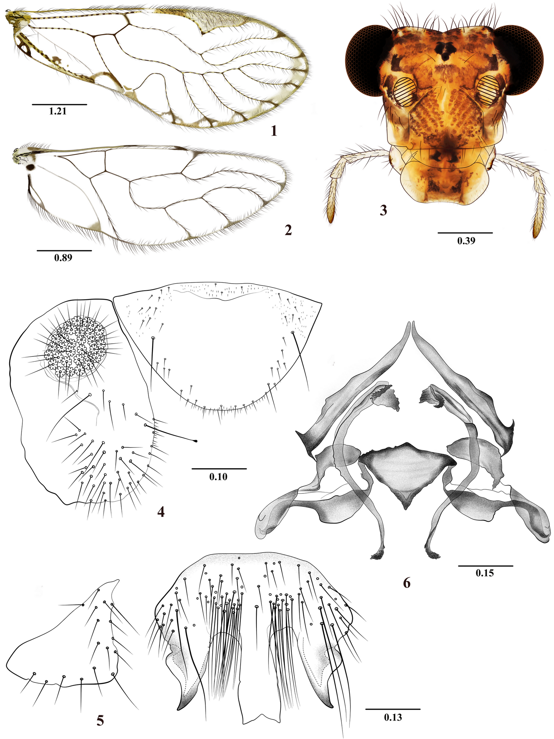
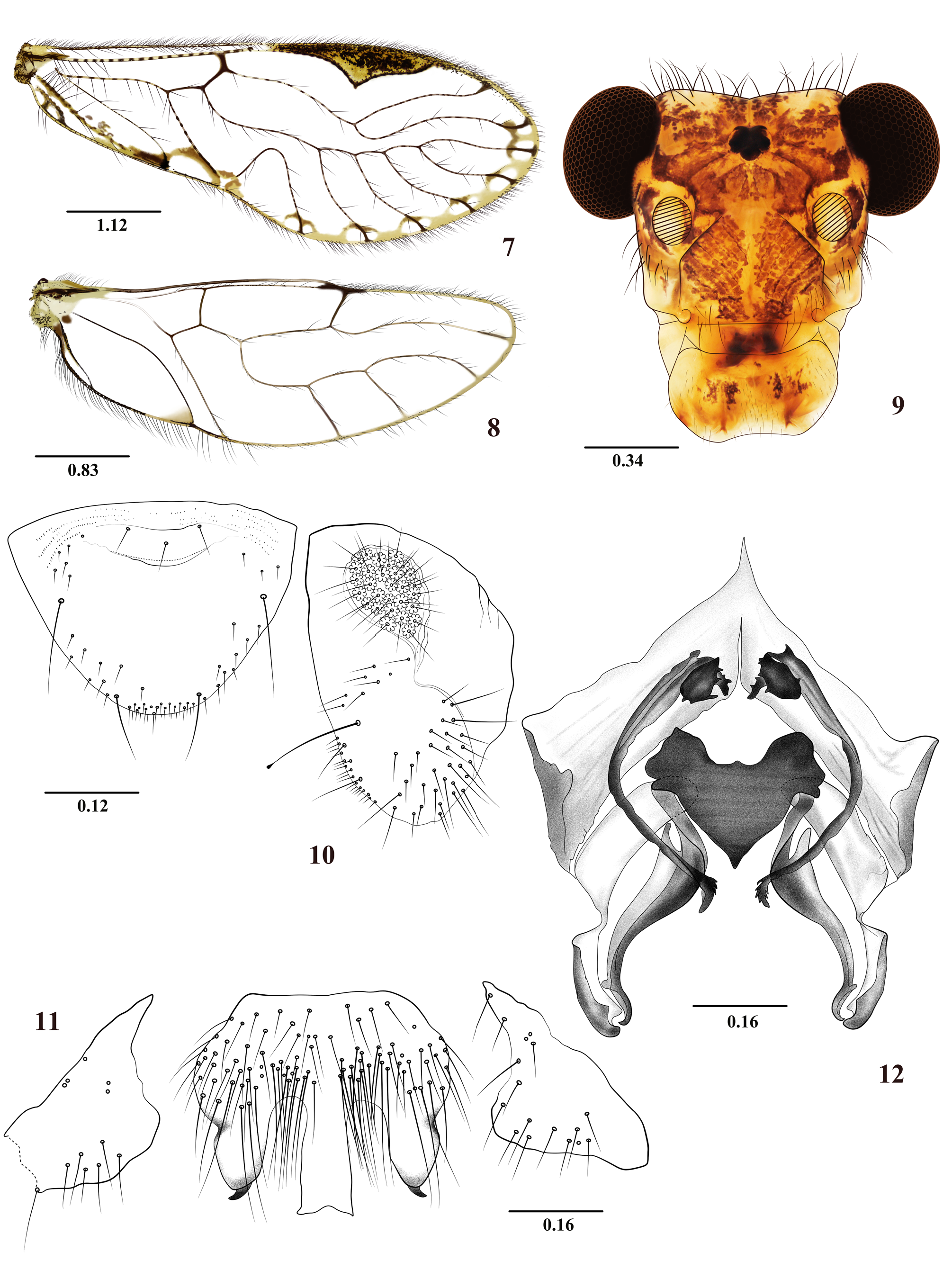
1. Forewings with pterostigma acutely extended in the middle towards Rs, with one crossvein between 2A and wing margin ( Figs 1 View FIGURES 1–6 and 7 View FIGURES 7–12 ); epiproct with basal spinose bulge; hypandrium of three sclerites, the central with three posterior processes, two lateral and one central....................................................................................... 2
- Forewings with pterostigma not acutely extended in the middle towards Rs, with or without crossvein between 2A and wing margin; epiproct without basal spinose bulge; hypandrium of three or five sclerites, if with three sclerites then the central one with variable number of posterior processes.......................... (See identification key in González et al. 2020).
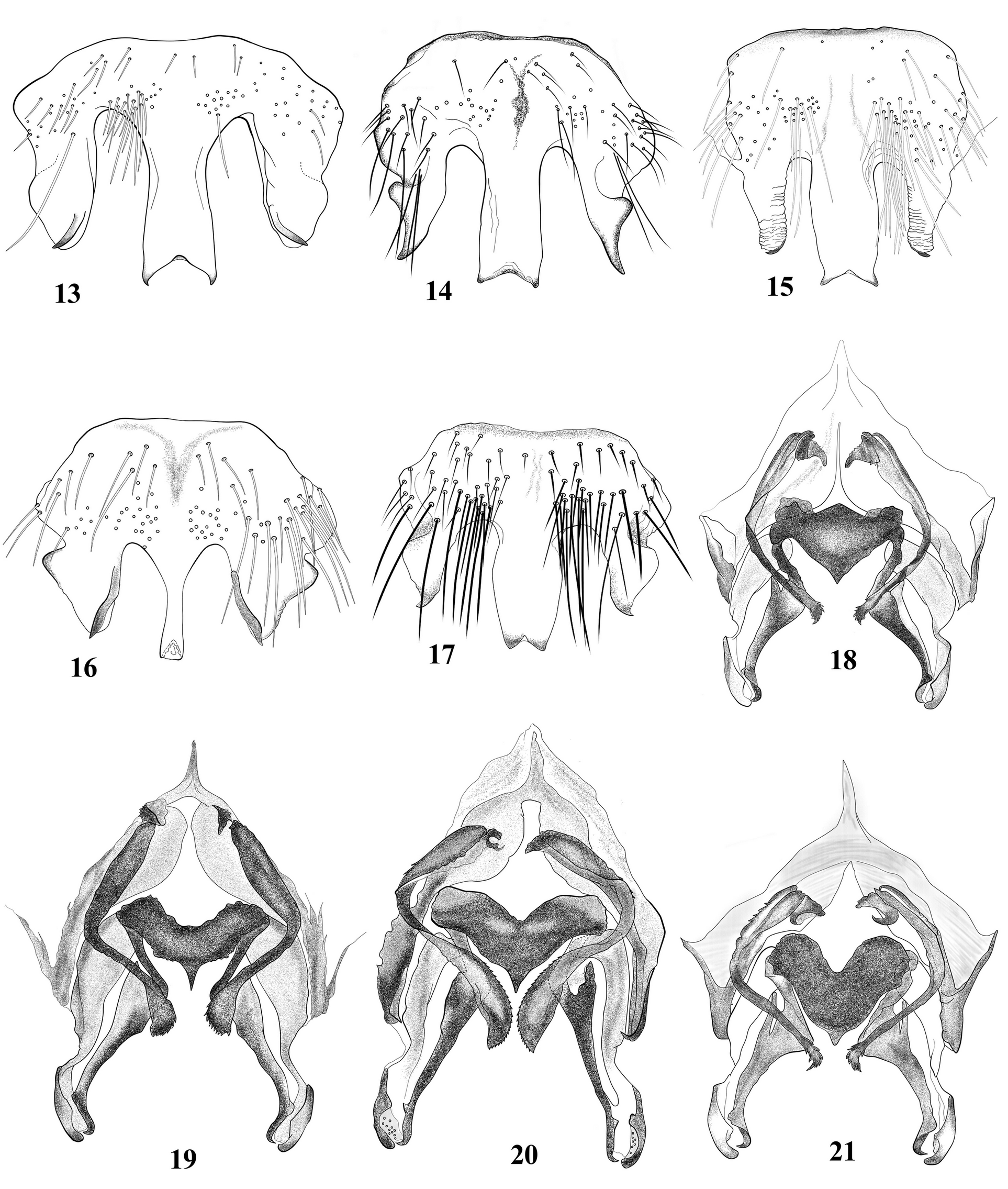
2. Central sclerite of the hypandrium with median process slender, posteriorly straight ( Fig. 16 View FIGURES 13–21 ), lateral processes with an outer bulge, inner border with a slender, sclerotized band............ P. ledesmai (García Aldrete, González-Obando & Carrejo) View in CoL
- Central sclerite of the hypandrium with median process stout, obtusely concave posteriorly ( Fig. 17 View FIGURES 13–21 ), lateral processes blunt ended or pointed..................................................................................... 3
3. Lateral endophallic sclerites (les) widened distally ( Fig. 20 View FIGURES 13–21 ); epiproct with a well-developed mesal, anterior bulge........ 4
- Lateral endophallic sclerites (les) not widened distally ( Figs 6 View FIGURES 1–6 , 12 View FIGURES 7–12 ); epiproct with variable developed mesal, anterior bulge. 5
4. Lateral endophallic sclerites with elongate distal widening ( Fig. 20 View FIGURES 13–21 ); median process of central sclerite of the hypandrium with apically convergent lateral margins ( Fig. 17 View FIGURES 13–21 ); postero-mesal sclerite preapically straight ( Fig. 20 View FIGURES 13–21 )........................................................................ P. colombianus (García Aldrete, González-Obando & Carrejo) View in CoL
- Lateral endophallic sclerites with short distal widening ( Fig. 19 View FIGURES 13–21 ); central sclerite of the hypandrium with median process of non-converging lateral margins ( Fig. 15 View FIGURES 13–21 ); postero-mesal sclerite preapically curved inward............................................................................ P. santanderinus (García Aldrete, González-Obando & Carrejo) View in CoL
5. Mesal endophallic sclerite without median concavity on anterior margin ( Figs 6 View FIGURES 1–6 and 18 View FIGURES 13–21 )............................. 6
- Mesal endophallic sclerite with median concavity on anterior margin ( Fig. 12 View FIGURES 7–12 , 19-21 View FIGURES 13–21 )............................... 7
6. Central sclerite of the hypandrium with lateral processes stout, blunt ended, bearing distally a slender, outwardly curved, sclerotized apophysis ( Fig. 13 View FIGURES 13–21 ); postero-mesal sclerites medially with continuous inner margin, smoothly rounded as illustrated ( Fig. 18 View FIGURES 13–21 )................................................................... P. venezolanus (García Aldrete) View in CoL
- Central sclerite of the hypandrium with lateral acuminate processes distally ( Fig. 5 View FIGURES 1–6 ), a bulge on outer borders; postero-mesal sclerites medially with abruptly widened inner margin as illustrated ( Fig. 6 View FIGURES 1–6 )........................ P. elduende n. sp.
7. Central sclerite of the hypandrium with lateral processes slender, distally acuminate ( Fig. 14 View FIGURES 13–21 ), a bulge on outer border........................................................ P. bretanaensis (García Aldrete, González-Obando & Carrejo) View in CoL
- Central sclerite of the hypandrium with lateral processes stout, blunt ended, bearing a short sclerotized and curved outward apophysis apically ( Fig. 11 View FIGURES 7–12 )............................................................. P. pichindensis n. sp.
No known copyright restrictions apply. See Agosti, D., Egloff, W., 2009. Taxonomic information exchange and copyright: the Plazi approach. BMC Research Notes 2009, 2:53 for further explanation.