
Rhombognathus amplus, Bartsch, 2013
|
publication ID |
https://doi.org/ 10.1051/acarologia/20132097 |
|
persistent identifier |
https://treatment.plazi.org/id/03C68797-0F63-4502-F74E-FEE0FC69FBE3 |
|
treatment provided by |
Marcus |
|
scientific name |
Rhombognathus amplus |
| status |
sp. nov. |
Rhombognathus amplus n. sp.
Figures 1-3 View FIGURE View FIGURE View FIGURE
Material examined — Holotype male, ZRC.ARA.01344, and paratype female, ZRC.ARA.01345, Singapore, Pulau Ubin, Chek Jawa, 1°24’N, 103°59’E, green algae ( Cladophorales ) on trunk of Rhizophora sp. 18 Oct 2012. Associated fauna: larvae of insects, oribatids ( Acari ). Paratype male, tritonymph, ZRC.ARA.01346, collection data as above. Paratype female, male, tritonymph, deutonymph, author’s collection; paratype male, deutonymph and protonymph, author’s collection, collection data as above.
Paratype male, tritonymph, protonymph, ZRC.ARA.01347, Singapore, Pulau Ubin, Chek Jawa, 1°24’N, 103°59’E, green algae ( Cladophorales ) on trunk of Rhizophora sp. 16 Oct 2012. Associated fauna: nematodes, terrestrial Acari , halacarids ( Agauopsis sp. ).
Two females, one deutonymph, ZRC.ARA. 01348, Singapore, mainland, Kranji, 1°21’N, 103°44’E, green algae ( Chaetomorpha sp. , Cladophorales ) and red algae (Ceraminalis) on trunk, 30 Oct 2012. Associated fauna: enchytraeids (Oligochaeta), larvae of insects, oribatids ( Acari ), halacarids ( Acarothrix grandocularis Chatterjee, Marshall, Guru, Ingole, Pešic, 2012 ). Female, tritonymph, deutonymph, author’s collection, collection data as above.
Diagnosis (adults) — Idiosoma wide (length / width 1.3/1); length of females 390 – 415 µm, of males 364 – 400 µm. Posterior margin of anterior dorsal plate triangular. Ocular plates almost triangular; both corneae close to lateral corner; two setae on ocular plate arranged along a median line. Posterior dorsal plate with two pairs of setae, both removed from margins of plate. Both female and male with long, slender perigenital setae arranged in a ring around genital opening, female with almost 60- 69 perigenital setae, male with approximately 38-47 setae. Gnathosoma short, its basis globular, rostrum conical. Legs almost equal in length, telofemora I to IV with 4(-5), (3-)4, 2, 2(-1) setae, genua with 4, (3-)4, (2-)3(-4), (2-)3 setae and tibiae I to IV with 5, 5, 5, 5 setae. Pair of ventral setae on tibiae III and IV long, slender, slightly plumulose. Paired claws smooth.
Etymology — The Latin word amplus means wide, enlarged. In contrast to most of the Rhombognathus species , which have a rather slender idiosoma and posterior dorsal plate, the new species and its posterior dorsal plate are wide.
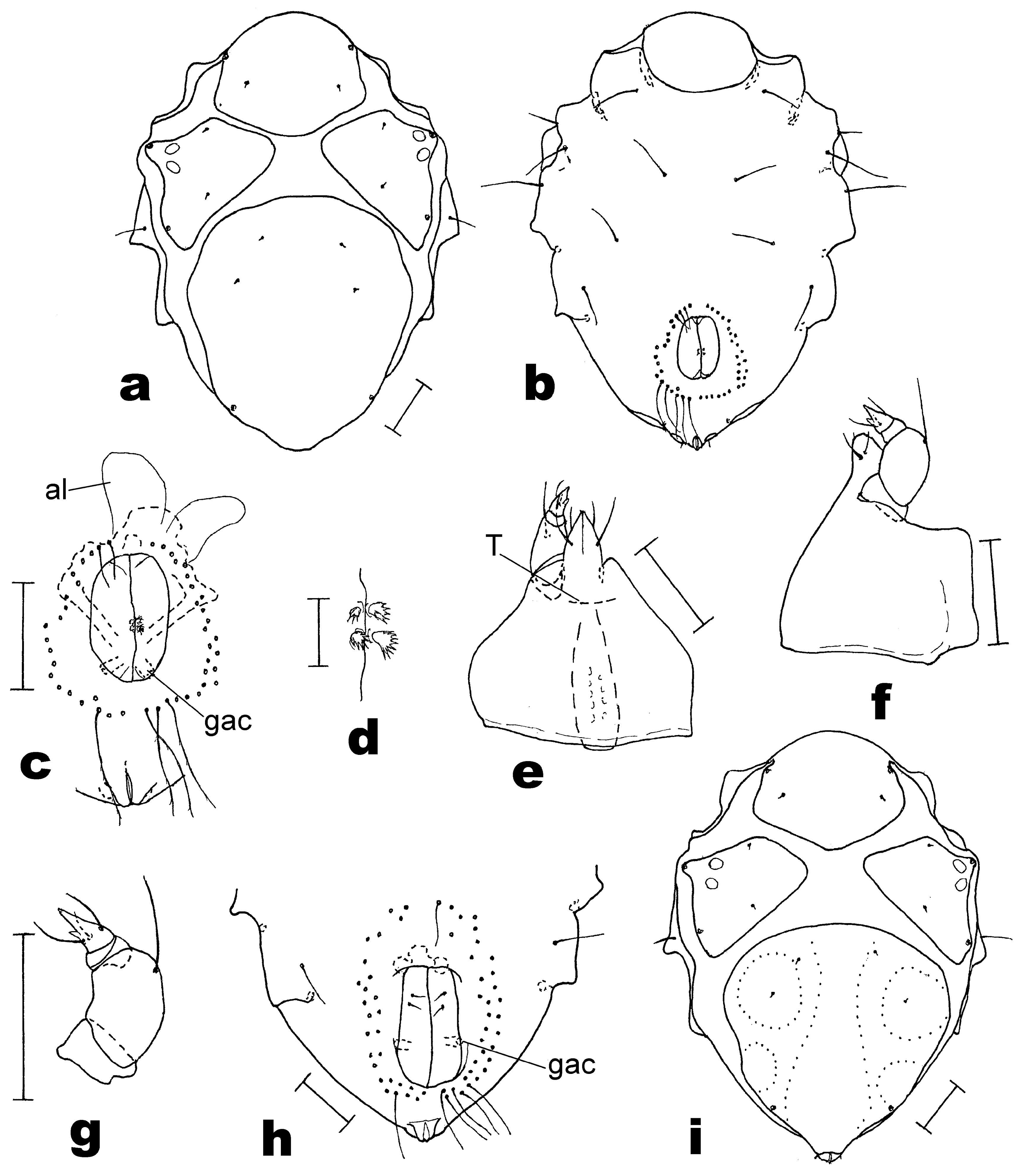
Description — Male. Idiosoma irregularly dark green, due to gut content shining through its transparent integument, and with three spots of dark-red eye pigment. Eye spots on ocular plates in almost marginal position. Gnathosoma and legs transparent. With an ovoid excretory body shining through dorsal integument.
Length of idiosoma 364 – 400 µm (holotype: 387 µm, and 290 µm wide). Surface of dorsal plates rather smooth, without markedly raised areas though with slight depressions. Anterior dorsal plate wider than long, length 110 µm, width 150 µm, anterior margin of plate slightly rounded, posterior margin almost triangular ( Figure 1a View FIGURE ). Ocular plates triangular in outline, with rounded medial and posterior corners. Length of ocular plates 119 – 127 µm, width 110 – 115 µm. Two corneae close to lateral margin, eye pigment immediately medial to corneae. Ocular plate with two gland pores, anterior pore on lateral protruding; posterior pore in lateral margin almost level with insertion of leg III. Posterior dorsal plate wide, anterior margin slightly arched; a median and two pairs of lateral areas slightly depressed (cf. Figure 1i View FIGURE ). Length of posterior dorsal plate 220 µm, width 202 µm. A pair of gland pores in posterior part of posterior dorsal plate. All dorsal setae small, peg-like, their length 4 µm. One pair of setae on anterior dorsal plate; two setae each on ocular plates, one in middle of anterior margin, one posterior to that seta; another two pairs of setae on posterior dorsal plate, both in anterior half of plate and distinctly removed from margins of plate. Adanal setae on anal cone, on small anal papillae.
Anterior and posterior epimeral plates, genital and anal plates fused into a ventral shield. Surface of shield smooth, length of shield 310 µm, width 290 µm. Area corresponding to anterior epimeral plate with three pairs of ventral setae and one pair of adjunct setae ( Figure 1b View FIGURE ); a pair of epimeral tubes within apodemes between epimera I and II. Area of posterior epimeral plate with one dorsal seta, one lateral and two ventral but no adjunct setae. Length of genital opening 60 µm, width 39 µm. Anterior margin of genital opening about level with insertion of leg IV. Perigenital setae arranged in a ring around genital opening, anterior setae smooth, posterior setae distinctly longer and very delicately plumose ( Figure 1c View FIGURE ). Holotype with 47 setae. Subgenital setae short, wide, with plumose outer edge ( Figure 1d View FIGURE ). Spermatopositor extending just beyond ring of perigenital setae, anteriorly flanked by a pair of alae. A single pair of genital acetabula distinctly seen; further internal structures obscured by spermatopositor and sperms. Anus small, anal sclerites narrow.
Gnathosoma – 107 µm long; only slightly longer than wide, basis globular, rostrum conical ( Figure 1e and f View FIGURE ). Palps four-segmented, distinctly shorter than gnathosomal base. P-2 with one dorsal seta, P- 4 with three basal setae and short spiniform process ( Figure 1g View FIGURE ).
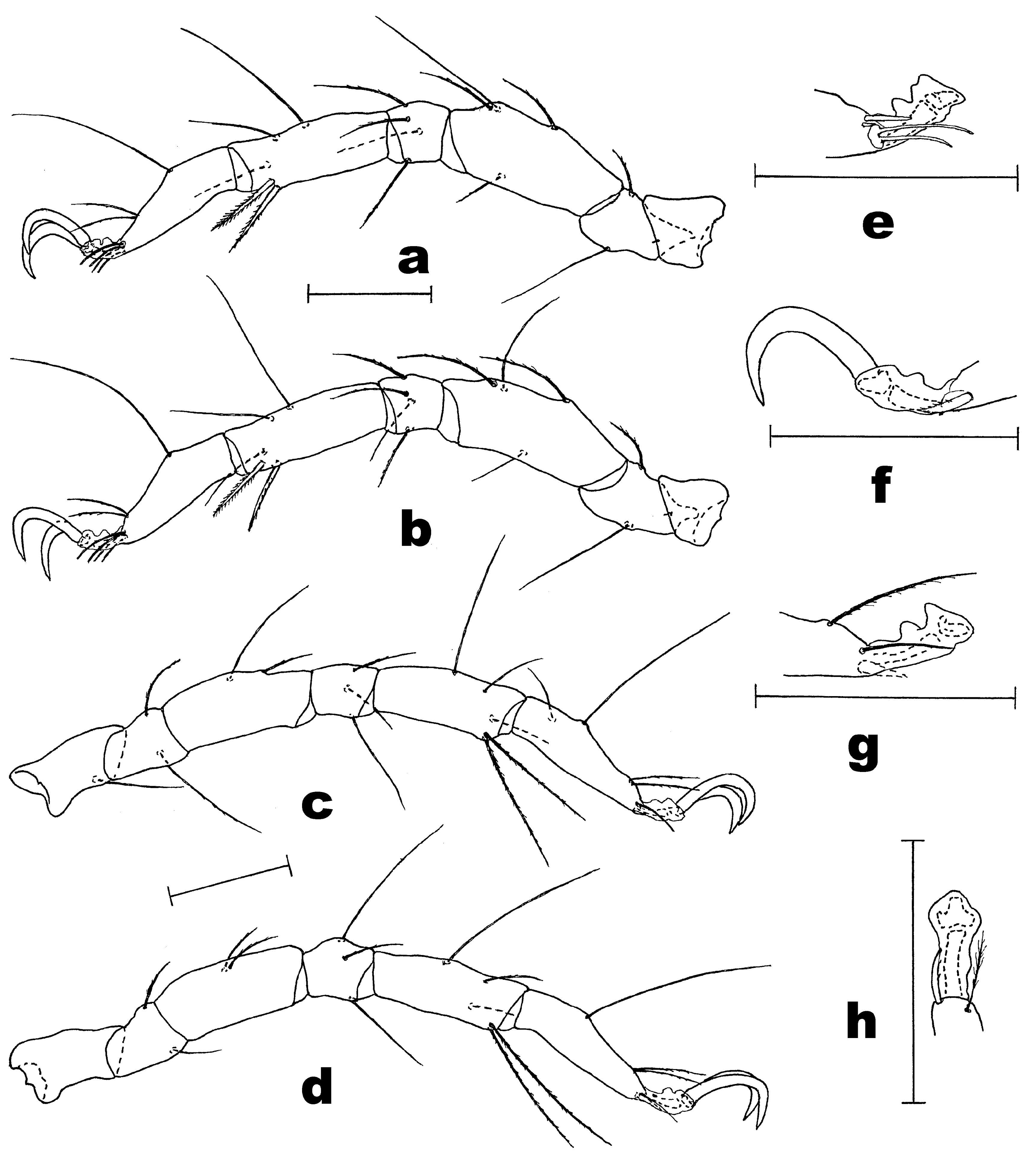
Legs slender, rather equal in length ( Figure 2 View FIGURE a- d). Telofemora I and II slightly longer than tibiae I and II, telofemora and tibiae III and IV almost equal in length. Telofemora I to IV longer than high (x2.3, x2.6, x2.4, x2.5 in respect). Leg chaetotaxy of holotype, from trochanter to tarsus (parambulacral setae and famulus excluded): legs I and II, 1, 2, 4, 4, 5, 4; leg III, 1, 2, 2, 3, 5, 4; leg IV, 0, 2, 2, 3, 5, 3. Several dorsal setae curved and delicately plumose. Setae on trochanters I and II peg-like. Both ventral setae on tibia I coarsely bipectinate. On tibia II, ventromedial seta bipectinate, ventrolateral seta with more delicate pectination. Two ventral setae of tibiae III and IV long, slender, slightly plumulose. Apical fossary setae paired, equal-sized and delicately plumose. Dorsolateral seta on tarsus III removed from dorsal fossary seta. Tarsus I with dorsolateral 9 µm- long solenidion and 3 µm- long famulus ( Figure 2e View FIGURE ), tarsus II with 9 µm- long solenidion, dorsolateral in position as on tarsus I. Tarsi I and II with pairs of doubled eupathid parambulacral setae; on tarsus III medial parambulacral seta eupathid, lateral seta spiniform and slightly pectinate ( Figure 2f View FIGURE ), on tarsus IV medial parambulacral seta setiform, delicately plumulose, lateral seta spiniform, similar to that on tarsus III ( Figure 2h View FIGURE ). Paired claws smooth, no accessory process seen. Median sclerite without dentiform process. Carpites I to IV 13 – 14 µm long.
Female – Length 390 – 415 µm. Dorsal aspect same as that of male. Often with an ovoid, stratified excretory body, 110 – 182 µm long, 62 – 77 µm wide. Ventral plates fused. Genital opening large, its length 105 µm, width 55 µm, extending anteriad beyond insertion of legs IV. Each genital sclerite with two subgenital setae. Ring of perigenital setae including 60 – 69 long, smooth setae; with ring extending anteriad to level of insertion of legs III ( Figure 1h View FIGURE ). Gnathosoma of 415 µm- long female 130 µm long, 115 µm wide (length / width 1.1/1). Shape of lateral parambulacral seta on tarsus III same as in male ( Figure 2g View FIGURE ), medial parambulacral seta on tarsus IV less plumulose than that of male. Eggs in ovigerous females spherical, 44 – 47 µm in diameter.
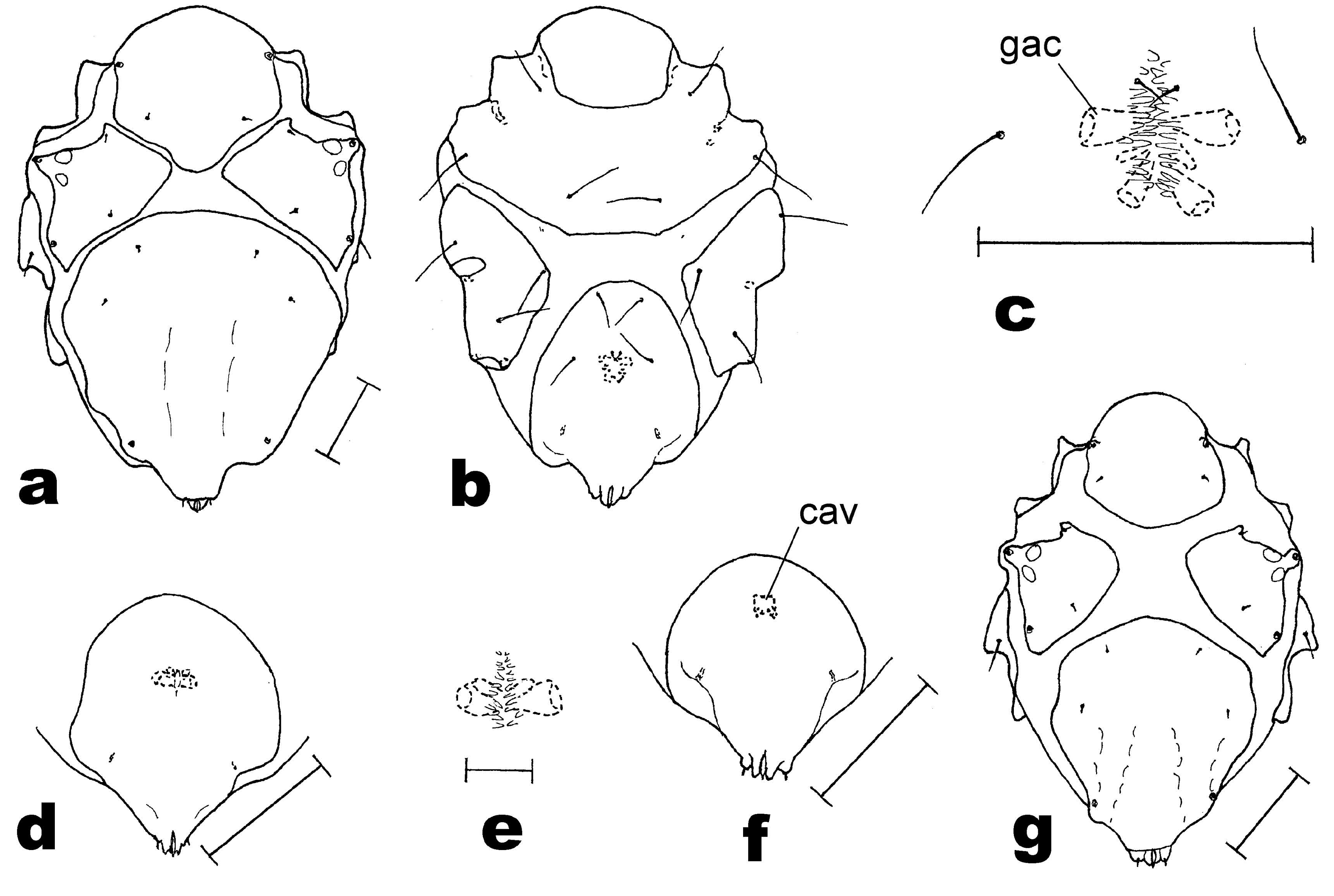
Tritonymph – Length 245 – 354 µm. Dorsal plates smaller but otherwise rather similar to those of adults. Ovoid excretory body 127 µm long, 30 µm wide. Position of dorsal setae as illustrated ( Figure 3a View FIGURE ). Anterior and posterior epimeral plates and genitoanal plate separated. Anterior epimeral plate with three pairs of ventral but no adjunct setae; posterior plate with one dorsal, one lateral and two ventral setae. Genital and anal plate fused ( Figure 3b View FIGURE ), this genitoanal plate with two pairs of perigenital setae and one pair of short subgenital setae. Primordial genital slit obscured by numerous papillae ( Figure 3c View FIGURE ). Genitoanal plate with two pairs of large-sized internal genital acetabula, anterior pair about 10 µm long and 6 µm wide, posterior pair 10 µm long and 4 – 5 µm wide. Between these pairs of acetabula a pair of small, flattened, blindly ending tubes present. Gnathosoma 1.1 times longer than wide, its length 93 – 95 µm, width 82 – 85 µm. Leg chaetotaxy: leg I, 1, 2, 4, 4, 5, 4; leg II, 1, 2, 4, 3-4, 5, 4; leg III, 1, 2, 2, 3, 5, 4; leg IV, 0, 2, 2, 3, 5, 3. Telofemora I to IV with 3/1, 3/1, 2/0, 2/0 dorsal/ventral setae.
Deutonymph – Length 242 – 284 µm. Excretory body 37 – 75 µm long, 25 – 30 µm wide. Anterior epimeral plate with three pairs of ventral setae; posterior plate with one dorsal, one lateral and two ventral setae. Genitoanal plate with one pair of large-sized genital acetabula, 6 – 7 µm long, 4 µm in diameter, adjacent a pair of short tube-like acetabula, 4 – 5 µm long, 2 – 3 µm in diameter. No perigenital setae present ( Figure 3d View FIGURE ). Primordial genital slit obscured by small papillae ( Figure 3e View FIGURE ). Gnathosoma 1.1 times longer than wide. Leg chaetotaxy: legs I and II, 1, 2, 3-4, 4, 5, 4; leg III, 1, 2, 2, 3, 5, 4; leg IV, 0, 1, 2, 3, 5, 3. Telofemora I to IV with 2-3/1, 2-3/1, 2/0, 2/0 dorsal/ventral setae. On tarsus III distance between dorsolateral seta and dorsal fossary seta equalling half the height of that tarsus.
Protonymph – Length 209 – 235 µm. In relation to idiosoma, posterior dorsal plate shorter and not as wide as in adults ( Figure 3g View FIGURE ). Anterior epimeral plate with three pairs of ventral setae; posterior epimeral plate with one dorsal and one lateral seta; no further ventral setae present. Genitoanal plate with small internal genital cavity, 6 µm long, 5 – 6 µm wide, and a pair of small tube-like structures, 2 µm long ( Figure 3f View FIGURE ). Integument close to that cavity with numerous papillae (not illustrated in Figure 3f View FIGURE ). Gnathosoma 1.1 times longer than wide. Leg chaetotaxy: legs I and II, 1, 2, 3, 4, 5, 4; leg III, 1, 1, 2, 3, 5, 4; leg IV, 0, 0+2 (basi- plus telofemur), 3, 5, 3. Telofemora I to IV with 2/1, 2/1, 2/0, 2/0 dorsal/ventral setae.
Variants — In contrast to many other halacarid genera, e.g., the above mentioned Isobactrus , the number of setae in Rhombognathus species often varies. The variants in adult Rhombognathus amplus are summarized in Table 1.
Remarks — Characters of Rhombognathus amplus are: idiosoma wide (length / width 1.3/1), ventral plates fused, gnathosoma short, hardly longer than wide (length / width 1.0-1.1/1), its base spherical, rostrum conical, females and males with long, slen- der, almost smooth perigenital setae, claws smooth. Amongst the 104 valid species known ( Bartsch 2009a, c), only two share this character combination, these are Rhombognathus verrucosus Bartsch ,
T-, 1992 and R. bulbosus Bartsch, 2005 . Both species are known from mangroves, from green algae growing in the upper tidal zone. Records of these two species are from Hong Kong ( R. verrucosus ) and Singapore ( R. bulbosus ) ( Bartsch 1992, 2005).
Differences between adults of R. amplus and these two species are summarized in Table 2. The juveniles can be distinguished on the basis of the shape of the dorsal plates and the position of the idiosomatic setae. As in R. amplus , the protonymph of R. bulbosus bears four setae on the left genua I and II (re-examined); this may be an anomaly. Adults, tritonymphs and deutonymphs of R. bulbosus have three setae on the genua I and II ( Bartsch 2005).
The idiosoma of Rhombognathus amplus is 1.3 times longer than wide, while in most of the other Rhombognathus species the idiosoma is more slen- der, 1.5 or more times longer than wide. The females of R. amplus have an unusual high number of perigenital setae. More than half of the presently known species bear five pairs of setae, only a few species have a lower or somewhat higher number of pairs of setae (cf. AbØ 1991). The numbers, shape and arrangement of the male perigenital setae are unusual. The most commonly found arrangement in Rhombognathus is, there are 7-14 pairs of short and distinctly plumose setae arranged trapezoidally.
Biology — The body of Rhombognathus amplus is covered by a biofilm which includes both organic (e.g. bacteria) and inorganic material (e.g. iron hydroxides). The fouling is most intense on adults but sparse on recently hatched protonymphs. Biofilms are commonly found on both littoral and deep-sea species ( Gillan et al. 2004; Corbari et al. 2008; Caro et al. 2012), and the integument of shallow brackish water as well as deep-sea hydrothermal vent halacarids may be covered with brown spots which most likely include iron hydroxide. Such films are often found on species of the genera Copidognathus and Halacarellus ( Bartsch 1994, 2011, unpublished). In addition to a biofilm several littoral halacarids may carry diatoms ( Cocconeis sp. , Synedra sp. ), minute green algae and protozoans ( Bartsch 1998, 2004). Several upper tidal rhomboganthines have the integument covered with delicate epicuticular villi or droplets ( Bartsch 2000, 2009b); these may prevent colonization but also reduce loss of water. The biofilm that covers R. amplus may similarly prevent desiccation.
The records of Rhombognathus amplus are from the upper tidal zone, from green algal patches growing on the trunks of trees. No R. amplus were extracted from such green algal patches on trees growing at the edge of a mud flat ( Avicennia sp. , Sonneratia sp. ). The mites were found in landward mangroves, also in a remote landward forest hardly reached by the tide and with a tidal creek which also served as rainwater runoff. Both the canopy of the trees, which affords shelter from sun radiation and severe aridity, and reduced silting may have favoured a successful colonization of this habitat.
The fauna within these spots of green algae is sparse. There are a few enchytraeid oligochaetes, larvae of insects and oribatid mites (Oribatida); halacarids were represented by Rhombognathus amplus , Acarothrix grandocularis and Agauopsis sp. ; R. amplus , although not very numerous, was found to be the dominant species. It is likely that R. amplus does not prefer the meagre green algal patches but can live here because of reduced competitive pres- sure in this habitat in respect to salinity, temperature and humidity. Compared to other meiobenthic aquatic arthropods, several upper littoral halacarids are known to be highly euryhaline and eurythermic ( Bartsch 1974). Rhombognathus amplus , as well, will certainly tolerate a wide range of such environmental parameters. As to the above mentioned silt fall, epibiontic halacarids are known to be sensitive to impact of sand and silt ( Bartsch 1974, 2003; Proches and Marshall 2002), and one can expect that silt between the algae will have a negative effect on a R. amplus population.
Individuals of Rhombognathus amplus contained an ovoid, stratified body of accumulated excretory material. In the majority of halacarid species such excretory bodies have an elongate, rod-like shape. Species with a similar ovoid excretory body, e.g. Halacarus excellens Lohmann, 1907 and all Limnohalacarus species , have a conspicuously small anus with unusual narrow anal sclerites ( Bartsch 2010, 2013). Rhombognathus amplus , too, has narrow anal sclerites.
No known copyright restrictions apply. See Agosti, D., Egloff, W., 2009. Taxonomic information exchange and copyright: the Plazi approach. BMC Research Notes 2009, 2:53 for further explanation.