
Scolopsomorpha boulardi, Constant, Jerome, 2009
|
publication ID |
https://doi.org/ 10.5281/zenodo.190028 |
|
DOI |
https://doi.org/10.5281/zenodo.6212914 |
|
persistent identifier |
https://treatment.plazi.org/id/03C6F818-D451-CD75-E5F2-F907FF51ACF1 |
|
treatment provided by |
Plazi |
|
scientific name |
Scolopsomorpha boulardi |
| status |
sp. nov. |
Scolopsomorpha boulardi View in CoL n. sp.
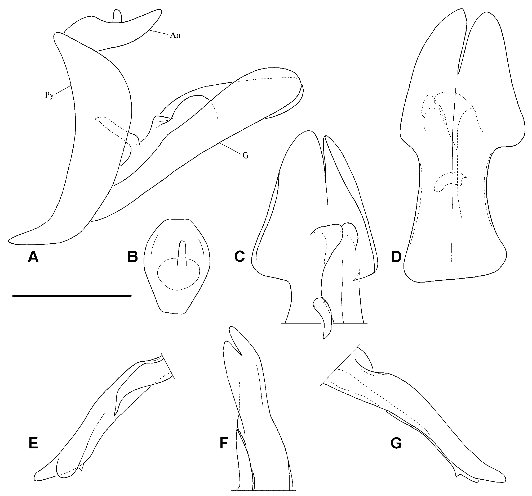
Figs. 3 A–G View FIGURE 3 A – G , 5 View FIGURE 5 , 6 E–H View FIGURE 6 A – L. A – D .
Etymology. Dedicated to its collector, Dr. Michel Boulard (MNHN, Paris, France).
Material examined. Holotype 3: [Coll. R.I.Sc.N.B., Boukoko – RCA, 14-iii-1969, Michel Boulard] [H. Synave det., 1973, Scolopsomorpha africana Mel. ] - dissected, genitalia in glycerine ( RBINS). Coordinates of Boukoko ( Central African Republic): 3°54'N 17°56'E.
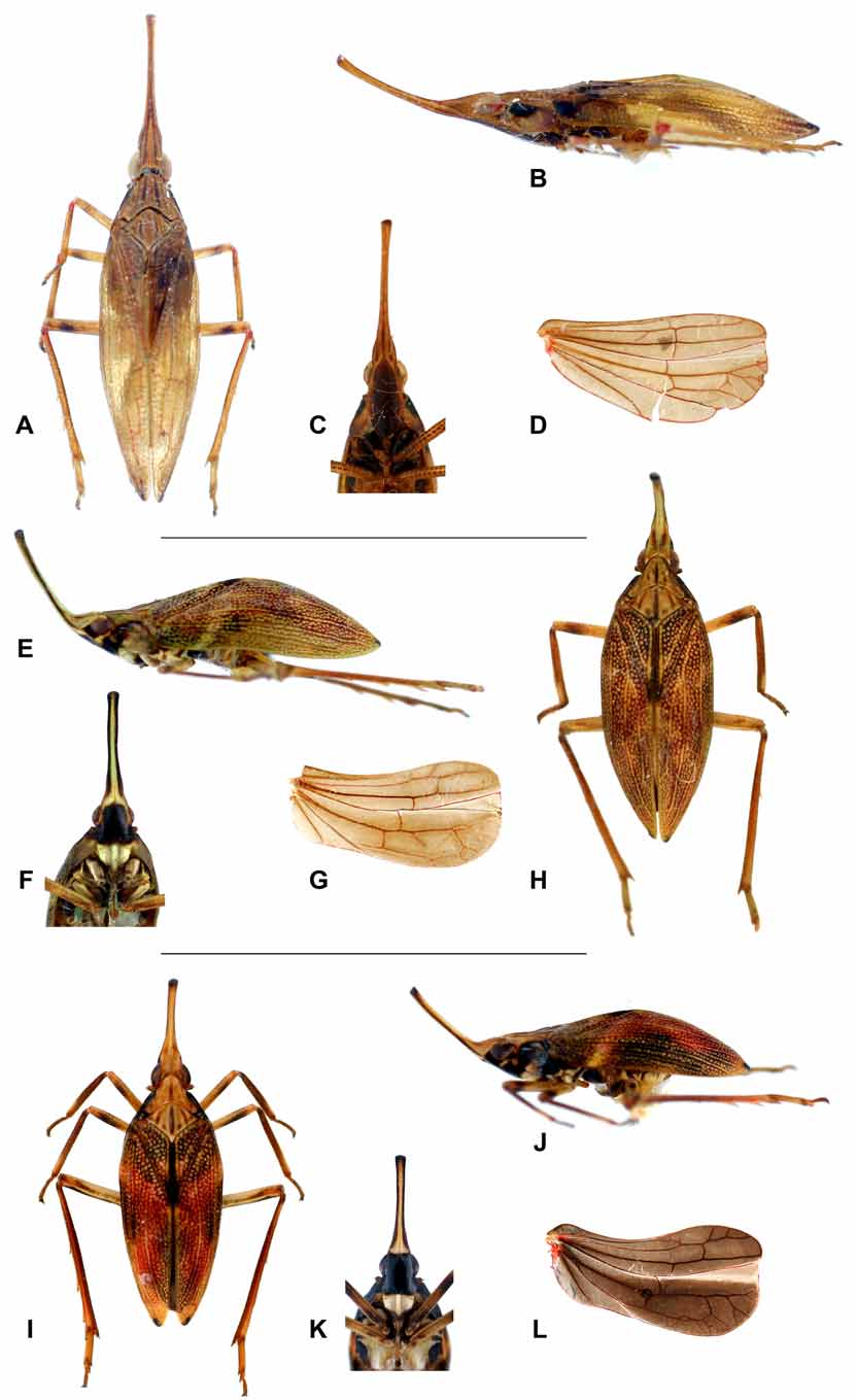
Diagnosis. The species is easily recognized by the white clypeus, the disc of frons black margined with whitish and anterior half ( Fig. 6 F View FIGURE 6 A – L. A – D ), the cephalic process strongly curved dorsad ( Fig. 6 E View FIGURE 6 A – L. A – D ), the black markings on the sides of the head ( Fig. 6 E View FIGURE 6 A – L. A – D ), the anterior and median coxae pale yellow-brown basally ( Fig. 6 F View FIGURE 6 A – L. A – D ) and the shorter and more convex tegmina (LTg/BTg = 3.1) ( Figs. 6 E, H View FIGURE 6 A – L. A – D ). Males should be identified by examination of the genitalia ( Figs. 3 A–G View FIGURE 3 A – G ).
Description. LT: 3 (n = 1): 11.7 mm. L [anterior margin of eye – apex of tegmina]: 8 mm
Head: pale yellow-brown tinged with green ( Fig. 6 H View FIGURE 6 A – L. A – D ); lateral sides with one black patch before eye and another under eye, including base of antenna ( Fig. 6 E View FIGURE 6 A – L. A – D ); frons dark shiny black-brown with anterior 1/5, sides anteriorly before antenna, small line before eye and baso-lateral angles pale yellow ( Fig. 6 F View FIGURE 6 A – L. A – D ); process projecting strongly antero-dorsad, with apex and lateral line black-brown ( Fig. 6 E View FIGURE 6 A – L. A – D ); median carina of vertex reaching apex of process; short brown line on vertex on each side of median carina ( Fig. 6 H View FIGURE 6 A – L. A – D ); clypeus very pale yellow green with apex brown ( Fig. 6 F View FIGURE 6 A – L. A – D ); antennae yellow-brown ( Fig. 6 F View FIGURE 6 A – L. A – D ); ratio BV/LV = 0.22; BF/LF = 0.24.
Thorax: pro- and mesonotum pale yellow-brown with carinae brown to dark brown; brown markings on disc of pronotum between carinae and on lateral angles of mesonotum ( Fig. 6 H View FIGURE 6 A – L. A – D ); sides of prothorax with black-brown patch behind eye ( Fig. 6 E View FIGURE 6 A – L. A – D ); tegulae brown with base pale yellow-green Figs. 6 E, H View FIGURE 6 A – L. A – D ); ratio LP+LM/BT = 1; LM/LP = 1.7.
Tegmina: tubercles pale yellow-brown; ground colour red-brown, black brown on sides on basal half and on clavus; apical black spot small, shiny and slightly elevated; apex of vein A1+A2 black ( Figs. 6 E, H View FIGURE 6 A – L. A – D ); ratio LTg/BTg = 3.125.
Hind wings: brown with veins and margins slightly suffused with red ( Fig. 6 G View FIGURE 6 A – L. A – D ).
Legs: (anterior legs missing after coxae on examined specimen) coxae I and II pale yellow-brown, brown apically ( Fig. 6 F View FIGURE 6 A – L. A – D ); coxae III pale brown; femora II yellow-brown with ante-apical black-brown ring; tibia II pale brown, yellow-brown basally and at apico-external angle, and with dorsal carina red; femora III yellowbrown dorsally, black-brown ventrally; tibiae III brown, lighter yellow apically, like tarsomeres; apices of spines black ( Fig. 6 H View FIGURE 6 A – L. A – D ).
Abdomen: pale yellow ventrally, dark brown dorsally.
Genitalia 3: pygofer narrow and curved in lateral view ( Fig. 3 A View FIGURE 3 A – G ); gonostyli elongate ( Figs. 3 A, D View FIGURE 3 A – G ), fused on basal 2/3, strongly emarginate laterally in middle ( Fig. 3 D View FIGURE 3 A – G ) and with digitate process dorsally ( Fig. 3 C View FIGURE 3 A – G ); right gonostylus with hook-shaped process dorsally near base and more strongly emarginate than left one ( Fig. 3 C View FIGURE 3 A – G ); anal tube dorso-ventrally compressed, suboval in dorsal view with apical margin cut straight ( Fig. 3 B View FIGURE 3 A – G ); phallic complex directed postero-ventrad ( Figs. 3 E–G View FIGURE 3 A – G ); basal pointed process and rounded preapical lobe on left side ( Fig. 3 E View FIGURE 3 A – G ); preapical small tooth ventrally ( Figs. 3 E, G View FIGURE 3 A – G ).
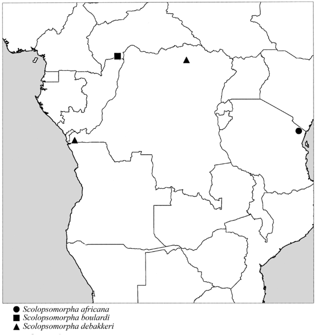
Biology. Nothing is known of this species which has been found only in the Central African Republic ( Fig. 5 View FIGURE 5 ).
| RBINS |
Royal Belgian Institute of Natural Sciences |
No known copyright restrictions apply. See Agosti, D., Egloff, W., 2009. Taxonomic information exchange and copyright: the Plazi approach. BMC Research Notes 2009, 2:53 for further explanation.