
Candelariodon barberenai, de Oliveira, Schultz, Soares & Rodrigues, 2011
|
publication ID |
https://doi.org/ 10.5281/zenodo.278705 |
|
DOI |
https://doi.org/10.5281/zenodo.6193917 |
|
persistent identifier |
https://treatment.plazi.org/id/03C7132A-FFF3-7822-FF54-187CFA9DF86E |
|
treatment provided by |
Plazi |
|
scientific name |
Candelariodon barberenai |
| status |
sp. nov. |
Candelariodon barberenai sp. nov.
Etymology. The specific word is a tribute to Prof. Mário Costa Barberena, a pioneer of research on Permian and Triassic Brazilian tetrapods.
Holotype. MMACR PV- 0001-T; right and left dentaries fused; right and left splenials approximately in articulated position; distal left lower incisor; both lower canines broken near the crown basis; first to fifth left and first to third right lower postcanines; isolated postcanine lying in the lateral surface of the left dentary, interpreted as a right maxillary tooth.
Specific diagnosis. Same as for the genus.
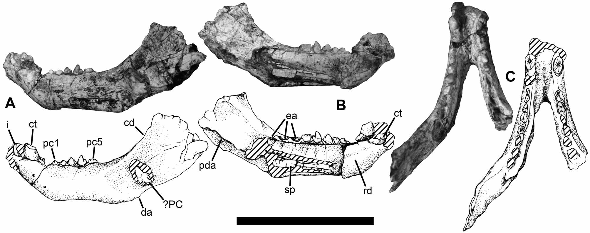
Description. The dentary of Candelariodon ( Fig. 4 View FIGURE 4 ) is greatly developed with its posterior end probably extending close to the craniomandibular articulation. The bone is considerably robust, i.e., the height and the thickness of its horizontal ramus are considerable large when compared with its total length. Two similarly sized mental foramina are present, one below the canine and the second placed slightly posterior and ventral to the first. The partially preserved coronoid process rises at the level of the sixth or seven postcanine and it is twice taller than the horizontal ramus of the dentary; laterally, the wide and shallow masseteric fossa extends over the complete process. Dentary without angular process. Medially in the most posterior region of the dentary, it is possible to observe a crest dorsally delimitating the area for the postdentary bones attachment. The dentaries are united by a long synostosed symphysis.
The splenial is slender and elongated, and although it is somewhat damaged it is possible to see that it starts posteriorly to the symphysis and reaches at least the level of the eighth postcanine ( Fig. 4 View FIGURE 4 B).
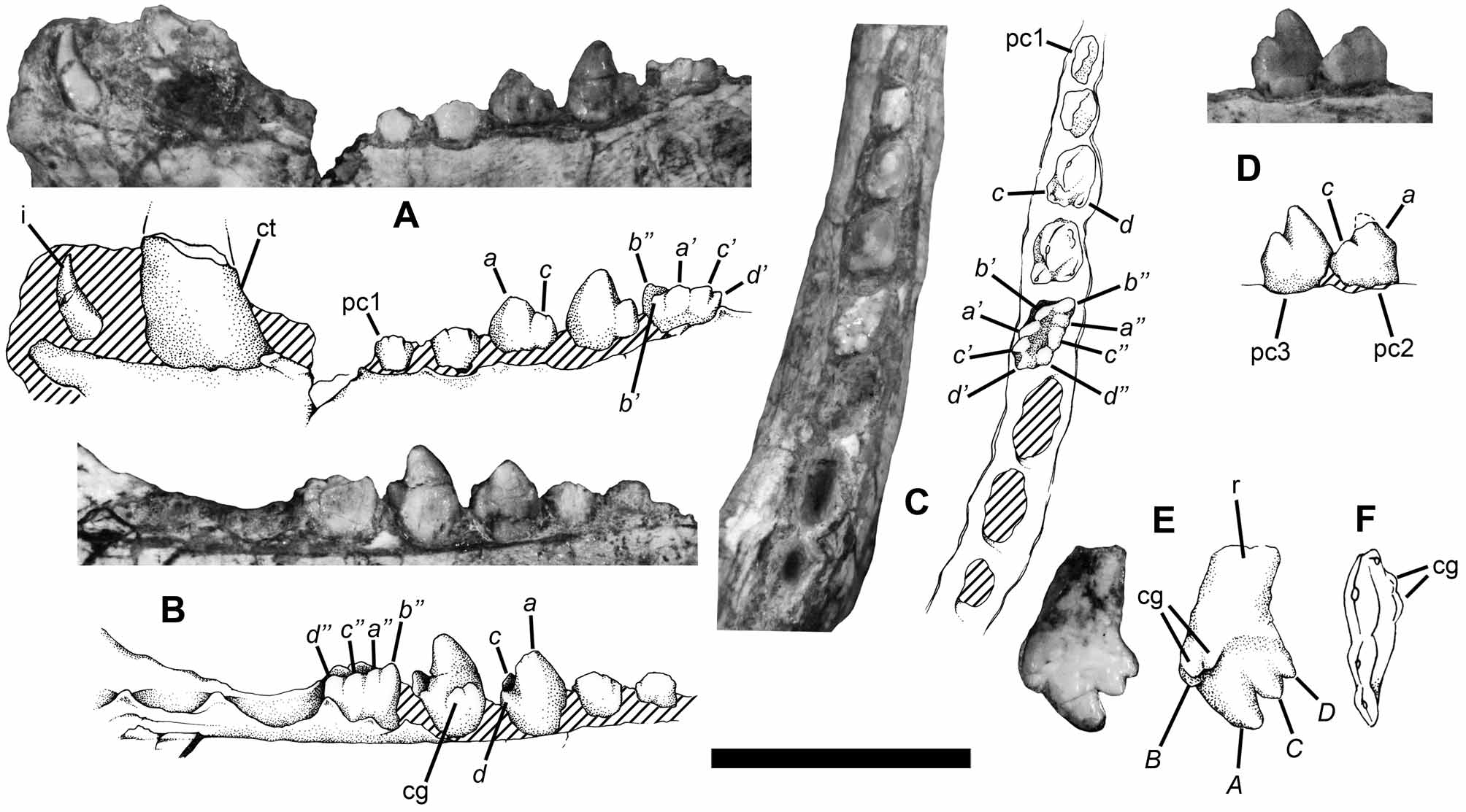
Only the left distal lower incisor is present in Candelariodon holotype ( Fig. 5 View FIGURE 5 A). It is considerably high when compared with the height of the postcanines, slightly procumbent and without serrations or denticulations along its edges. Both canines are broken near the crown basis, but the condition of the preserved portions suggests a considerably large tooth ( Fig. 5 View FIGURE 5 A). As in the incisor, there is no evidence of serration or denticulation in the canine margins.
In the left dentary, the first to fifth postcanines (pc) are preserved; in addition, there are still three distal empty alveoli ( Fig. 5 View FIGURE 5 A–C). The pc1 is damaged in both sides but apparently it has only two cusps, the main (cusp a) and the distal accessory (cusp c); the pc2 shows the same cusp pattern ( Fig. 5 View FIGURE 5 D). The pc3 has its main cusp followed by two distal accessory cusps, one buccal (cusp c) and another lingual (cusp d), that gives the impression of a more expanded appearance to the distal half of the tooth ( Fig. 5 View FIGURE 5 A–C). The pc4 shows this same pattern but there are also two small cusps placed lingually to the main one, forming an incipient cingulum ( Fig.5 View FIGURE 5 B). The pc5 is very peculiar, lacking the sharp cusps present in the anterior teeth and being buccolingually more expanded. In this tooth, the cusps are low and distributed in two longitudinal rows, in buccal and lingual position, separated by a shallow “occlusal basin” or central valley ( Fig. 5 View FIGURE 5 A–C). Each row has four cusps, named here as b’, a’, c’ and d’, in the buccal row, and b’’, a’’, c’’ and d’’, in the lingual row ( Fig. 5 View FIGURE 5 A–C). Buccally, the cusps a’ and c’ are the taller, whereas in the lingual row the cusp b’’ is the most eminent. The use of a noncommittal terms for these cusps is because the peculiar morphology of Candelariodon teeth does not allow the determination of exact homology between its cusps and the cusps of other non-mammaliaform cynodonts.
The lower postcanine teeth change their morphology from subsectorial teeth in the anterior region to a low, multicuspidate tooth in the posterior half of the mandible ( Figs. 5 View FIGURE 5 A–C). Some cynodonts, as Andescynodon Bonaparte 1969 (see Goñi 1986) and Diademodon Seeley 1895 (see Martinelli et al. 2009) have sectorial (and/or subsectorial) and buccolingually teeth in their mandibles, although not in the exact pattern seen in Candelariodon . The most relevant fact is that the change of buccolingually expanded teeth to sectorial postcanines in these cynodonts is not reversed in none known specimen, i.e. once the teeth reach a sectorial morphology, none tooth posterior to them became expanded again. This was the reason that led us to consider the tooth preserved on the lateral surface of the left dentary ( Fig. 4 View FIGURE 4 A) as an upper postcanine and not as a tooth that could to occupy one of the empty alveoli of the left dentary. It is more sectorial than the lower postcanines, with four mesiodistally aligned cusps (B, A, C, and D; Fig. 5 View FIGURE 5 E–F); there are also two small cingular cusps that we consider lingual [as occurs in e.g. Procynosuchus Broom 1937 (see Kemp 1979)]. Consequently the tooth is from the right side of the cranium.
No known copyright restrictions apply. See Agosti, D., Egloff, W., 2009. Taxonomic information exchange and copyright: the Plazi approach. BMC Research Notes 2009, 2:53 for further explanation.
|
Kingdom |
|
|
Phylum |
|
|
Class |
|
|
Order |
|
|
Genus |