
Psychropotes semperiana Théel, 1882
|
publication ID |
https://doi.org/10.5281/zenodo.173337 |
|
DOI |
https://doi.org/10.5281/zenodo.6255860 |
|
persistent identifier |
https://treatment.plazi.org/id/03C787A1-4D49-D224-E43A-FA14FB4CFE4F |
|
treatment provided by |
Plazi |
|
scientific name |
Psychropotes semperiana Théel, 1882 |
| status |
|
Psychropotes semperiana Théel, 1882 View in CoL
(Figs 2B–E, 5)
Psychropotes semperiana Théel, 1882: 100 View in CoL –101, pl. 34 (figs 10–11); Hansen, 1975: 102 –105, figs 41–42 [synonymy]
Psychropotes kerhervei Hérouard, 1902: 27 View in CoL –30, pl. 4 (figs 1–9) Euphronides anchora Hérouard, 1912: 6 –7, fig. 5
Material. M 48/1321: 2 specimens ( ZSM 20043075).
Description. Anteriorly, the specimens are more or less flat, while the posterior part of the body is elevated and gives rise to the unpaired dorsal appendage. The larger specimen is 79 mm long and 22 mm wide (across ventral sole), while the smaller specimen measures 53 mm in length and 17 mm across the ventral sole. Both are of a purple colour (preserved). The mouth and the anus are ventral and confined to the anterior and the posterior end of the body. There are 16 tentacles, with conspicuous, rounded discs. The ventral sole is delimited on both sides by a narrow brim (damaged for the most part), formed by basally fused tube feet. Midventral tube feet are conical and restricted to a double row, which seems to be present (partly damaged) throughout the length of the ventral sole. The unpaired dorsal appendage arises from a dorsal bulge, which is situated oneseventh body length in the large specimen and onefourth body length in the small specimen from the posterior end of the body. In both specimens, the dorsal appendage is more or less contracted and thus nothing can be said about the original size. The almost complete dorsal appendage of the large specimen ends in two long slender papillae, each about 18 mm long. Anterior to the dorsal bulge of the small specimen, are three pairs of short conical papillae (indistinct in the larger specimen).
Calcareous deposits of the dorsal body wall are crosses of two types (Figs 2B–D). The four arms of the larger type are smooth in their proximal parts and are equipped with irregularly placed spines close to their distal ends (Fig. 2C). The high central apophysis is smooth and ends in three or four downwardly bent hooks (Fig. 2B). Crosses of the second type are considerably smaller, have irregularly placed spines along the arms and a low and also spinous central apophysis (Fig. 2D). The calcareous deposits of the ventral body wall are rods and crosses with irregularly placed spines (Fig. 2E).
Remarks. The two specimens described herein conform to the detailed description given by Hansen (1975) for this species. Very characteristic are the large crossshaped deposits of the dorsal body wall with a high and smooth central apophysis, ending in three or four downwardly bent hooks, which are unique within the genus ( Hansen 1975).
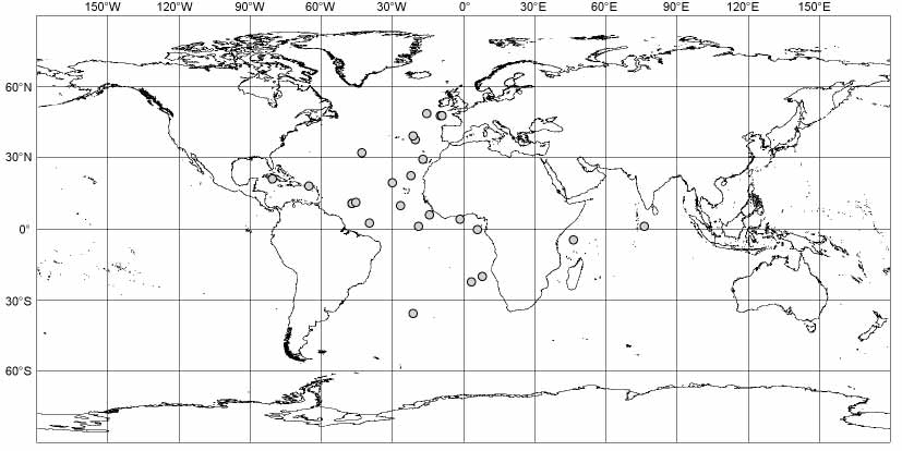
Distribution. ( Fig. 5 View FIGURE 5 ) Northern and southern Atlantic Ocean, northern and western Indian Ocean, 2695–5610 m ( Deichmann 1930, 1940; Hansen 1975; Hérouard 1902, 1923; IFREMER BIOCEAN; Madsen 1953; Sibuet 1977; Théel 1882; herein).
| ZSM |
Bavarian State Collection of Zoology |
No known copyright restrictions apply. See Agosti, D., Egloff, W., 2009. Taxonomic information exchange and copyright: the Plazi approach. BMC Research Notes 2009, 2:53 for further explanation.
|
Kingdom |
|
|
Phylum |
|
|
Class |
|
|
Order |
|
|
Family |
|
|
Genus |
Psychropotes semperiana Théel, 1882
| Bohn, Jens Michael 2006 |
Psychropotes kerhervei Hérouard, 1902 : 27
| Herouard 1912: 6 |
| Herouard 1902: 27 |
Psychropotes semperiana Théel, 1882 : 100
| Hansen 1975: 102 |
| Theel 1882: 100 |