
Alcha sinensis Wang & Hu, 2019
|
publication ID |
https://doi.org/ 10.11646/zootaxa.4695.4.3 |
|
publication LSID |
lsid:zoobank.org:pub:1CF2E78F-8B1A-4E53-9BB7-FAD3D5F06FDD |
|
DOI |
https://doi.org/10.5281/zenodo.5587737 |
|
persistent identifier |
https://treatment.plazi.org/id/03C787A1-FFBD-FF8B-B9D0-FDEFFAF14675 |
|
treatment provided by |
Plazi |
|
scientific name |
Alcha sinensis Wang & Hu |
| status |
sp. nov. |
Alcha sinensis Wang & Hu n. sp.
( Figs. 1–3 View FIGURE 1 View FIGURE 2 View FIGURE 3 )
Locality. Specimens were collected in the intertidal zone of Eastern Shenzhen City, Guangdong Province, China (22°28′N, 114°32′E) by LH Zhong in the March of 2018.
Material examined. Holotype: PLA-Po091 , whole-mounted specimen. Paratypes: PLA-Po092–093, wholemounted specimens; PLA-Po094-095 , whole-mounted slides of stylet; PLA-Po096–102, serially sectioned (sagittal) specimens.
Etymology. The species is named for its discovery in China.
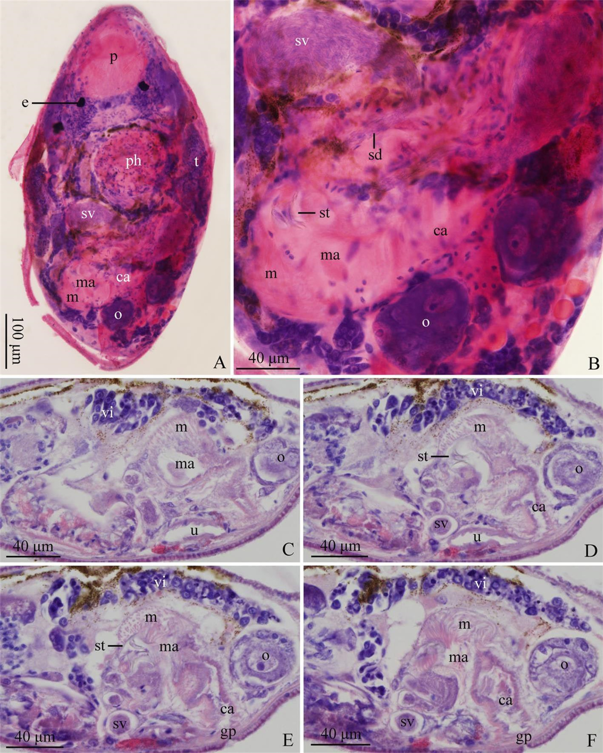
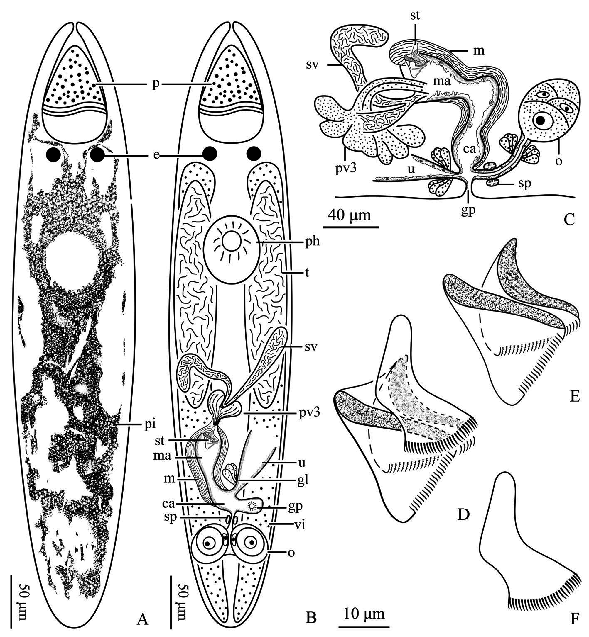
Description. Matured individuals are 557–794 μm in length, 100–140 μm in width (n=3). It is in fusiform while swimming. Between the parenchymal cells, the yellow perivisceral fluid makes its body yellowish-brown, while dark-blue pigment can be observed in the center of its body ( Fig. 1A View FIGURE 1 & C–D, 3A). Proboscis cone-shaped, 99–135 μm in length and 51–69 μm in diameter of the base (n=3, Fig. 1 View FIGURE 1 A–B, 2A, 3A–B). The paired circular eyes, 16–19 μm in diameter (n=3), are situated posterior to the pharynx. Distance between eyes is 40–55 μm (n=3, Fig. 1 View FIGURE 1 A–B, 2A, 3A–B). The pharynx (38–43 μm in diameter, n=3) is located ventrally at 29% of the body ( Fig. 1B View FIGURE 1 , 2A View FIGURE 2 , 3B View FIGURE 3 ), while the pharyngeal pore has the four sclerotic knobs typical of Polycystidae .
A pair of rod-shaped testes (245–373 μm in length, 35–50 μm in width, n=3) are situated at middle of body ( Fig. 1B View FIGURE 1 , 2A View FIGURE 2 , 3B View FIGURE 3 ). A pair of long vesicular seminal vesicles (55–73 μm in length, n=3) in middle of body ( Fig. 1 View FIGURE 1 B–E, 2A & D–F, 3B–C). Seminal duct formed by the fusion of two posterior ends of the seminal vesicles. It enters male atrium along with the prostate vesicle type III (pv 3), which contains two kinds of glands with different secretions ( Fig. 1 View FIGURE 1 B–E, 3B–C). Thus, the characters of the pv 3 are similar to those of Alcha evelinae described by Artois & Schockaert 2003. Stylet located at male atrium and consists of two symmetrical triangular plates and one lamellar plate. All three parts of the stylet jagged at their posterior ends. The triangular plates (25–30 μm in length and 24–28 μm in width, n=3, Fig. 1 View FIGURE 1 B–E & F–I, 2B & D–E, 3B & D–F) covered by a thick muscular layer at their anterior end ( Fig. 1E View FIGURE 1 , 2 View FIGURE 2 B–F, 3B–C). The lamellar plate (33–36 μm in length, n=3) is a mantle above the triangular plates. Male atrium connected to the common atrium, which has a thick muscular layer. Genital pore at 60% of the body ( Fig. 1 F View FIGURE 1 , 2 View FIGURE 2 E–F, 3B–C).
The two rod-shaped vitellaria situated dorsally at both sides of the body, extending from the pharynx to the posterior end ( Fig. 3B View FIGURE 3 ). Ovaries paired and spherical, 22–27 μm in diameter (n=3, Fig. 1 View FIGURE 1 B–D & F, 2A–F, 3B–C). Two oviducts fuse to form female duct, which is surrounded by glands and enters the common atrium. Uterus connects to common atrium via a duct, with glands at its opening ( Fig. 2 View FIGURE 2 C–D, 3B–C).
Remarks. To our knowledge, only one Alcha species, namely A. evelinae Marcus, 1949 , has been recorded so far.
The genus Alcha is characterized by paired gonads, seminal vesicles and ovaries. The species in this genus has a ventral common genital pore, and the male copulatory apparatus has a cuticular organ consisting of three distal plates. The seminal duct and a bundle of glands open into the male atrium close to the cuticular organ ( Marcus, 1949). Based on the characteristics mentioned in Alcha sinensis n.sp., it is apparent that this species belongs to the genus Alcha .
Marcus (1949) described the stylet of A. evelinae as three curved pointed blades. According Karling's observation ( Karling & Schockaert 1977), the overall structure of the stylet is triangular. It is a single membrane structure, which was called the skirt based on an irregularly triangular frame. The skirt consists of three different and irregularly folded plates with some ridges resembling hooks ( Karling & Schockaert 1977). However, the three-plate structure of Alcha sinensis n. sp. obviously differs from that of A. evelinae , in that its lamellar plate covers two equally symmetrical triangular plates and the distal hooks of these three plates are smaller than those of A. evelinae . Besides, the "hem" of A. evelinae ’s skirt is laterally drawn out to a "tongue" and there is a slit in its skirt ( Karling & Schockaert 1977), which is absent in this new species. To sum up, the stylet structure of A. sinensis n. sp. in this study is less complicated and differs from that of A. evelinae . As such, it is evident that A. sinensis n. sp. is a new species.

No known copyright restrictions apply. See Agosti, D., Egloff, W., 2009. Taxonomic information exchange and copyright: the Plazi approach. BMC Research Notes 2009, 2:53 for further explanation.