
Echinoderes imperforatus Higgins, 1983
|
publication ID |
https://doi.org/ 10.5852/ejt.2020.730.1197 |
|
publication LSID |
lsid:zoobank.org:pub:857A9432-9083-46B3-B0BF-B34D619EB350 |
|
DOI |
https://doi.org/10.5281/zenodo.4419005 |
|
persistent identifier |
https://treatment.plazi.org/id/03C79270-FFB6-5707-B16D-FA4518F2FAF4 |
|
treatment provided by |
Plazi |
|
scientific name |
Echinoderes imperforatus Higgins, 1983 |
| status |
|
Echinoderes imperforatus Higgins, 1983
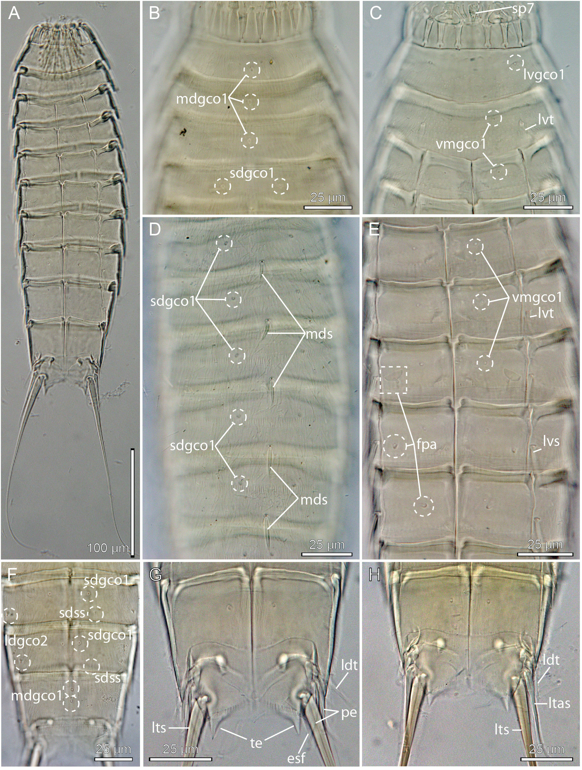
Fig. 7 View Fig
Emended diagnosis
Echinoderes with short middorsal spines on segments 4 to 8, and lateroventral spines on segments 6 to 9, never extending beyond the posterior margin of their respective segments. Tubes present in lateroventral positions on segments 2 and 5, and in laterodorsal positions on 10. Minute glandular cell outlets type 2 in laterodorsal positions on segments 8 and 9. Tergal extensions of segment 11 short, pointed and well-spaced; sternal extensions short with ventrolateral seta-like tuft of extended fringe tips. Females with ventromedial female papillae resembling glandular cell outlets type 2 on segments 6 to 8. Trunk segments with cuticular hairs, but perforation sites are indistinct.
Material examined
Holotype
BELIZE • 1 ♀; Carrie Bow Cay ; 16°50′ N, 088°06′ W; 14 m b.s.l.; 8 Apr. 1977; R.P. Higgins leg.; RH 442, fine coral sand with mangrove and seagrass detritus; USNM-69972 . Specimen mounted for LM. GoogleMaps Paratypes
BELIZE – Carrie Bow Cay • 1 ♀; same collection data as for holotype; USNM-69974 GoogleMaps • 1 ♀, 1 ♂; 500 m southwest of type locality; 3 m b.s.l.; 8 Apr. 1977; R.P. Higgins leg.; RH443, coralline mud with mangrove and seagrass detritus; USNM-69975 • 1 ♀; 500 m south of RH443; 3 m b.s.l.; 8 Apr. 1977; R.P. Higgins leg.; RH444, very fine coralline mud with mangrove and seagrass detritus; USNM-69976 .
All specimens mounted for LM. See Table 1 View Table 1 for an overview.
Description
The appearance of the species generally follows the description provided by Higgins (1983), hence the following notes only provide additional information not included in the original description.
Introvert with six trichoscalids attached to trichoscalid plates (four dorsal and two ventral). Tubes present in lateroventral positions on segments 2 and 5 ( Fig. 7C, E View Fig ), and in laterodorsal positions on segment 10 ( Fig. 7 View Fig G–H). Spines are present in middorsal positions on segments 4 to 8 ( Fig. 7D View Fig ), and lateroventral positions on segments 6 to 9 ( Fig. 7E View Fig ); spines never extend beyond the posterior margins of their respective segments. Sensory spots could not be observed on all segments, but it is positively confirmed that sensory spots are present in following positions: paradorsal positions on segments 6 to 8, subdorsal positions on segments 4 to 9 ( Fig. 7F View Fig ), and ventromedial positions on segment 1. Glandular cell outlets type 1 are present in following positions: middorsal position on segments 1 to 3 ( Fig. 7B View Fig ), subdorsal positions on segments 4 to 9 (closer to paradorsal positions on segment 9) ( Fig. 7B, D, F View Fig ), lateroventral positions on segment 1 ( Fig. 7C View Fig ), and ventromedial positions on segments 2 to 10 ( Fig. 7C, E View Fig ). Minute glandular cell outlets type 2 present in laterodorsal positions on segments 8 and 9 ( Fig. 7F View Fig ). Females with ventromedial female papillae in ventromedial positions on segments 6 to 8 ( Fig. 7E View Fig ); outlet of papillae on segments 6 and 7 close to the ventrolateral positions, whereas outlets on segment 8 are closer to the midventral line. The intracuticular structures of the papillae on segment 6 form a semicircle with a small protuberance in the curved part of the structure; substructure of segment 7 and 8 papillae forms very short intracuticular tubes. Tergal extensions triangular and well-spaced ( Fig. 7 View Fig G–H). Posterior margins of sternal plates of terminal segment obliquely straight towards a ventrolateral point; pectinate fringe well-developed, with differentiated fringe tips forming seta-like extensions ( Fig. 7G View Fig ). Perforation sites of cuticular hairs are not invisible on most segments, however, they can be visualized as indistinct dots on segments 1 and 2.
No known copyright restrictions apply. See Agosti, D., Egloff, W., 2009. Taxonomic information exchange and copyright: the Plazi approach. BMC Research Notes 2009, 2:53 for further explanation.
|
Kingdom |
|
|
Phylum |
|
|
Class |
|
|
Order |
|
|
Family |
|
|
Genus |