
Witchelinamiris, Namyatova, Anna A., Elias, Michael & Cassis, Gerasimos, 2011
|
publication ID |
https://doi.org/ 10.5281/zenodo.277994 |
|
DOI |
https://doi.org/10.5281/zenodo.6191944 |
|
persistent identifier |
https://treatment.plazi.org/id/03C7D746-FFFE-4018-FF6E-7B195FDDFCD4 |
|
treatment provided by |
Plazi |
|
scientific name |
Witchelinamiris |
| status |
gen. nov. |
Witchelinamiris gen. nov.
Etymology. This genus is named after the Witchelina Reserve, from which most of the material was collected. Type species. Witchelinamiris mchughi Namyatova, Elias & Cassis , by original designation.
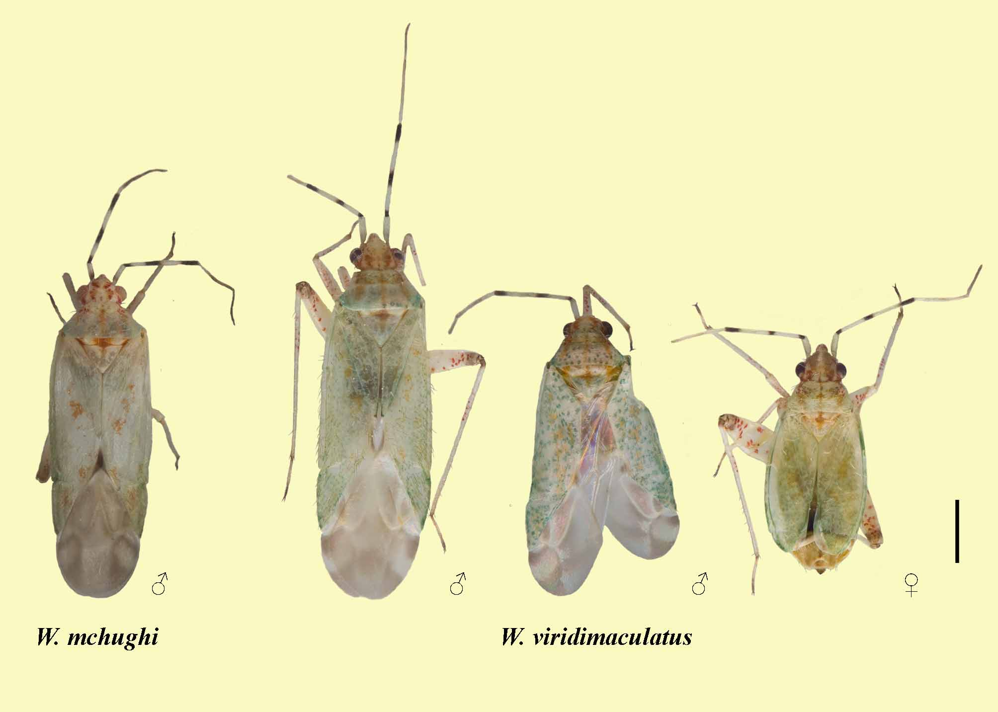
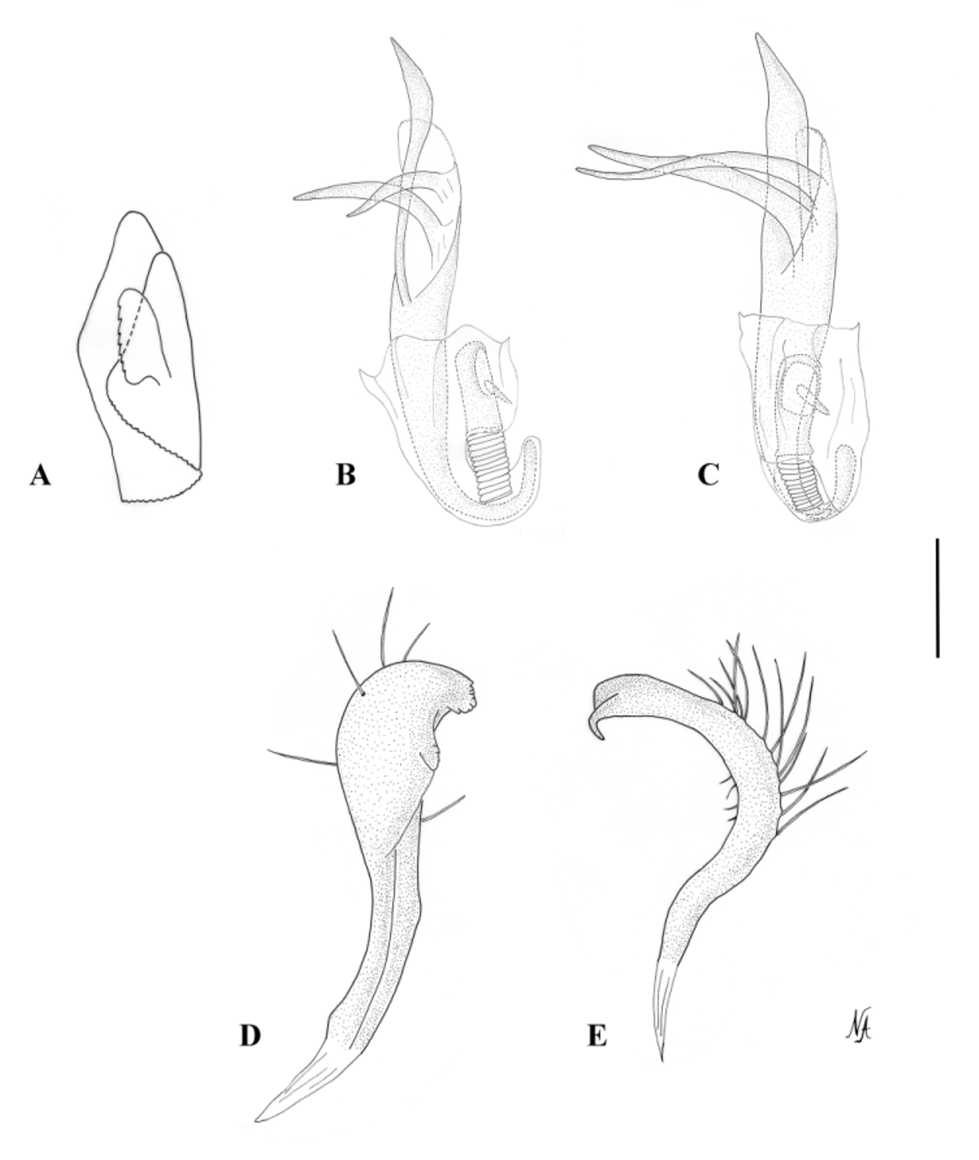
Diagnosis. Witchelinamiris is recognised by the following combination of characters: body with pale green colouration ( Fig. 1 View FIGURE 1 ); banded AII; elongate head in lateral view ( Fig. 2 View FIGURE 2. A B); phallotheca with a wedge shaped process on dorsal surface ( Figs. 3 View FIGURE 3 A, 6A); right paramere club shaped ( Figs. 3 View FIGURE 3 D, 6D); left paramere C-shaped ( Figs. 3 View FIGURE 3 E, 6E) with bifurcate apex ( Figs. 3 View FIGURE 3 E, 6E); aedeagus with two endosomal spicules, a large one with two or three long smooth processes and a small one placed near the secondary gonopore ( Figs. 3 View FIGURE 3 B, C, 6B, C). It is distinguished from all other described Australian Orthotylinae by the patterned forewing membrane and male genitalia.
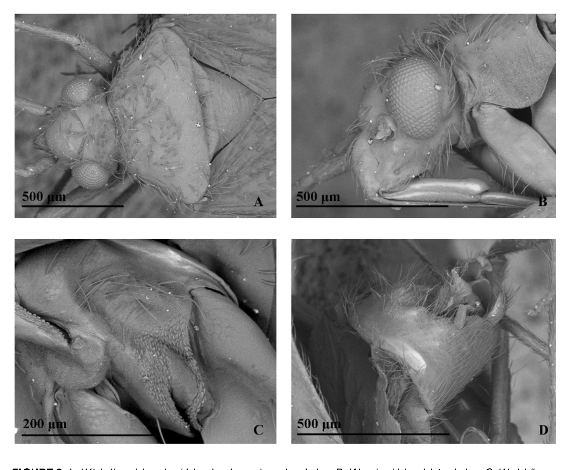
Description. Male. Macropterous. COLOURATION ( Fig. 1 View FIGURE 1 ): Head: yellow or pale green dorsally with medial reddish stripe and reddish or reddish brown marking along inner margin of eye, sometimes with reddish marking along posterior margin of eye; yellow laterally with reddish marking along eye and reddish stripe along inferior margin of maxillary plate; mandibular and maxillary plates yellow with reddish stripe between them; buccula yellowish; uniformly yellow ventrally; frontal region yellow with reddish markings, forming semicircle at each side, and orange transverse stripes between semicircles; frons with few small markings surrounding each eye; clypeus yellow with reddish markings. Eye: pale brown to brownish with reddish tinge, sometimes uniformly reddish. Antennae: AI uniformly pale green or with pale brown or brown band at base; AII yellow or pale green, with brown bands basally, medially and apically, basal band about 2x shorter than medial band, medial and apical bands subequal in length; AIII pale green basally and pale brown apically; AIV pale brown, pale green at base. Labium: LI pale green; LII uniformly yellow or pale green; LIII yellow, sometimes apically darkened; LIV brownish with yellow or pale brown base. Pronotum: mostly pale green, with three longitudinal yellow stripes dorsally and single red stripe at lateral margin, dorsal stripes sometimes very pale; calli yellowish to pale green, paler than posterior part of pronotum, with reddish markings mostly at sides; posterior part of pronotum sometimes with dense bright green markings. Scutellum: whitish or pale green with pale brown or reddish marking at base medially. Mesoscutum: whitish or pale green with reddish, pale brown or yellowish markings medially and laterally. Thoracic pleura: yellow or pale green with small reddish markings at sides, often with inferior part reddish; metapleuron pale green. MTG: pale green. Hemelytron: pale green, with yellowish markings, sometimes entire hemelytron clothed with bright green small markings; membrane mostly pale brown with distinct brownish spot at base and indistinct whitish areas. Legs: coxae yellow with reddish markings at base; femora yellow with reddish markings mostly apically, hind femur pale brown apically; tibiae yellow, sometimes with reddish markings, often pale brown apically; tarsi yellow or pale brown, tarsal segment III and sometimes segment I brown. Abdomen: pale green with reddish and yellow markings basally, laterally and dorsally. VESTITURE: Dorsum clothed mostly with greyish suberect or adpressed short setae, paler on head and pronotum, darker on posterior part of corium and cuneus; setae mostly shorter than width of AI, setae on head posteriorly, pronotum anteriorly and anterior margin of corium as long as or longer than width of AI; setae on ventral side of head pale, suberect or adpressed, mostly as long as width of AI; setae on thoracic pleura pale, adpressed, very short and rare; setae on antennae and legs mostly pale, dense, very short and adpressed, distinctly shorter than AI; AI with two medial pale spine-like setae; tibiae with rows of pale spines; abdomen clothed with short dense pale setae, shorter than width of AI. STRUCTURE: Head: elongate, frons and clypeus slightly convex with distinct depression between them ( Figs. 2A View FIGURE 2. A , B). Eye: semioval in frontal view, with medial margin straight, oval in lateral view, removed from anterior margin of pronotum by one third of eye diameter ( Fig. 2A View FIGURE 2. A ); distance from inferior margin of eyes to clypeus apex as long as diameter of eye in frontal view, and longer than diameter of eye in lateral view ( Fig. 2 View FIGURE 2. A B). Antennae: antennal segments of similar width, AI-II slightly wider than segments AIII-IV; AI short, about two third length of vertex width; AII about 4- 5 x longer than AI; AIII about 1.5x as short as AII; AIV about 2x shorter than AIII. Labium: elongate, surpassing hind coxa, reaching between abdominal sternum III and middle of pregenital abdomen; LI 2x as wide as L2 and L3, L4 slightly thinner than other segments; L1 slightly longer than ventral side of head; LII slightly longer or as long as LI; LIII as long as or longer than LII, LIV slightly shorter than LIII. Pronotum ( Fig. 2A View FIGURE 2. A ): trapeziform; collar very narrow; posterior margin about 2x as long as anterior margin; calli shallow, separated medially by very shallow suture; lateral margins rounded; posterior margin slightly concave. Scutellum: flat, aproximately as long as posterior part of pronotum, mesoscutum exposed. MTG: triangular, anterior and posterior margins concave. Legs: hind femur broad, about 2x wider than mesofemur; tarsi narrow, hind tarsal segment I short, segment II about 3x longer than segment I, segment III slightly shorter than segment II; pretarsus with convergent lamellate parempodia, pulvilli absent. Male genitalia: pygophore symmetrical, trapeziform, without processes, genital opening dorsal ( Fig. 2 View FIGURE 2. A D); right paramere club-shaped with small serrations on mesial surface ( Figs. 3 View FIGURE 3 D, 6D); left paramere C-shaped with bifurcate apex ( Figs. 3 View FIGURE 3 E, 6E); phallotheca with dorsal process medially ( Figs. 3 View FIGURE 3 A, 6A); aedeagus with two endosomal spicules, first spicule large, basally arcuate, with two or three long smooth processes, second spicule very small, placed close to secondary gonopore ( Figs. 3 View FIGURE 3 B, C, 6B, C); endosomal membrane only at base of spicules ( Figs. 3 View FIGURE 3 B, C, 6B, C); distal part of ductus seminis sclerotised ( Figs. 3 View FIGURE 3 B, C, 6B, C); secondary gonopore oval, about ½ length of sclerotised part of ductus seminis, directed laterally ( Figs. 3 View FIGURE 3 B, C, 6B, C).
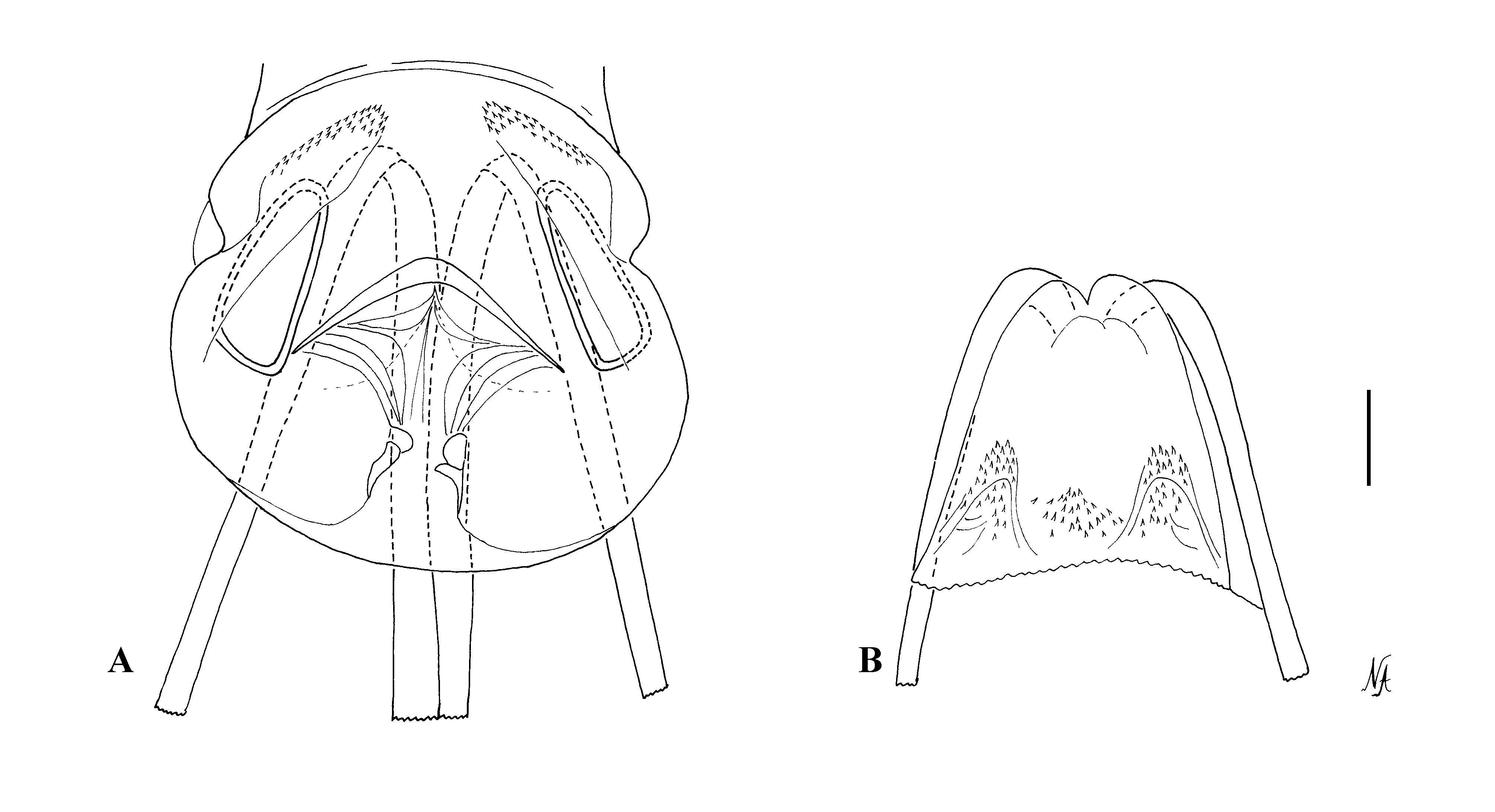
Female (only W. viridimaculatus ). Brachypterous. COLOURATION ( Fig. 1 View FIGURE 1 ): Mostly as in male. Head: vertex greenish yellow with markings near inner margin of eyes reddish brown, and with marking along posterior margins of eyes reddish; frons with pale brown markings, no orange transverse stripes between reddish semicircles. Pronotum: posterior part of pronotum with yellow stripes distinct, without bright green markings. Scutellum: mostly yellowish with pale green apex, stripes and markings as in male. Mesoscutum: yellow, stripes and markings as in male. Thoracic pleura: uniformly pale green. MTG: efferent system pale green with reddish spot laterally. Hemelytron: without bright green markings. Legs: tibiae yellow with reddish markings, pale brown apically; tarsi yellow, segments I and III brown. Abdomen: yellow, with sparse reddish markings basally and dorsally. VESTITURE: as in male. STRUCTURE: similar to male. Antennae: AII about 3- 4 x as long as AI; AIII about ½ AIII length; AIV slightly shorter than AIII. Labium: reaching middle of abdomen; LII longer than LI; LIII slightly longer than LII; LIV shorter than LII. Pronotum: calli very shallow, almost flat. Female genitalia: dorsal labiate plate with elongate sclerotized rings, single weakly sclerotized semicircular sclerite ( Fig. 7 View FIGURE 7 A); posterior wall of genital chamber with two processes, finely tuberculate ( Fig. 7 View FIGURE 7 B); vestibular area symmetrical, membranous.
Host plants. Host plant records are known only for W. viridimaculatus , which is known from two chenopod species. Mirids have been commonly collected from chenopods in arid and semi-arid Australia, with many of them new to science ( Tatarnic & Cassis 2008). However, much of the chenopod-inhabiting mirid fauna, particularly new species belonging to the Orthotylini and Phylini are awaiting description.
Remarks. The systematic position of Witchelinamiris is uncertain. On colouration characters the species shares some similarities with members of the Zanchius genus group (sensu Schuh 1974), with W. viridimaculatus sp. nov. having a green spotted dorsum. However, the head of both species of Witchelinamiris is elongate and the postocular margins are straight. In comparison, the head of members of the Zanchius genus group is short and not elongate in front, and the postocular margins are convex. In addition, the endosomal membrane is reduced, and both species have an elongate smooth endosomal spicule. The spicule structure differs considerably from Austromirini , typified by the Lattinova complex, which have two elongate spicules with limited endosomal membrane (Cassis 2008). It is also unlike examined Australian members of the Orthotylini , such as Harveycapsus (Cassis et al. 2010) and an undescribed genus of myrtaceaous inhabiting orthotylines (Cheng & Cassis in preparation), which have three endosomal spicules, which like austromirines have elongate spicules. Unlike these genera, the spicules of Witchelinamiris are smooth, and do not possess the sawlike margins of the spicules in the other abovementioned orthotylines. In addition, the parameres of both species of Witchelinamiris are distinctive, with the left paramere Cshaped with a small bifurcation at its apex, and the right paramere is simple, and is somewhat club-shaped. The female genitalia are simple, with the posterior wall of the genital chamber with simple paired processes that are unlike the inter-ramal lobes of other orthotylines (cf. Slater 1950; Cassis et al. 2010). Moreover, the opening of the vestibulum is symmetrical, which is akin to what is found in the Halticini (Tatarnic & Cassis in press) and species of the Hadronema complex ( Forero 2008), and unlike asymmetry found in many Austromirini and Orthotylini .
At present, it is difficult to assess the systematic position of Witchelinamiris because of the inadequacy of the tribal classification of the Orthotylinae . We tentatively place it within the Ortothylini pending further investigation and any future formalisation of the Zanchius genus group at the tribal level.
No known copyright restrictions apply. See Agosti, D., Egloff, W., 2009. Taxonomic information exchange and copyright: the Plazi approach. BMC Research Notes 2009, 2:53 for further explanation.