
Unionicola Haldeman, 1842
|
publication ID |
https://doi.org/ 10.5281/zenodo.282494 |
|
DOI |
https://doi.org/10.5281/zenodo.6166622 |
|
persistent identifier |
https://treatment.plazi.org/id/03C82744-C412-FFC5-FF64-FE9FD8A19301 |
|
treatment provided by |
Plazi |
|
scientific name |
Unionicola Haldeman, 1842 |
| status |
|
Genus Unionicola Haldeman, 1842 View in CoL
Unionicola (Pentatax) figuralis (Koch, 1836) ( Figs 1–11 View FIGURES 1 – 2 View FIGURES 3 – 6 View FIGURES 7 – 11 )
Material examined. Larvae (n = 8) were reared from single female collected in D’yakonovskoe Lake, Ust’- Kamchatsky District, Kamchatka Province, 8 August 1983. The duration of the embryonic period was 14 days at room temperature.
Diagnosis. Dorsal plate elongated (L/W ratio 1.65–1.75), setae Oe shorter than Hi, He, Sci and Sce; excretory pore plate much longer than wide; setae Ai relatively long and reaching posterior margin of excretory pore plate; basal segments of chelicerae nearly as long as wide; P-5 with two very long setae; II—Leg-4 with a single greatly thickened seta.
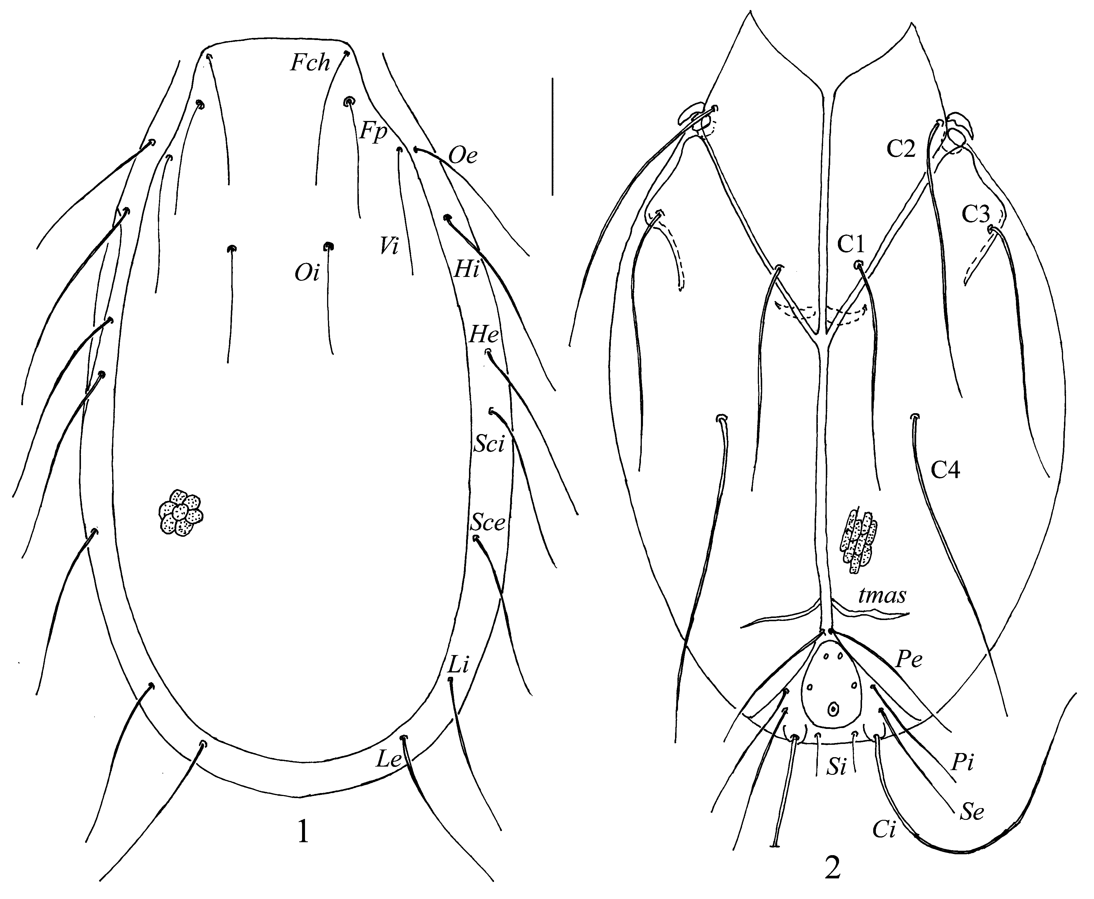
Description. Idiosoma flat, dorsal plate in unengorged larvae covering almost the whole dorsum ( Fig. 1 View FIGURES 1 – 2 ), with slightly convex lateral margins, its anterior margin straight or slightly convex, posterior margin rounded, with short and punctuate scale-like patterns. All four pairs of setae on the dorsal plate thin and approximately equal in length. Setae Oe, Hi, He, Sci, Sce, Li and Le situated in soft integument: Oe, Li and Le subequal, shorter than other setae situated in the integument.
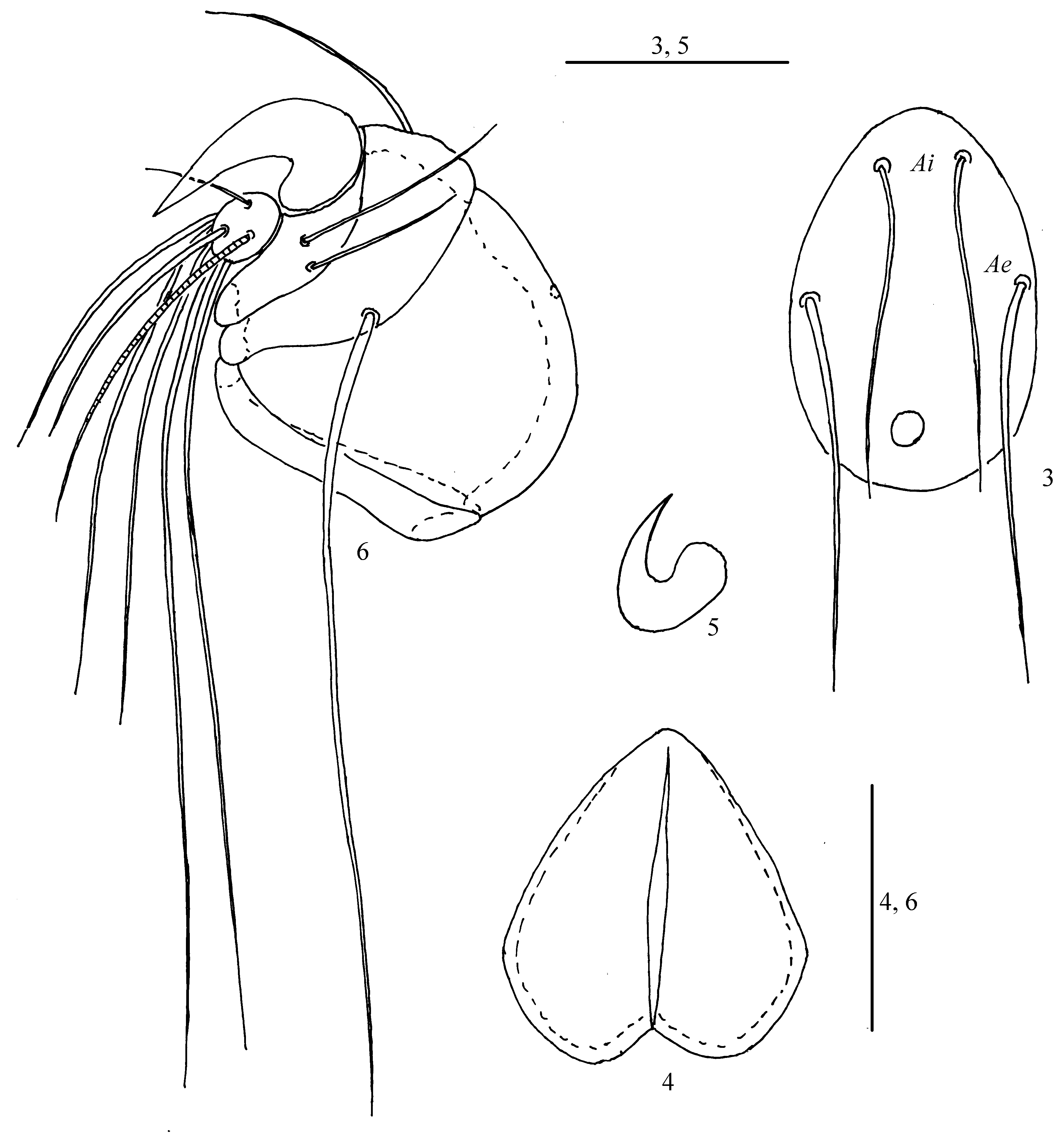
Coxal plates moderately large and elongate, plate II–III with a posteromedial muscle attachment scar on each side ( Fig. 2 View FIGURES 1 – 2 ). Setae C1 relatively short and extending beyond bases of C4; the latter setae longest among the coxal setae and extending to posterior margin of the coxal plates II–III. Setae C2 and C3 subequal, longer than C1 but shorter than C4. Well developed tmas situated in posteromedial corner of coxa III on each side. Setae Ci very long, well thickened, located on small tubercles; Si very short; Pi longer than Pe and Se. Excretory pore plate oval ( Fig. 3 View FIGURES 3 – 6 ), much longer than wide (L/W ratio 1.4–1.5), rounded posteriorly, narrow anteriorly; setae Ai relatively short, thin located anteriorly close to each other, and reaching or slightly extending to posterior margin of excretory pore plate; setae Ae longer and thicker than Ai, located laterally nearly in middle of plate, and extending well beyond posterior margin of excretory pore plate; excretory pore placed posteromedial to Ae near posterior edge of plate; distance between setae Ae–Ae almost three time longer than distance between Ai–Ai. Setae Pi located very close to each other anterior to excretory pore plate. Setae Pe and Se moderately long and approximately equal in length.
Chelicerae short and very heavy, nearly as long as wide; basal segments of chelicerae with broad incision posteromedially ( Fig. 4 View FIGURES 3 – 6 ); cheliceral stylets small and hidden in the anterior region of chelicerae, crescent-shaped ( Fig. 5 View FIGURES 3 – 6 ).
Pedipalps very short and stocky ( Fig. 6 View FIGURES 3 – 6 ): P-1 short; P-2 large with convex dorsal margin, dorsal seta usually absent or its location visible as alveole; P-3 with very long, thick lateroproximal seta and short, fine dorsodistal one; P-4 with two rather long unequal setae and massive, dorsodistal claw; P-5 small, with single solenidion and seven unequal simple setae, two of them very long.
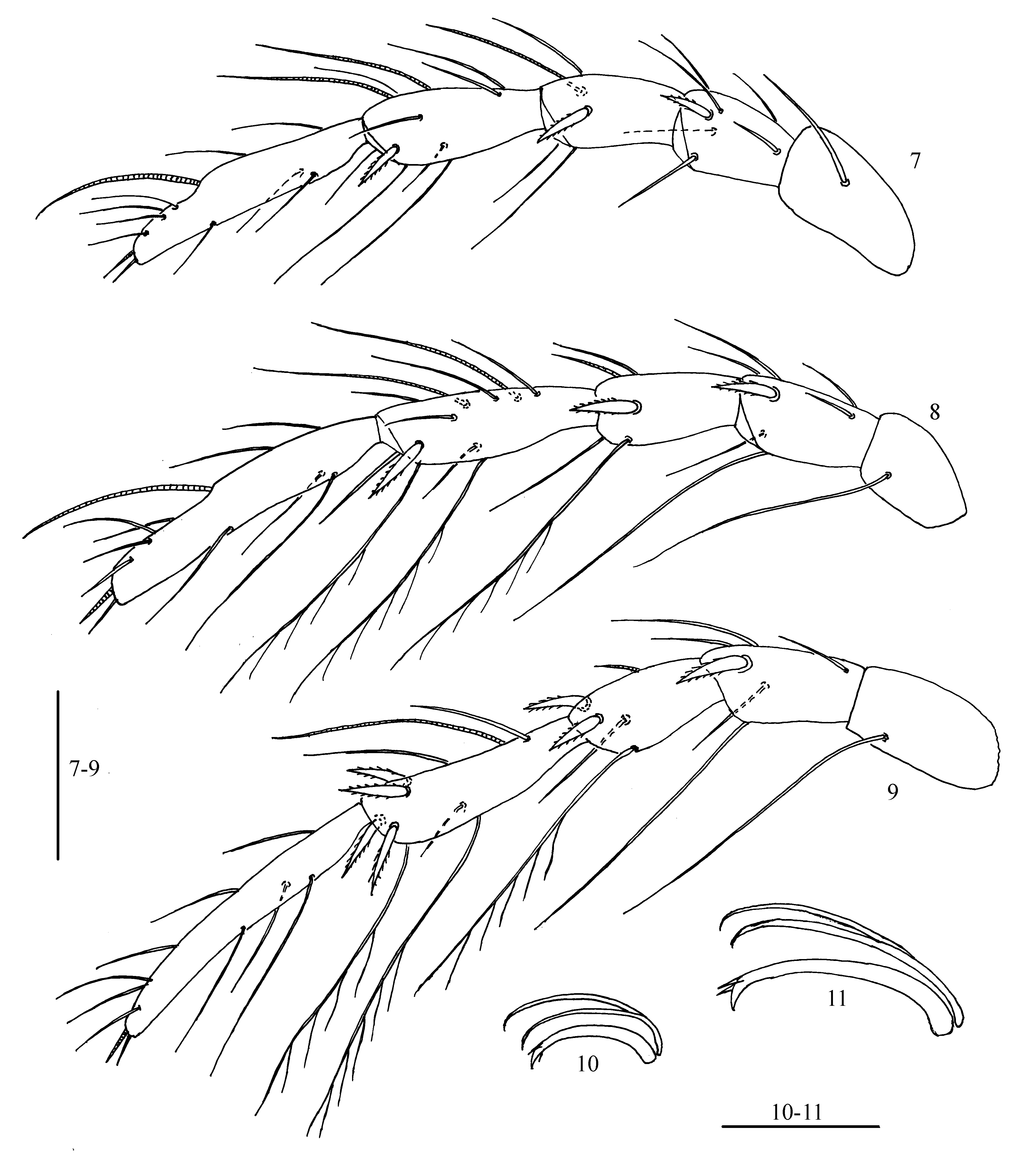
Legs 5—segmented. Shape and arrangement of setae on legs segments as shown in Figs 7–9 View FIGURES 7 – 11 . Total number of leg setae, excluding eupathidia, as follows (specialized setae indicated in parentheses): I—Leg-1–5: 1, 7, 5 (s), 11 (2s), 12 (s, ac); II—Leg-1–5: 1, 7, 5 (s), 11 (2s), 12 (s, ac); III—Leg-1–5: 1, 6, 5 (s), 10 (s), 11 (ac). Number of thickened setae from trochanter to tarsus: I—Leg: 0, 1, 1, 1, 0; II—Leg: 0, 1, 1, 1, 0; III—Leg: 0, 1, 2, 4, 0. I—Leg- 1 with relatively short seta, II—Leg-1 and III—Leg-1 each with rather long seta. Solenidion on I—Leg-3 and I—Leg-5, and both solenidia on I—Leg-4 approximately equal in length; solenidion on II—Leg-3 and III—Leg-3 very short; solenidion on III—Leg- 4 and both solenidia on II—Leg-4 long and approximately equal in length. Acanthoid seta on tarsi I to III comparatively long and setose. Claws of legs III ( Fig.11 View FIGURES 7 – 11 ) larger than claws of legs I and II ( Fig. 10 View FIGURES 7 – 11 ). Lateral claws nearly as long as central claw, all claws are sickle shaped.
Measurements, n=8. L of dorsal plate 285–305, W 165–175; L of setae Fch, Fp, Vi, Oi, Oe 40–55; L of setae Hi 55–65; L of setae He, Sci, Sce, Li, 40–55; L of setae Le 55–60, L of setae Si 15–25, L of setae Se 45–55, L of setae Ci 160–170, L of setae Pi 40–50, L of setae Pe 45–60, L of setae C1 80–85, L of setae C2 95–105, L of setae C3 95–110, L of setae C4 130–145; L of medial edge of coxae I 95 –100, L of medial edges of coxae II–III 150–160; L of excretory pore plate 38–40, W 25–30; L of basal segments of chelicerae 63–66, W 64–67; L of cheliceral stylet 20–23; L of pedipalpal segments (P–1–5): 5–6, 35–39, 16–19, 8–11, 6–8; L of legs segments: I—Leg-1–5: 30–35, 33–40, 40–45, 50–60, 75–80; II—Leg-1–5: 35–40, 35–42, 40–45, 60–65, 94–98; III—Leg- 1–5: 44–48, 35–42, 40–45, 65–72, 95–100.
Remarks. The larva of the present species slightly differs from others known larvae of the subgenus Pentatax by the shape of the posterior coxal plates. Coxal plates II-III in the larvae of this subgenus pointed posteriorly ( Hevers 1980, Wainstein, 1980); in contrast, coxal plates II-III in the larva of U. figuralis are rounded posteriorly.
Interestingly, U. figuralis is only the third known Unionicola -species where larvae could be reared outside of mussel or sponge tissue; other reported cases are U. (U.) crassipes (Müller, 1776) and U. (U.) dresscheri Besseling, 1946 respectively ( Wainstein 1980, Tuzovskij 1985).
No known copyright restrictions apply. See Agosti, D., Egloff, W., 2009. Taxonomic information exchange and copyright: the Plazi approach. BMC Research Notes 2009, 2:53 for further explanation.