
Coelana Kramer, 1964
|
publication ID |
https://doi.org/ 10.5281/zenodo.178025 |
|
DOI |
https://doi.org/10.5281/zenodo.6249785 |
|
persistent identifier |
https://treatment.plazi.org/id/03C8879C-E830-FFA2-91BC-07700D5EAFBC |
|
treatment provided by |
Plazi |
|
scientific name |
Coelana Kramer, 1964 |
| status |
|
Coelana Kramer, 1964 View in CoL
( Figures 1–29 View FIGURES 1 – 9 View FIGURES 10 – 25 View FIGURES 26 – 29 )
Coelidiana (Coelana) DeLong, 1953: 95 View in CoL (key to genera) [nomen nudum]; Metcalf, 1964: 112 (catalogue, citation as an invalid name). Type species not designated.
Coelana DeLong View in CoL ; Oman et al., 1990: 201, 305 (catalogue, citation as an invalid name); Freytag & Sharkey, 2002: 254 (citation, number of species); Dietrich, 2003: 701 (citation, distribution).
Coelana Kramer, 1964: 261 View in CoL , 269–270 (key to genera, description, key to species). Type species: Neocoelidia modesta Baker, 1898 View in CoL . Designated by Kramer, 1964: 269.
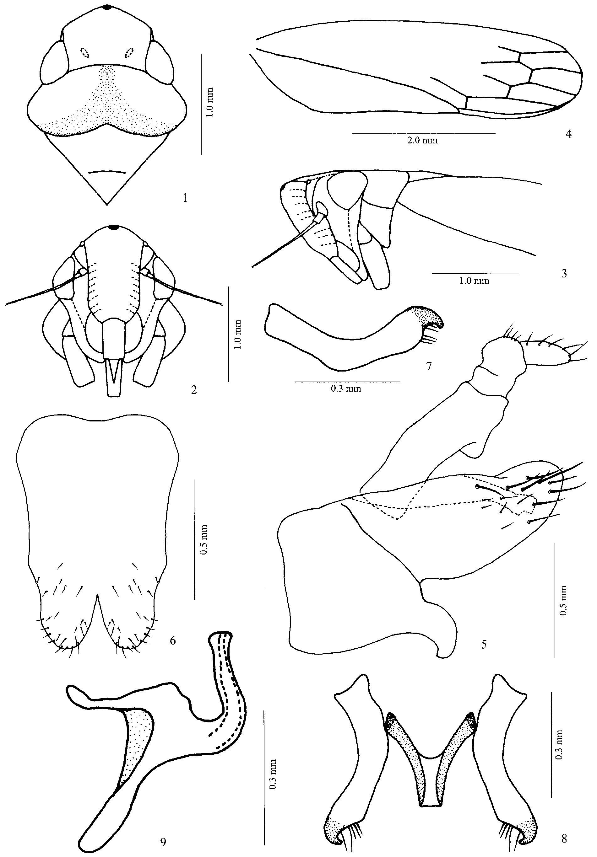
Diagnosis. Color stramineous to yellow with small black spot at apex of crown ( Figs. 1 View FIGURES 1 – 9 , 10 View FIGURES 10 – 25 ); carina present in transition between crown and frons; posterior margin of pronotum emarginated, broadly indented ( Figs. 1 View FIGURES 1 – 9 , 10 View FIGURES 10 – 25 ); forewing with venation indistinct, except for claval suture and apical cells ( Figs. 4 View FIGURES 1 – 9 , 13 View FIGURES 10 – 25 ); male genitalia: pygofer greatly inflated, with large ventral hook, a pair of long inner processes dorsally, and laterally with a long ventrodorsal suture ( Figs. 5 View FIGURES 1 – 9 , 14 View FIGURES 10 – 25 ); connective Y-shaped ( Figs. 8 View FIGURES 1 – 9 , 17 View FIGURES 10 – 25 ) and aedeagus simple, without processes ( Figs. 9 View FIGURES 1 – 9 , 18 View FIGURES 10 – 25 ); anal tube with a pair of ventral processes ( Figs. 5 View FIGURES 1 – 9 , 14 View FIGURES 10 – 25 ).
Description. In dorsal view, body approximately cylindrical, not flat.
Head. Crown almost quadrangular, slightly projected anteriorly and slightly wider than long; median length of crown approximately one half of transocular width and approximately equal to interocular width; anterior margin broadly angular, with carina in the transition between crown and frons; lateral margins, adjacent to compound eyes, elevated (not at the same level of the dorsum of compound eyes) and carinate; surface flat, smooth, without grooves, with weak coronal maculae at the base of crown, close to compound eyes; coronal suture indistinct ( Figs. 1 View FIGURES 1 – 9 , 10 View FIGURES 10 – 25 ); ocelli located on anterior margin of head, in the transition between crown and frons, closer to compound eyes than to midline, above antennal ledges; in lateral view, antennal ledges oblique and carinate ( Figs. 3 View FIGURES 1 – 9 , 12 View FIGURES 10 – 25 ); antennae setaceous, scape and pedicel enlarged, flagellum longer than basal half of forewings ( Figs. 2–3 View FIGURES 1 – 9 , 11–12 View FIGURES 10 – 25 ); frons with length approximately two times basal width, profile convex, muscle impressions generally indistinct, not swollen on the central portion of base; frontogenal sutures reaching ocelli; apical margin of maxillas not extending beyond apex of clypeus; lorum small, approximately halfmoon shaped, inferior margin not reaching apical margin of clypeus; gena partially concealing proepisternum, only apex visible in anterior view; epistomal suture distinct and complete, slightly curved; clypeus almost rectangular, lateral margins parallel, generally without a gibbosity in lateral view, and apical margin straight ( Figs. 2–3 View FIGURES 1 – 9 , 11–12 View FIGURES 10 – 25 ).
Thorax. Pronotum slightly wider than head, width between humeri slightly larger than transocular width; median length approximately one third of width between humeri; lateral margins rounded and posterior margin emarginated, broadly indented ( Figs. 1 View FIGURES 1 – 9 , 10 View FIGURES 10 – 25 ); dorsal surface with transverse grooves; dorsopleural carina complete and evident; in ventral view, mesothorax moderately swollen; scutellum (sensu Young 1968) as long as maximum width, with distinct preapical fold ( Figs. 1 View FIGURES 1 – 9 , 10 View FIGURES 10 – 25 ). Forewing ( Figs. 4 View FIGURES 1 – 9 , 13 View FIGURES 10 – 25 ) with rounded apex, without punctuations, hyaline, about 3–3.5 times longer than greatest width; venation indistinct, except for claval suture and apical cells; four apical cells, all almost rectangular, first larger than second, third slightly enlarged apically; second and fourth apical cells approximately same distance to base, closer to forewing base than third cell; anteapical cells distinct or not; appendix narrow, extending only along first apical cell. Hindwing with three apical cells, R4+5 and M1+2 apically convergent, fused, forming a single vein. Hindleg: femoral setal formula 2+2+0; tibial row PD with long setae little separated amongst themselves, distributed throughout length of tibia, intercalary setae absent; row AD with long and robust setae in form of spines, with two or more small intercalary setae present; row AV with setae moderately long, distributed only on apical two thirds, intercalary setae absent; row PV densely bristly, with short setae at base, becoming progressively longer toward apex; first tarsomere as long as combined length of two distal ones, plantar surface with two parallel rows of short setae.
Abdomen. Male genitalia: pygofer greatly inflated, longer than subgenital plates, with large ventral hook, a pair of long inner processes dorsally, and laterally with a long suture, extending from ventral margin to near dorsal margin ( Figs. 5 View FIGURES 1 – 9 , 14 View FIGURES 10 – 25 ); subgenital valve entirely fused to subgenital plates; subgenital plates fused on the basal and median thirds, separated only on apical third ( Figs. 6 View FIGURES 1 – 9 , 15 View FIGURES 10 – 25 ); styles simple, not bifurcated, with sclerotized hook-like apex, curved ventrally ( Figs. 7–8 View FIGURES 1 – 9 , 16–17 View FIGURES 10 – 25 ); connective Y-shaped, articulated to base of aedeagus ( Figs. 8 View FIGURES 1 – 9 , 17 View FIGURES 10 – 25 ); aedeagus simple, without processes ( Figs. 9 View FIGURES 1 – 9 , 18 View FIGURES 10 – 25 ); anal tube with a pair of ventral processes ( Figs. 5 View FIGURES 1 – 9 , 14 View FIGURES 10 – 25 ).
Total length (body + forewings). 6.0– 7.5 mm.
Geographical distribution. Argentina, Bolivia and Brazil.
Comments. This genus is similar to Coelidiana Oman regarding the external morphology and general color of the species. It possesses some diagnostic characters that easily separate it from the other genera: general color stramineous to yellow with a small black spot at apex of crown and the characteristic male genitalia, mainly the morphology of the pygofer and anal tube.


No known copyright restrictions apply. See Agosti, D., Egloff, W., 2009. Taxonomic information exchange and copyright: the Plazi approach. BMC Research Notes 2009, 2:53 for further explanation.
|
Kingdom |
|
|
Phylum |
|
|
Class |
|
|
Order |
|
|
Family |
Coelana Kramer, 1964
| Marques-Costa, Ana Paula & Cavichioli, Rodney Ramiro 2007 |
Coelana
| Dietrich 2003: 701 |
| Freytag 2002: 254 |
| Oman 1990: 201 |
Coelana
| Kramer 1964: 261 |
| Kramer 1964: 269 |
Coelidiana (Coelana)
| Metcalf 1964: 112 |
| DeLong 1953: 95 |