
Camptosauridae, Marsh, 1885
|
publication ID |
https://doi.org/10.5252/geodiversitas2022v44a25 |
|
publication LSID |
urn:lsid:zoobank.org:pub:EA12DCB7-A5BE-4763-B805-25087EBD726D |
|
DOI |
https://doi.org/10.5281/zenodo.6928908 |
|
persistent identifier |
https://treatment.plazi.org/id/03C887B9-FFA8-FFCB-76C8-A509FAFEFE61 |
|
treatment provided by |
Felipe |
|
scientific name |
Camptosauridae |
| status |
|
Camptosauridae indet.
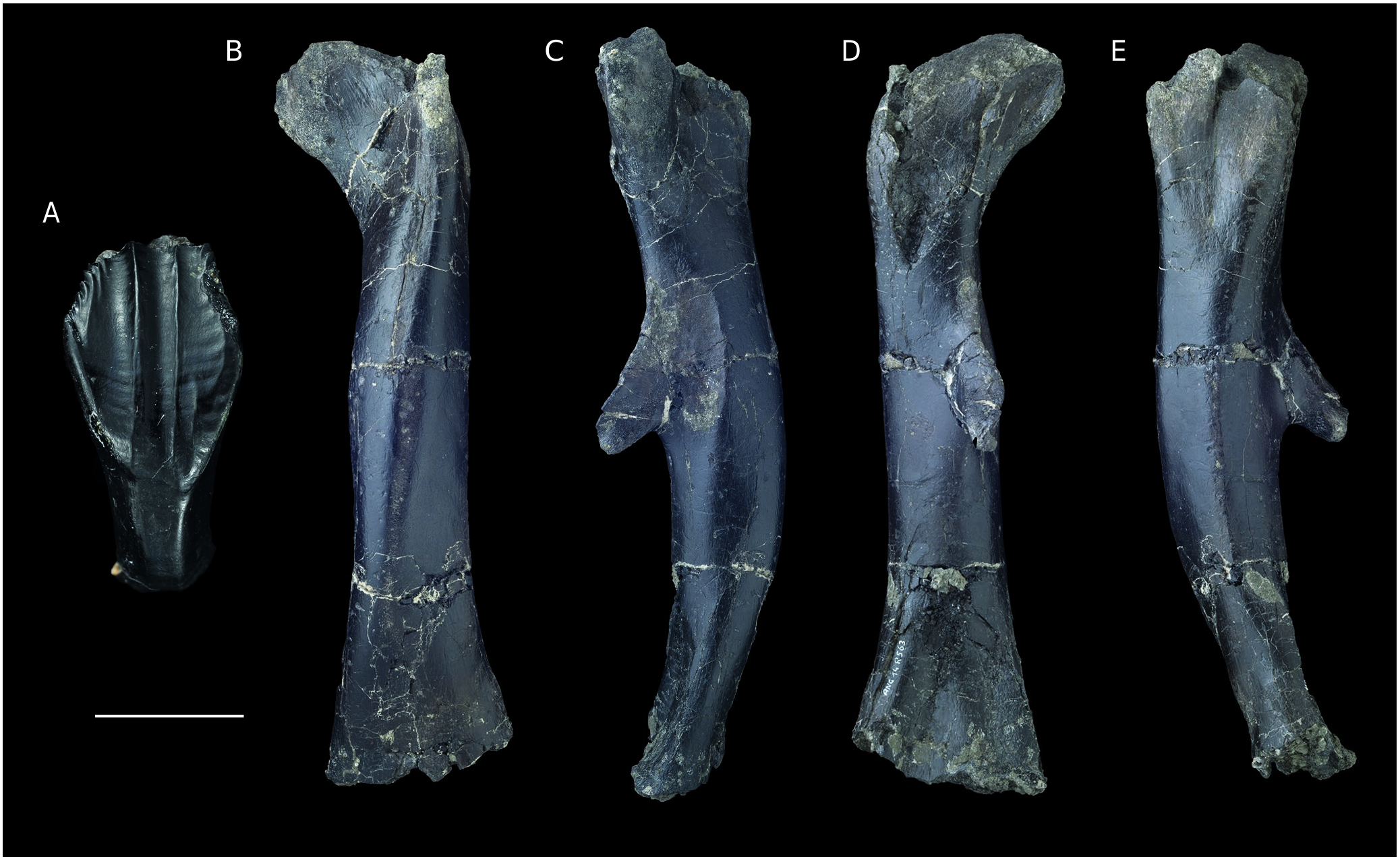
( Fig. 22 View FIG )
DESCRIPTION
Several diagnostic features allow the referral of the material described below to Camptosauridae , including: 1) lozengeshaped teeth with prominent vertical keel more strongly developed on maxillary teeth than on dentary teeth, several secondary vertical ridges on sculptured surface of crown with anterior and posterior ridges bordering the non-denticulate half of maxillary tooth crown, posterior ridge on equivalent part of dentary teeth ( Galton & Powell 1980); and 2) femur robust, with a dorsally arched shaft, a midshaft positioned and pendant fourth trochanter, and with a dorsally open, troughlike anterior intercondylar groove ( Norman & Barrett 2002).
Dentary tooth
One extremely well-preserved, isolated camptosaurid tooth (ANG11-1120) exhibits an almost complete crown and a root broken at approximately mid-length ( Fig. 22E View FIG ). The crown is straight folidont ( Hendrickx et al. 2015a), moderately elongated, and relatively robust. The mesiodistal basal length is 7 mm and the preserved crown height is 17 mm. It appears very similar to dentary teeth from the Kimmeridgian of England described by Galton & Powell (1980) and to the Owenodon sp. material figured by Galton (2009). The crown is labiolingually compressed, flat lingually, and strongly convex labially, with a D-shaped cross-section at mid-height. In lingual view, the main axis of the crown is straight, with a crown apex that was most likely pointing strictly dorsally. Both the mesial and the distal margins are symmetrical, with a marked angle at mid-height, conferring to the crown a diamond-shaped aspect in lingual and labial views. On the distal border, the lower edge of the crown is flexed mesiolingually, forming a pseudo-cingulid, as seen in Camptosaurus prestwichii ( Norman & Barrett 2002: fig. 5) and Camptosaurus medius ( Galton 2009: fig. 10D). The carinae extend from the apex to half of the length of the crown, until the mesial and distal angulations. They appear shorter than in some Camptosaurus specimens, in which the carinae extend along almost two-thirds of the edges ( Norman & Barrett 2002: fig. 5A; Galton 2009: fig. 10D). Both carinae are straight and oblique. Denticles are visible on the entire length of the preserved carinae. The mesial carina is damaged, but it apparently exhibits the same density of denticles as the distal carina with 3 denticles per mm. The denticles are fine, slightly mesially and distally hooked, and apicobasally oriented. They present a marked increase in size, the largest being the most apical. Ventral to the denticles, small ridges extend from their base towards the crown base. Most are approximately 1 mm in length but the longest is almost 5 mm in length and extends from the third well-developed denticle. The longitudinally concave lingual crown surface bears two strongly developed central and apicobasally oriented ridges separated by a flute. In contrast to the condition observed in some dentary teeth of the holotype of C. prestwichii ( Norman & Barrett 2002: fig. 5A; Galton 2009: fig. 9K) and C. medius ( Galton 2009: fig. 10D), but as seen in Owenodon sp. ( Galton 2009: fig. 18J), the ridges are parallel throughout their length. The crown enamel extends more basally in the mesiolingual part of the crown than in the distolingual part. The enamel has a smooth texture and its surface bears transverse undulations contacting both carinae. They are more marked on the basal half of the crown.
A pronounced constriction occurs at the base of the crown forming a cervix. Breakage of the root renders it impossible to assess whether or not it was longer than the crown. It is, however, clearly narrower. The width of the root is the same throughout the preserved portion, and it is roughly equivalent to the mesiodistal basal length measured at the level of the cervix. The root is labiolingually narrow and exhibits subparallel mesial and distal borders. On its lingual surface, a shallow concavity is visible. At the fracture point, it has a suboval cross section.
Femur
One subcomplete left femur is part of the material from Angeac-Charente referred to Camptosauridae . It is well-preserved, but both ends are crushed and eroded, and a small part of the distal end is missing ( Fig. 22 View FIG B-E). The preserved length of the femur is 265 mm. The diaphysis is relatively stout. In anterior and posterior views, the femur is straight ( Fig. 22B, D View FIG ). In lateral and medial views, the femur is curved with convex anterior and concave posterior margins.
The femoral head is positioned in the same plane relative to the transverse axis of the distal condyles. The femoral head is discrete, unlike that of C. dispar ( Galton & Powell 1980: fig.2F) and oval in anterior and posterior views. In those views, it is inclined at approximately 40° with respect to the main axis of the bone. The neck is large and it merges obliquely with the femoral shaft. In some Camptosaurus specimens, the neck looks more constricted ( Norman & Barrett 2002: fig. 6; Galton 2009: fig. 5T).
The lesser trochanter is located on the anterior surface of the femur, on the proximal extremity. It is eroded, but it still appears as a strong and high protrusion extending proximodistally in the anterolateral angle of the femur. Its proximal extremity is lower than the proximal margin of the femoral head, but it still appears more strongly developed than in most described Camptosaurus species ( Galton & Powell 1980; Norman & Barrett 2002; Carpenter & Wilson 2008; Galton 2009). There is no deep cleft visible between the lesser trochanter and the greater trochanter, in contrast to the condition observed in C. aphanoecetes ( Carpenter & Wilson 2008) . The greater trochanter is difficult to discern, probably as a result of erosion.
The femur diaphysis has a quadrangular cross section. It is as robust as in other camptosaurids. Under the fourth trochanter, the diaphysis is slightly narrower transversely than anteroposteriorly. On the posterior surface of the diaphysis, the pendent fourth trochanter is located on the medial edge of the bone, just above midshaft ( Fig. 22 View FIG C-E). It is strongly developed, as in most species of Camptosaurus ( Carpenter & Wilson 2008; Galton 2009), and it is blade-shaped in lateral view ( Fig. 22E View FIG ). In posterior view, the base of the trochanter is straight ( Fig. 22D View FIG ). The proximal margin of the trochanter is elongated and gradually rises from the diaphysis, at an angle of approximately 115° relatively to the main axis of the shaft. The distal margin of the fourth trochanter is shorter and steeper.
The distal extremity of the femur is incomplete.A longitudinal bulge is visible above the distal condyles, but the condyles themselves are not preserved. On the anterior surface of the femur, a very shallow intercondylar groove is present. On the posterior surface, the popliteal fossa is visible.
No known copyright restrictions apply. See Agosti, D., Egloff, W., 2009. Taxonomic information exchange and copyright: the Plazi approach. BMC Research Notes 2009, 2:53 for further explanation.
|
Kingdom |
|
|
Phylum |
|
|
Class |
|
|
Order |
|
|
Family |