
Hypsilophodontidae, Dollo, 1882
|
publication ID |
https://doi.org/10.5252/geodiversitas2022v44a25 |
|
publication LSID |
urn:lsid:zoobank.org:pub:EA12DCB7-A5BE-4763-B805-25087EBD726D |
|
DOI |
https://doi.org/10.5281/zenodo.6928906 |
|
persistent identifier |
https://treatment.plazi.org/id/03C887B9-FFAA-FFF5-76E6-A48AFB23F93D |
|
treatment provided by |
Felipe |
|
scientific name |
Hypsilophodontidae |
| status |
|
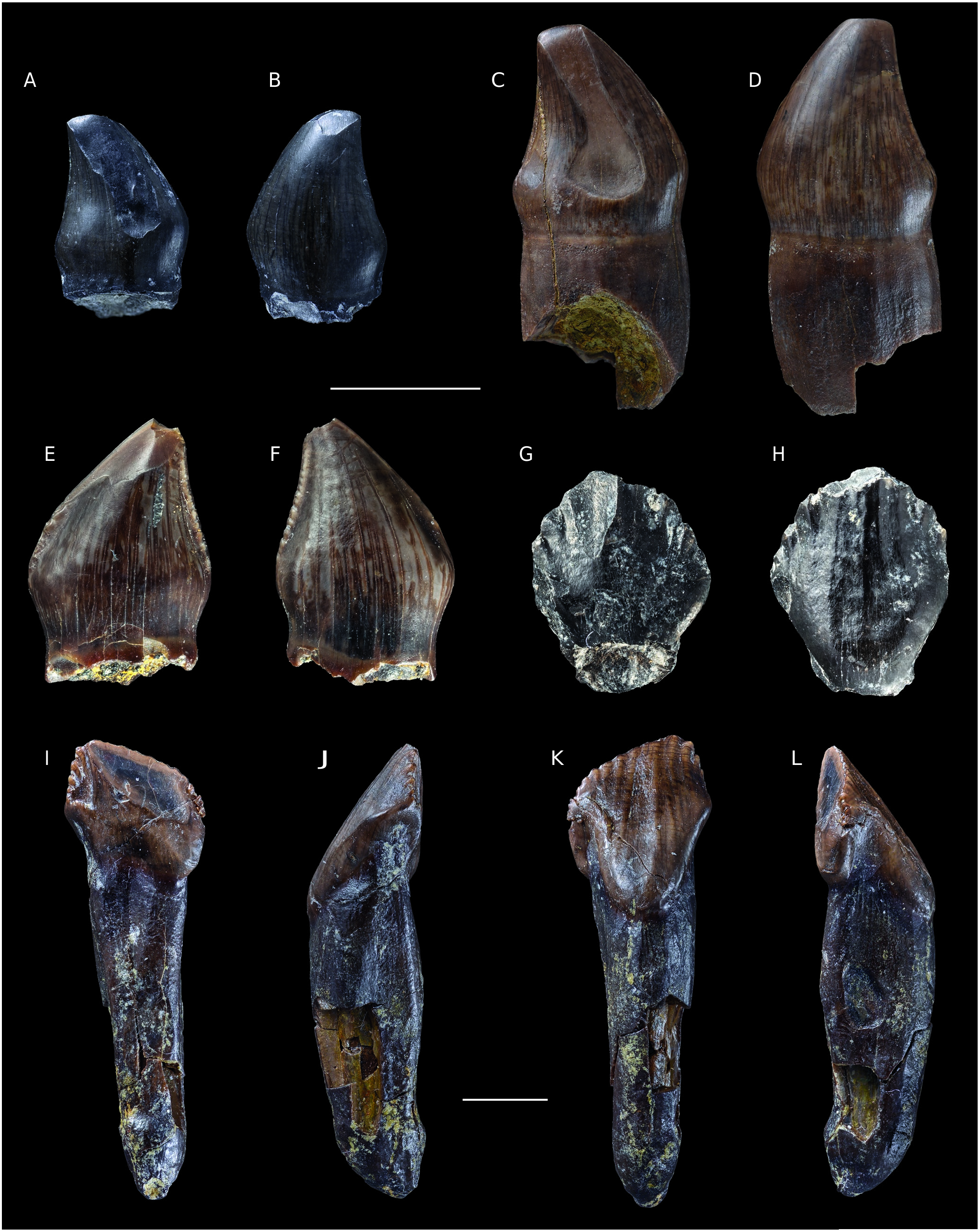
Hypsilophodontidae indet.
( Fig. 21E, F View FIG )
DESCRIPTION
Premaxillary tooth
A well-preserved isolated hypsilophodontid crown (ANG M-119) only lacks its apex ( Fig. 21E, F View FIG ). In general shape, it is very similar to the aforementioned heterodontosaurid premaxillary teeth, but has a less bulbous morphology, bears denticles and lacks a wear facet on the lingual surface of the crown. Based on its shape, it is identified as a premaxillary tooth, the maxillary and dentary teeth being more quadrangular in shape ( Norman et al. 2004: fig. 18.3). The crown is recurved folidont ( Hendrickx et al. 2015a) and short, with a mesiodistal basal length of 3 mm and a preserved crown height of 4 mm. It appears quite similar to the premaxillary teeth of H. foxii figured by Galton (2009: fig. 2G, L-P). The crown is compressed labiolingually and has a slightly convex surface on both sides. At mid-height, the cross-section is elliptical. The main axis of the crown is recurved, so that in lingual and labial views, the apex appears distal to the midpoint of the base of the crown. Both the mesial and distal margins are mesially and distally expanded respectively at their base. However, the mesial margin is convex, whereas the distal margin is concave for most of its length. The carinae extend from the apex along two-thirds of the crown and bear a series of fine, bulbous denticles. The mesial carina is damaged, but all the denticles are visible on the distal border. The density of the denticles is approximately 6 per mm. The crown surfaces have a relatively smooth texture. The crown enamel does not extend further basally on one particular side as for dentary teeth (see below). There is a visible constriction forming a cervix, at the base of the root.
Maxillary tooth
One well-preserved and complete isolated hypsilophodontid maxillary tooth (ANG10-153) ( Fig. 21 View FIG I-L) has been recovered from Angeac-Charente. It is 27 mm long with the root being twice as long as the crown. The crown is sub-quadrangular and short, with a mesiodistal basal length of 6 mm and a crown height of 10 mm. It looks similar to the maxillary crown of H. foxii ( Galton 1974: fig. 14a, b). The crown is expanded mesiodistally, compressed labiolingually, and has a sub-oval cross section at mid height. The lingual surface is relatively flat, whereas the labial surface bears a longitudinal concavity in the central area of the crown and is convex mesiodistally. In lingual and labial views, the main axis of the crown is straight, but the apex is displaced distally relatively to the midpoint of the crown base. The rounded apex is however not distally bent, but strictly ventrally oriented. The mesial and distal margins are subparallel, even if the mesial margin is slightly convex, whereas the distal one is straight with a small angle at mid-height. The carinae are located on the upper portions of the mesial and distal edges, and on the apical border of the crown. The tooth carinae are covered by small denticles throughout their length, in contrast to the H. foxii tooth figured by Galton (1974: fig. 14a, b), in which the apical border is worn and, therefore lacks denticles. The mesial carina is damaged, but probably exhibited denticles. The distal and apical carinae exhibit large and regular denticles, at a frequency of approximately 1 per mm. They are rounded in shape, apicobasally oriented and their size slightly increases towards the apex. The denticles on the apical margin are smoothly worn and, consequently, an extended single and oblique wear facet is visible on the dorsal part of the lingual surface. There is no wear on the labial surface, but small ridges extend ventrally to denticles, towards the crown base.
The longest visible is almost 6 mm long and is directly mesial to the apex. The enamel texture is relatively smooth, except towards the base of the crown where it is more irregular. The crown enamel is clearly more basally extended on the labial side than on the lingual side of the tooth. Consequently, the base of the crown appears swollen on the labial surface and forms an incipient cingulum, as in most basal euornithopods ( Norman et al. 2004).
At the level of the cervix, there is a constriction in labial and lingual views. It is not particularly pronounced, in which respect it differs from the condition observed in H. foxii ( Galton 1974: fig. 14a, b). The root is long, tubular and narrower than the crown. Its mesiodistal width decreases towards the apex, whereas its labiolingual thickness is more or less equivalent on most of the root length and only decreases slightly towards the apex.
Dentary tooth
An isolated hypsilophodontid dentary crown, only lacking the apex, has also been recovered at Angeac-Charente, (ANG R-927; Fig. 21 View FIG G-H). The crown is straight folidont ( Hendrickx et al. 2015a) and short, with a mesiodistal basal length of 4 mm and a preserved crown height of 5 mm. At mid-length, it has a semicircular cross-section. In lingual and labial views it is diamond-shaped, and the proportions and shape are quite similar to the dentary teeth of H. foxii ( Galton 1974: fig. 15; 2009: fig. 3). The crown is compressed labiolingually, with a convex labial surface and a slightly concave lingual surface. The main axis of the crown is straight, and the apex was most likely directed strictly dorsally. The mesial and distal borders are strongly convex, forming an angle of approximately 100° at mid-height of the crown. The carinae extend from there towards the apex. They bear a series of large denticles. The denticles are regular and semicircular, and have an apicobasal orientation. Ventral to each denticle, a blunt ridge extends towards the crown base on the lingual surface. It seems that the same ridges are also present on the labial surface, but this cannot be assessed with certainty because the tooth is eroded. On the lingual surface, the longest and most pronounced ridge is positioned below the apex. On the labial surface, three subvertical and subparallel ridges are visible on the central area of the crown. The enamel texture is irregular. The crown enamel extends further basally on the labial side than on the lingual side. A marked constriction is visible at the base of the crown, as in the dentary teeth of H. foxii ( Norman et al. 2004: fig. 18.3E).
No known copyright restrictions apply. See Agosti, D., Egloff, W., 2009. Taxonomic information exchange and copyright: the Plazi approach. BMC Research Notes 2009, 2:53 for further explanation.
|
Kingdom |
|
|
Phylum |
|
|
Class |
|
|
Order |
|
|
Family |