
OECANTHIDAE, Brunner von Wattenwyl, 1873
|
publication ID |
https://doi.org/10.1093/zoolinnean/zlac066 |
|
publication LSID |
lsid:zoobank.org:pub:4BB4333D-64F0-4485-9C2B-47546ECFE65F |
|
DOI |
https://doi.org/10.5281/zenodo.7803518 |
|
persistent identifier |
https://treatment.plazi.org/id/03C98796-BF6F-FFE4-8C5D-9B6D9602FAB2 |
|
treatment provided by |
Plazi |
|
scientific name |
OECANTHIDAE |
| status |
|
IDENTIFICATION KEY TO OECANTHIDAE View in CoL SUBFAMILIES, SUPERTRIBES AND TRIBES
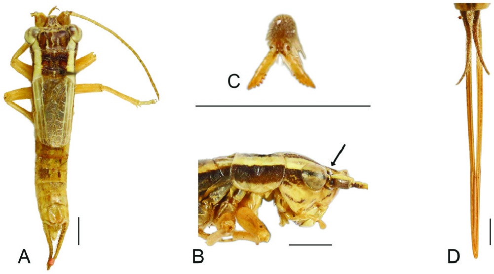
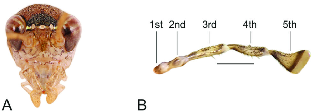
1. Fastigium truncated in frontal and lateral views ( Fig. 5B View Figure 5 ; Supporting Information, Fig. S1G View Figure 1 ), male anterolateral region of metanotum not inflated, tarsal claws inner margin serrulated ( Fig. 5C View Figure 5 ; Supporting Information, Fig. S6 View Figure 6 Gb), TIII bearing six or more inner subapical spurs (Supporting Information, Fig. S6 View Figure 6 Gb) ............................................................................................................................................... Euscyrtinae View in CoL
- Fastigium not truncated in frontal and lateral views (Supporting Information, Fig. S1A–C, I View Figure 1 ), male anterolateral region of metanotum inflated (Supporting Information, Fig. S4C–E View Figure 4 ), TIII bearing maximum six inner subapical spurs (except some Podoscirtinae View in CoL taxa bearing more than six), claws inner margins not serrulated (Supporting Information, Fig. S6 View Figure 6 Ga) ..............................................................................................2
2. Ventral inner apical spur of TIII reduced or absent (Supporting Information, Fig. S6E View Figure 6 ), FWs lateral field with an angle less than 90° related to dorsal field in posterior view (Supporting Information, Fig. S2J2 View Figure 2 ), ovipositor straight in lateral view (Supporting Information, Fig. S4 View Figure 4 Jb), tip of ovipositor dorsal valves forked (Supporting Information, Figs S4 View Figure 4 Ja, S5C). Male genitalia: rami two or more times longer than pseudepiphallic sclerite ............................................................................................................ Oecanthinae View in CoL …4
- Ventral inner apical spur of TIII developed (Supporting Information, Fig. S6D View Figure 6 ), FWs lateral field with an angle ~90° related to dorsal field in posterior view (Supporting Information, Fig. S2J View Figure 2 1 View Figure 1 ), ovipositor up or downcurved in lateral view (Supporting Information, Fig. S4K, L View Figure 4 ), tip of ovipositor dorsal valves single. Male genitalia: rami slightly longer or shorter than pseudepiphallic sclerite ...............................................3
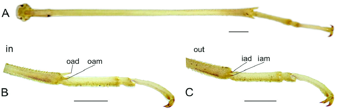
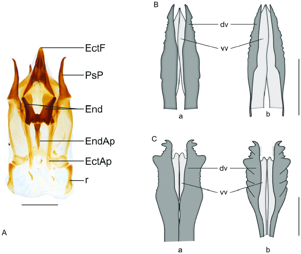
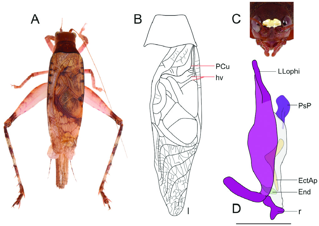
3. Posterior margin of pronotum convex in the middle (Supporting Information, Fig. S4 View Figure 4 Aa). TIII with 5/5 subapical spurs or more. Ovipositor not flattened (Supporting Information, Fig. S4J, L View Figure 4 ), apex of dorsal valves covering ventral valves laterally and sometimes ventrally ( Fig. 9B, C View Figure 9 ; Supporting Information, S5A, D, E, G), generally strongly sclerotized, laterals strongly serrulate, laterals of apex of ventral valves generally smooth. Male genitalia: inner margin of LLophi not membranous; ectophallic fold single or bilobate, posterior projection of endophallic sclerite, when present, bilobate ( Fig. 9A View Figure 9 ); endophallic apodeme, when present, flattened laterally ( Fig. 9A View Figure 9 , 12E View Figure 12 ; Supporting Information, Fig. S9A View Figure 9 )........... Podoscirtinae View in CoL …6
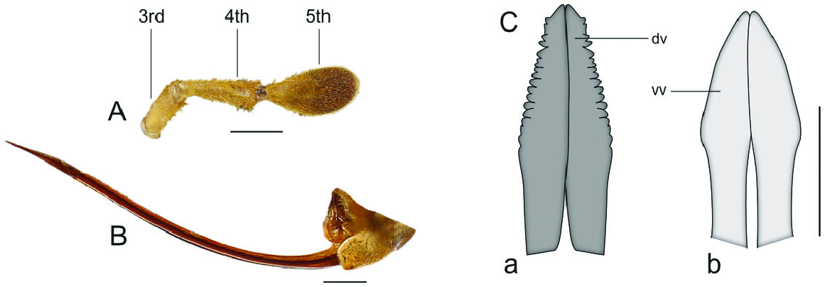
- Posterior margin of pronotum entirely convex (Supporting Information, Fig. S4 View Figure 4 Ab). TIII with 5/4 subapical spurs (4/ 3 in Perutrella View in CoL and 4/ 4 in some Paroecanthini View in CoL ). Ovipositor flattened dorso-ventrally ( Fig. 16B View Figure 16 ; Supporting Information, Fig. S4K View Figure 4 ), apex of dorsal valves above ventral valves ( Fig. 16C View Figure 16 ; Supporting Information, Fig. S5B, F, H View Figure 5 ), same sclerotization as the entire ovipositor, laterals slightly or not serrulate; laterals of apex of ventral valves generally serrulate. Male genitalia: inner margin of LLophi generally membranous ( Figs 18C View Figure 18 , 19D View Figure 19 ; Supporting Information, Fig. S7A View Figure 7 ); ectophallic fold single, posterior projection of endophallic sclerite, when present, single; endophallic apodeme, when present, not flattened laterally (Supporting Information, Fig. S8L–N View Figure 8 ).................................................................................... Tafaliscinae …12
4. Specimens hypognathous ( Fig. 8B View Figure 8 ); TI and TII ventral inner apical spurs present (Supporting Information, Fig. S5O, P View Figure 5 ); FIII proximal portion wider than distal portion in lateral view ( Figs S4K, S View Figure 4 6A View Figure 6 ); first tarsomere of leg III with dorsal spines ( Fig. 12C, D View Figure 12 ); tarsal claws not bifurcated ( Fig. S6 View Figure 6 Ga). Tip of ventral valves of ovipositor forked. Male FW: mirror not occupying more than half of FW (Supporting Information, Fig. S2E, G View Figure 2 ), with one dividing vein; apical field present (Supporting Information, Figs S5J, S View Figure 5 6C, D View Figure 6 ). Male genitalia: ectophallic arc curved posteriorly; ventral valves present, larger than the entire genitalia (Supporting Information, Fig. S9C View Figure 9 ); endophallic sclerite two times or longer than pseudepiphallic sclerite (Supporting Information, Fig. S7D View Figure 7 ). Neotropical distribution ........................................................... Diatrypidi /Diatrypini
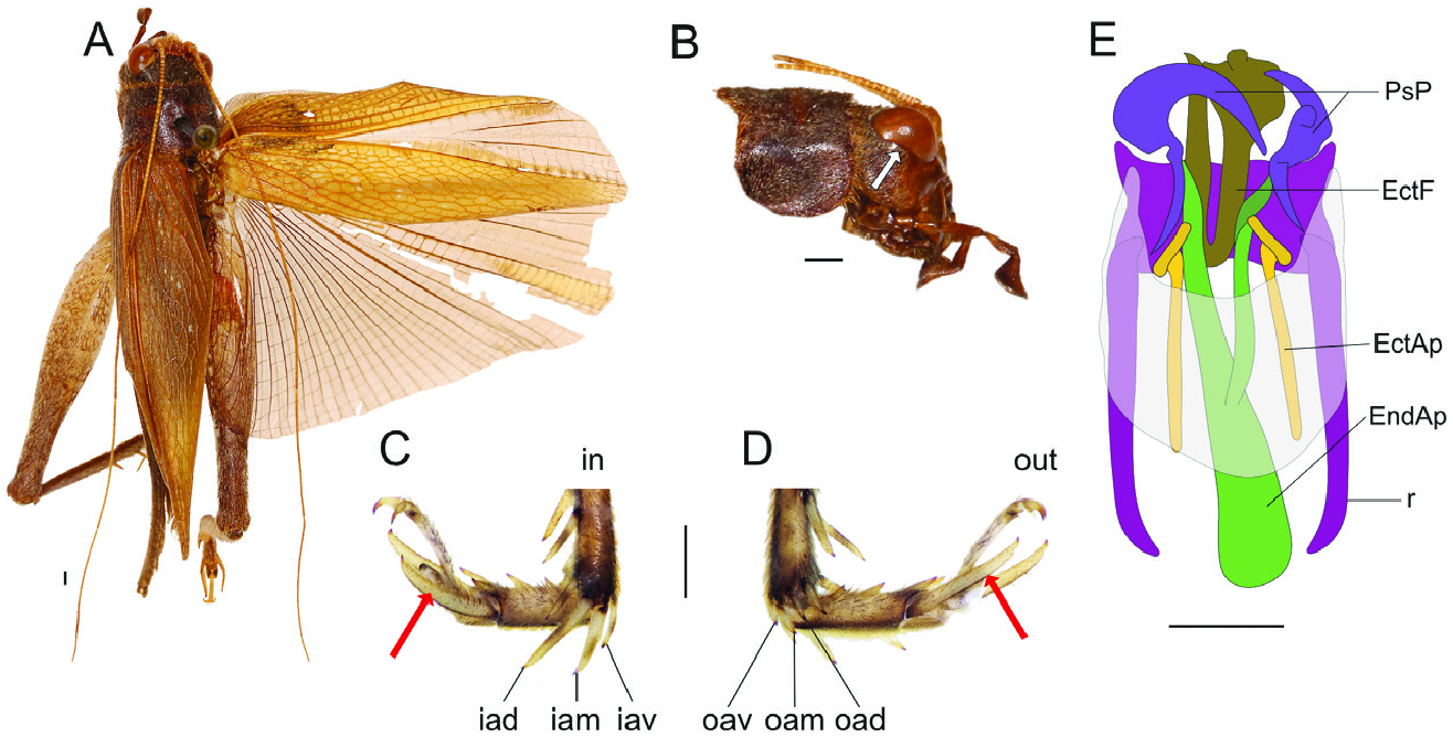
- Specimens prognathous ( Fig. 6A View Figure 6 ; Supporting Information, S1K); TI and TII ventral inner apical spurs absent; FIII proximal portion similarly thin to distal portion in lateral view; first tarsomere of leg III without spines ( Fig. 7B, C View Figure 7 ); tarsal claws bifurcated ( Fig. 6C View Figure 6 ; Supporting Information, Fig. S6 View Figure 6 Gc). Tip of ventral valves of ovipositor not forked. Male FW (when developed): large mirror occupying almost or more than half of FW, with two dividing veins ( Fig. 6B View Figure 6 ; Supporting Information, Fig. S3H View Figure 3 ); apical field absent or almost no discernible (Supporting Information, Fig. S3H View Figure 3 ). Male genitalia: ectophallic arc curved anteriorly (Supporting Information, Fig. S8G View Figure 8 ); ventral valves poorly developed or absent; endophallic sclerite short (Supporting Information, Fig. S8G View Figure 8 ). Worldwide distribution .................................................... Oecanthidi …5
5. Cerci short, shorter than FII (Supporting Information, Fig. S4G View Figure 4 ); ventral outer apical spur of TII present; subapical spurs of TIII absent ( Fig. 7A View Figure 7 ); ventral outer and inner apical spurs of TIII absent (2/2) ( Fig. 7B, C View Figure 7 ), first tarsomere apical spurs of leg III absent. Male genitalia: distal prolongation of ectophallic arc absent ................................................................................................................................................ Xabeini View in CoL
- Cerci long, longer than FII (Supporting Information, Fig. S4H View Figure 4 ); ventral outer apical spur of TII absent; subapical spurs of TIII present, generally 3/3; ventral outer and inner apical spurs of TIII present (3/3); first tarsomere apical spurs of leg III present. Male genitalia: distal prolongation of ectophallic arc present (Supporting Information, Fig. S8G View Figure 8 ) ................................................................................................. Oecanthini
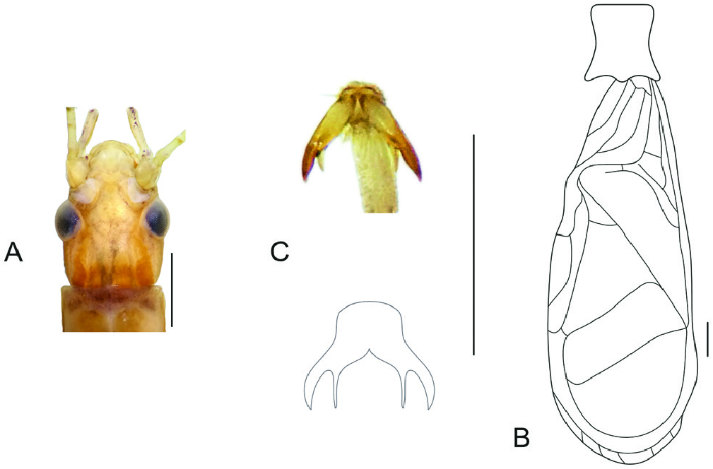
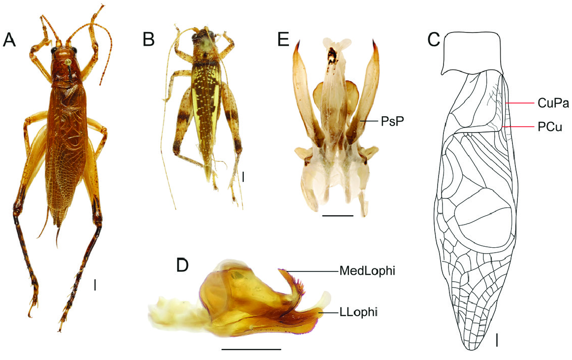
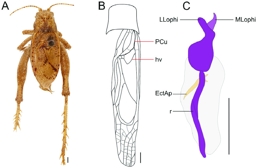
6. Median and lateral ocelli generally aligned ( Fig. 11A View Figure 11 ; Supporting Information, Fig. S1B View Figure 1 ); fifth article of maxillary palpi upcurved 90° ( Figs 11B View Figure 11 , 12B View Figure 12 ; Supporting Information, Fig. S1 View Figure 1 Lc). Male FWs: PCu vein curved more than 90°, sinuous ( Figs 14B View Figure 14 , 15B View Figure 15 ; Supporting Information, Fig. S3 View Figure 3 Bb, G); CuPa not parallel to proximal region of PCu ( Figs 14B View Figure 14 , 15B View Figure 15 ; Supporting Information, Fig. S3G View Figure 3 ); harp veins, when present, parallel to PCu vein ( Figs 14B View Figure 14 , 15B View Figure 15 ; Supporting Information, Fig. S3G View Figure 3 ). New world distribution ........................................................................................................................................................ Hapithidi …7
- Median and lateral ocelli not aligned; fifth article of maxillary palpi slightly upcurved (Supporting Information, Fig. S1 View Figure 1 Lb). Male FWs: PCu vein curved 90°, not sinuous ( Fig. 10C View Figure 10 ; Supporting Information, Fig. S3 View Figure 3 Ba, F); CuPa parallel to proximal region of PCu ( Fig. 10C View Figure 10 ; Supporting Information, Fig. S3F View Figure 3 ); harp veins, when present, diagonal ( Fig. 10C View Figure 10 ; Supporting Information, Fig. S3F View Figure 3 ). Old world distribution .................................................................................................................................................. Podoscirtidi …10
7. Veins of lateral field parallel to dorsal field (Supporting Information, Fig. S2 View Figure 2 Ia). Male FWs: stridulatory apparatus absent (Supporting Information, Fig. S3A View Figure 3 ); apical field not delimited .........................................8
- Veins of lateral field perpendicular to dorsal field (Supporting Information, Fig. S2 View Figure 2 Ib). Male FWs: stridulatory apparatus present; apical field delimited (Supporting Information, Fig. S3F, G View Figure 3 ) .....................9
8. Eyes posterior margin concave in lateral view ( Fig. 12B View Figure 12 ; Supporting Information, Fig. S1 View Figure 1 Eb), with one horizontal band or without bands; TI outer tympana generally present, inner absent; first tarsomere apical spurs of leg III longer than the tarsomere ( Fig. 12C, D View Figure 12 ). Male genitalia: ectophallic arc not connected ( Fig. 12E View Figure 12 ; Supporting Information, Fig. S9A View Figure 9 ); endophallus U-shaped ( Fig. 12E View Figure 12 ; Supporting Information, Fig. S9A View Figure 9 ), endophallic apodeme two or more times longer than PsP ( Fig. 12E View Figure 12 ; Supporting Information, Fig. S9A View Figure 9 ); rami not connected......................................................................................................... Aphonomorphini View in CoL
- Eyes posterior margin straight in lateral view (Supporting Information, Fig. S1 View Figure 1 Ea); eyes with two or three horizontal bands ( Figs 11A View Figure 11 , 13A View Figure 13 ; Supporting Information, Fig. S1F View Figure 1 ); TI outer tympana absent, inner present; apical spurs of first tarsomere of leg III generally same size or shorter than the tarsomere. Male genitalia: ectophallic arc connected ( Fig. 13B View Figure 13 ; Supporting Information, Fig. S8I View Figure 8 ); endophallus not U-shaped, endophallic apodeme same shorter or slightly longer than PsP; rami connected ( Fig. 13B View Figure 13 ; Supporting Information, Fig. S8I View Figure 8 ) .................................................................................................................... Cearacesaini
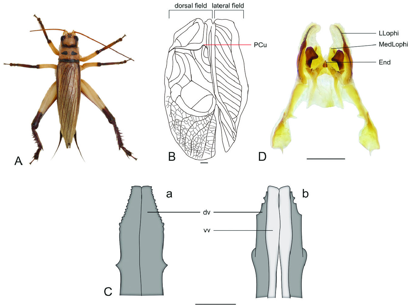
9. Median and lateral ocelli distant, not connected. FWs lateral and dorsal field same-sized (Supporting Information, Fig. S2H View Figure 2 1 View Figure 1 ). Pronotum caudal margin entirely convex ( Fig. 14A View Figure 14 ; Supporting Information, Fig. S4 View Figure 4 Ab). Dorsal valves of ovipositor lateral margins slightly serrulate, same colour as the entire ovipositor. Male genitalia: MedLophi present ( Fig. 14C View Figure 14 ; Supporting Information, Fig. S8C View Figure 8 ); anterior margin of pseudepiphallic sclerite not folded; ectophallic arc reduced or absent ............................................ Hapithini View in CoL
- Median and lateral ocelli remarkably close, sometimes connected ( Fig. 15C View Figure 15 ; Supporting Information, Fig. S1B, D View Figure 1 ). FWs lateral field shorter than dorsal field (Supporting Information, Fig. S2H2 View Figure 2 ). Dorsal valves of ovipositor lateral margins strongly serrulate, darker than the entire ovipositor (Supporting Information, Fig. S5A, E View Figure 5 ). Pronotum caudal margin convex on the middle ( Figs 15A View Figure 15 ; Supporting Information, Fig. S4 View Figure 4 Aa). Male genitalia: MedLophi absent; anterior margin of pseudepiphallic sclerite folded dorsally ( Fig. 15D View Figure 15 ; Supporting Information, Fig. S8F View Figure 8 ); ectophallic arc present ....................................................... Phyllogryllini
10. Male FWs without stridulatory apparatus, only longitudinal veins, anal area not delimited; male genitalia PsP not cylindrical; endophallus asymmetric; apex of dorsal valves of ovipositor generally wider than entire ovipositor in dorsal and ventral views................................................................................ Aphonoidini View in CoL
- Male FWs generally bearing stridulatory apparatus sometimes harp veins or mirror lacking (or both), anal area delimited; male genitalia PsP frequently cylindrical; endophallus frequently symmetrical; apex of dorsal valves of ovipositor generally same width of the entire ovipositor in dorsal and ventral view .......11
11. TI inner tympanum usually not covered by a sclerotized tab; pronotum DD not flattened in lateral view; male genitalia generally capsular (Supporting Information, Fig. S7F View Figure 7 ); arc and ventral projection of ectophallic invagination short, endophallic sclerite well developed .............................................Podoscirtini
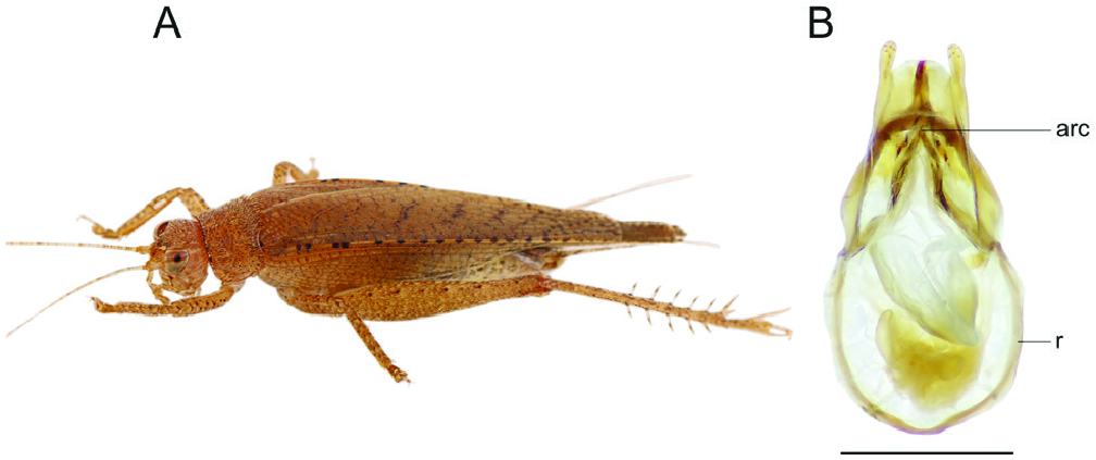
- TI inner tympanum covered by a sclerotized tab; pronotum DD not flattened in lateral view; male genitalia not capsular; arc and ventral projection of ectophallic invagination elongated ( Fig. 10E View Figure 10 ), endophallic sclerite sometimes regressed or absent ............................................................................................ Truljaliini
12. Body not robust, pronotum longer than wide in dorsal view. Apex of ovipositor pointed or rounded ( Fig. 16C View Figure 16 ; Supporting Information, Fig. S5F View Figure 5 ). Male genitalia: MedLophi absent; endophallus flattened dorso-ventrally (Supporting Information, Fig. S8H View Figure 8 ) or strongly reduced (Supporting Information, Fig. S8B View Figure 8 ), lateral margins not folded; endophallic apodeme absent ......................................... Paroecanthidi …13
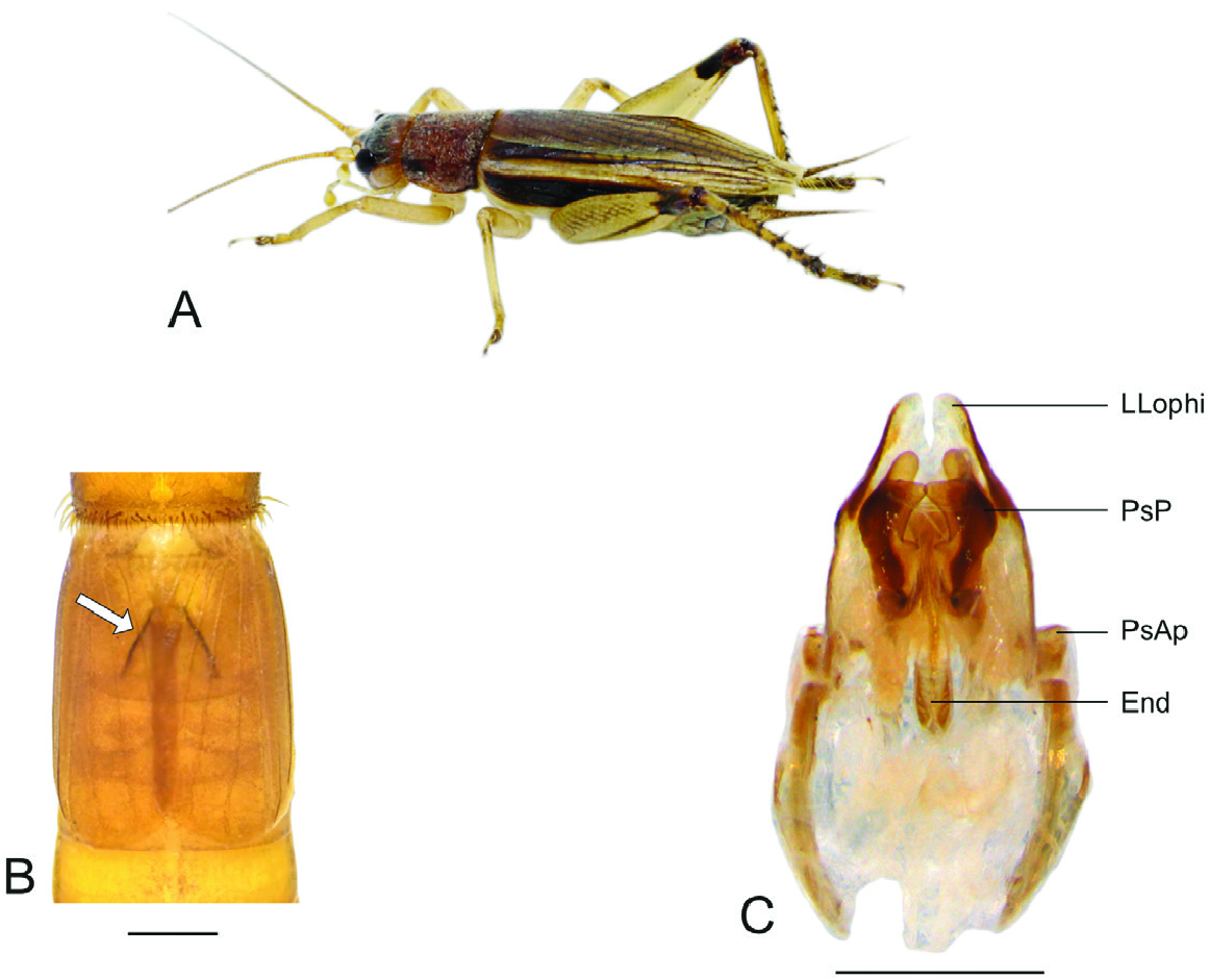
- Body robust, pronotum as wide as long or wider than long in dorsal view. Apex of ovipositor truncated ( Fig. 19C View Figure 19 ; Supporting Information, Fig. S5B, H View Figure 5 ). Male genitalia: MedLophi present; endophallus not flattened, lateral margins folded ( Fig. 19D View Figure 19 ; Supporting Information, Fig. S8L View Figure 8 ); endophallic apodeme generally present .................................................................................................................................. Tafaliscidi /Tafaliscini…14
13. TI tympana present. FWs covering abdomen partially or totally, stridulatory apparatus present; PCu vein curved, generally bisinuous, rarely sinuous (Supporting Information, Fig. S3C View Figure 3 ); apical field delimited. Structures of male genitalia generally regressed, mainly from ectophallic invagination and endophallus (except Adenophallusia View in CoL and Ectotrypa View in CoL ) (Supporting Information, Fig. S8B View Figure 8 ) .................... Paroecanthini View in CoL …19
- TI tympana absent. FWs absent (Supporting Information, Fig. S2A View Figure 2 ), only covering the metanotum (Supporting Information, Fig. S2B View Figure 2 ) or covering the abdomen with longitudinal veins ( Fig. 18A View Figure 18 ), stridulatory apparatus absent; PCu vein not curved or absent; apical field, when present, not delimited. Structures of male genitalia not regressed ................................................................................................ Neometrypini …25


No known copyright restrictions apply. See Agosti, D., Egloff, W., 2009. Taxonomic information exchange and copyright: the Plazi approach. BMC Research Notes 2009, 2:53 for further explanation.
|
Kingdom |
|
|
Phylum |
|
|
Class |
|
|
Order |
|
|
SubOrder |
Ensifera |
|
SuperFamily |
Grylloidea |
|
Family |
|
|
SubFamily |
Tafaliscinae |
|
SuperTribe |
Tafaliscidi |