
Martensolasma jocheni, Shear, William A., 2006
|
publication ID |
https://doi.org/ 10.5281/zenodo.174089 |
|
DOI |
https://doi.org/10.5281/zenodo.6495790 |
|
persistent identifier |
https://treatment.plazi.org/id/03C987D6-FFCC-3C79-FEA3-FEADFD704DBF |
|
treatment provided by |
Plazi |
|
scientific name |
Martensolasma jocheni |
| status |
sp. nov. |
Martensolasma jocheni View in CoL n. sp.
Figs 1–14 View FIGURES 1 – 5 View FIGURES 6 – 14
Types: Male holotype and male paratype from Ciudad Aguascalientes, Aguascalientes, Mexico, collected under stones in a garden by James C. Cokendolpher, 5 January 1977, deposited in Museum of Comparative Zoology, Harvard University, Cambridge MA, USA.
Etymology: The species epithet honors Jochen Martens on the occasion of his retirement, in recognition of his many contributions to our knowledge of the systematics and evolution of the Opiliones , and with pleasant memories of excursions in the Great Smoky Mountains of North Carolina and in the Italian Dolomites.
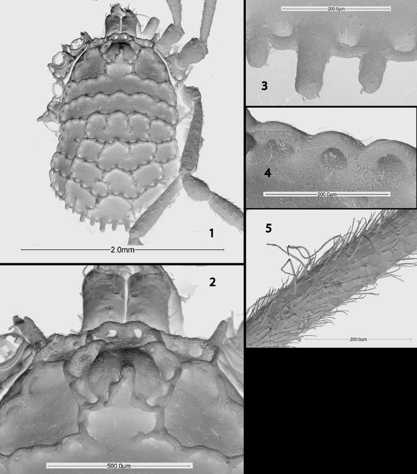
Male holotype: About 2 mm long. Dorsum ( Fig. 1 View FIGURES 1 – 5 ) glossy black, unmarked; venter black with some dark brown areas at sternal margins. Leg articles black, but most articles yellow to chestnut brown proximally, giving effect of light banding. Scutum magnum present (dorsal shield of prosoma, thoracic tergites and opisthosomal scute fused into single dorsal shield). Eye tubercle without hood, circumocular keels small, eyes visible dorsally ( Fig. 2 View FIGURES 1 – 5 ). Single lateral process on each side of dorsal shield connected to anterior marginal keel. Cells defined by keels (some cells not completely enclosed) as follows: Two large cells lateral to eye tubercle, single smaller cell (open posteriorly) directly posterior to eye tubercle. More posterior cells aligned roughly in six transverse rows. First two rows with incomplete cells; first row with slightly indicated boundaries of two small lateral cells, larger median cell, second row without clear division of cells. Third row with five roughly subequal cells, boundaries sometimes incomplete. Fourth row with single large median cell about five times as wide as long, partially divided by two keels, two lateral cells each about onethird width of median cell. Fifth row with median cell divided by keels into three smaller cells, these subequal to two lateral cells. Sixth row of five approximately subequal cells. Keel pegs with projecting spines becoming more prominent posteriorly, eventually forming row of strong, subacute processes on posterior margin of scute ( Fig. 3 View FIGURES 1 – 5 ); keel arms strongly recurved, fused to dorsum and arms of adjacent pegs, producing series of loops most obviously seen in dorsal view along lateral margins of scute ( Fig. 4 View FIGURES 1 – 5 ). Keel pegs typically with two or three such keel arms. Genital operculum apically narrowed, tonguelike. Sterna with rows of tubercles, but lacking keels and keel pegs.
Chelicera ( Fig. 6 View FIGURES 6 – 14 ) with basal article distinctly humped, bearing acute, curved mesal tooth; distal article as usual for subfamily, bearing mesoproximal curved, acute tooth, area of slightly denser setation mesoproximal to tooth indicating presence of cheliceral epigamic gland (not possible to determine in absence of comparison with females, see also Discussion below).
Pedipalpus ( Fig. 7 View FIGURES 6 – 14 ) small, short, with reduced vestiture of glandular hairs. Femur slender, basally with three tubercles bearing blunt ordinary setae, less than 15 glandular setae scattered over ventral surface; patella slightly shorter than femur, thickened ventrally in its ventrodistal half, thickened region with dense vestiture of acute nonglandular setae, probably indicating presence of palpal epigamic gland (females not available for comparison); tibia about four fifths length of patella, basally swollen, with setae that indicate possible epigamic gland, vestiture of glandular setae more dense; tarsus about half length of tibia, distally rounded, claw absent, much denser vestiture of glandular setae than tibia, glandular setae mixed with ordinary setae toward distal end.
Legs ( Figs 8–11 View FIGURES 6 – 14 ) in order of length: 2, 4, 3, 1. Coxae with distal marginal keel pegs and keels, coxae 1 and 4 with marginal keels on anterior and posterior surfaces, respectively. Trochanters with ventral tubercles bearing blunt setae. Femora distally expanded, third and fourth femora with single pseudoarticulation about onesixth their length from articulation with trochanters. Patellae tending to subglobular, no more than twice as long as wide. Tibiae short, one and one half to twice as long as patellae. Metatarsi longest leg segments, nearly cylindrical, without pseudoarticulations, about twice length of tibiae except on second legs, where almost four times length of tibiae. Tarsal articles of first and third legs four, second legs with five; third article of fourth leg tarsi with single pseudoarticulation, giving appearance of five articles. Leg vestiture ( Figs 5 View FIGURES 1 – 5 , 12 View FIGURES 6 – 14 ) complex; femora basally smooth in pale colored region, more distally with closely appressed, acute, flattened tubercles evenly scattered over surface, small blunt setae on low, rounded cones.
On patellae and tibiae, tubercles reduced, suppressed, setae present. Metatarsi lacking tubercles, densely covered with fine, short, hairlike trichomes and scattered, longer, thin, nearly filamentous setae, curved or curled distally. Tarsal articles with same dense, short setae, on fourth legs also with few curled, longer setae basally.
Penis ( Figs 13–14 View FIGURES 6 – 14 ) long, narrow, shaft slightly widened distally, glans without any obvious dorsal membranous area, narrowing somewhat abruptly to stylus; stylus slightly sigmoid at tip, not strongly curved or hookshaped, opening terminal. Base of stylus with six small setae, otherwise glans without setation.
Physical dimensions of the described specimen may be ascertained from the figures, using the appropriate scale lines.

No known copyright restrictions apply. See Agosti, D., Egloff, W., 2009. Taxonomic information exchange and copyright: the Plazi approach. BMC Research Notes 2009, 2:53 for further explanation.