
Joinvillichthys kriweti, Taverne & Capasso, 2014
|
publication ID |
https://doi.org/10.5852/ejt.2014.101 |
|
publication LSID |
lsid:zoobank.org:pub:C320F2F8-E9BB-46FC-9A0C-56ECF4B1CE2B |
|
DOI |
https://doi.org/10.5281/zenodo.3852234 |
|
persistent identifier |
https://treatment.plazi.org/id/B8C4822F-44CA-4776-8455-452C5DBDCA1C |
|
taxon LSID |
lsid:zoobank.org:act:B8C4822F-44CA-4776-8455-452C5DBDCA1C |
|
treatment provided by |
Tatiana |
|
scientific name |
Joinvillichthys kriweti |
| status |
sp. nov. |
Joinvillichthys kriweti sp. nov.
urn:lsid:zoobank.org:act:
Figs 12-16 View Fig View Fig View Fig View Fig View Fig
Diagnosis
Joinvillichthys with a body depth equal to 46.0 % of the standard length. A dorsal prominence present on the frontal. Maxilla elongated. Large parietal. Dermosupraoccipital sutured to the parietal and not to the dermopterotic. Dermopterotic much longer than deep. Small dermosphenotic. Comma-shaped opercle. Anterior ventral branch of the cleithrum lost. Broad and short pectoral spine articulated on the ventral margin of the cleithral posterior process. Caudal fin double emarginated.
Etymology
The name of the new species is dedicated to Dr Jürgen Kriwet (Vienna) who has greatly improved our knowledge of the pycnodontiform fishes.
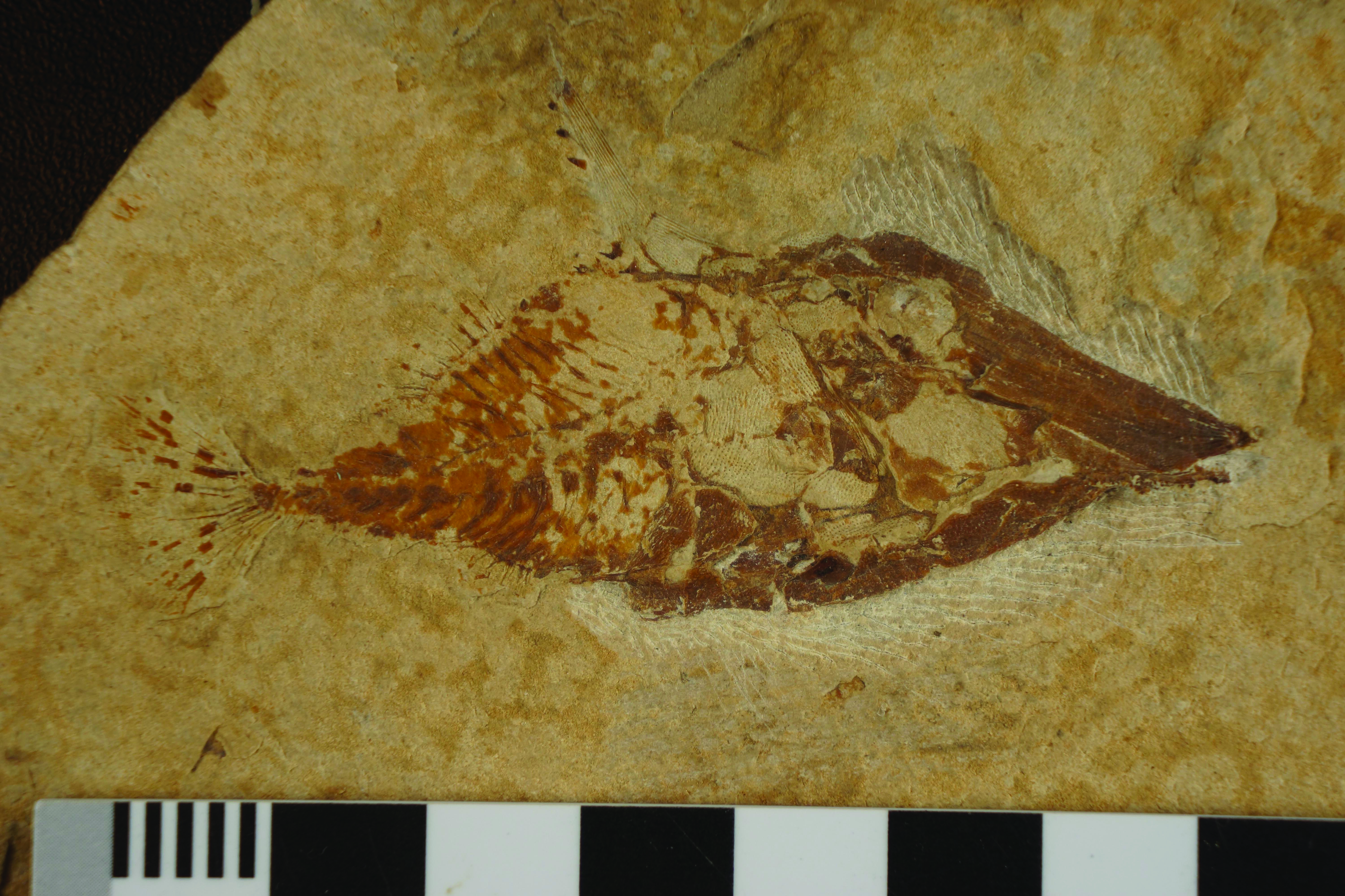
Holotype
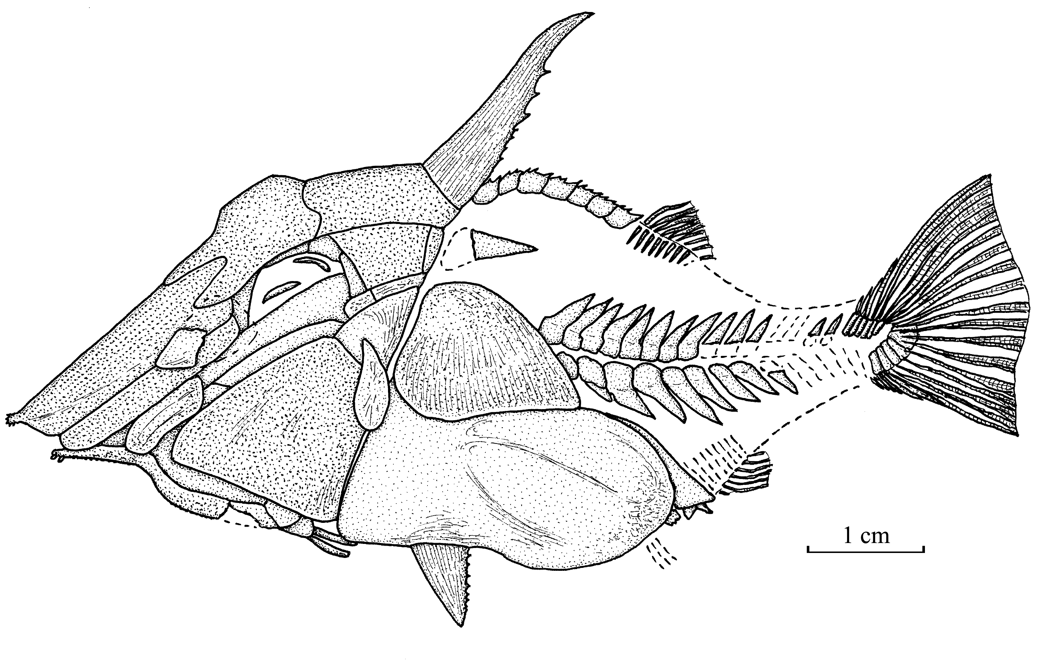
LEBANON: sample CLC S-137 , a complete specimen from Haqel ( Fig. 12 View Fig ), total length: 91 mm.
Paratype
LEBANON: sample AMNH 4517a (3698) and counterpart, an incomplete specimen from Hgula ( Hay 1903: pl. 29, fig. 1 View Fig ); only the head and the beginning of the body are preserved, length: 63 mm.
Formation and locality
Marine Upper Cenomanian, Haqel and Hgula, Lebanon.
Morphometric data ( Fig. 13 View Fig )
The morphometric data are given in % of the holotype standard length ( 76 mm)
Length of the head (dermosupraoccipital included) …………………………………………… 54.2 % Length of the cephalo-thorax (cleithrum included) ………………………………………… 75.3 % Depth of the head (without the nuchal horn) ……………………………………………… 43.8 % Length of the nuchal horn …………………………………………………………………… 27.7 % Maximum depth of the body (just behind the nuchal horn) ………………………………… 46.0 % Predorsal length ………………………………………………………………………………… 76.6 % Basal length of the dorsal fin …………………………………………………………………… 9.8 % Preanal length …………………………………………………………………………………… 80.4 % Basal length of the anal fin ………………………………………………………………………… 6.4 % Depth of the caudal peduncle ……………………………………………………………………… 9.8 %
Osteology
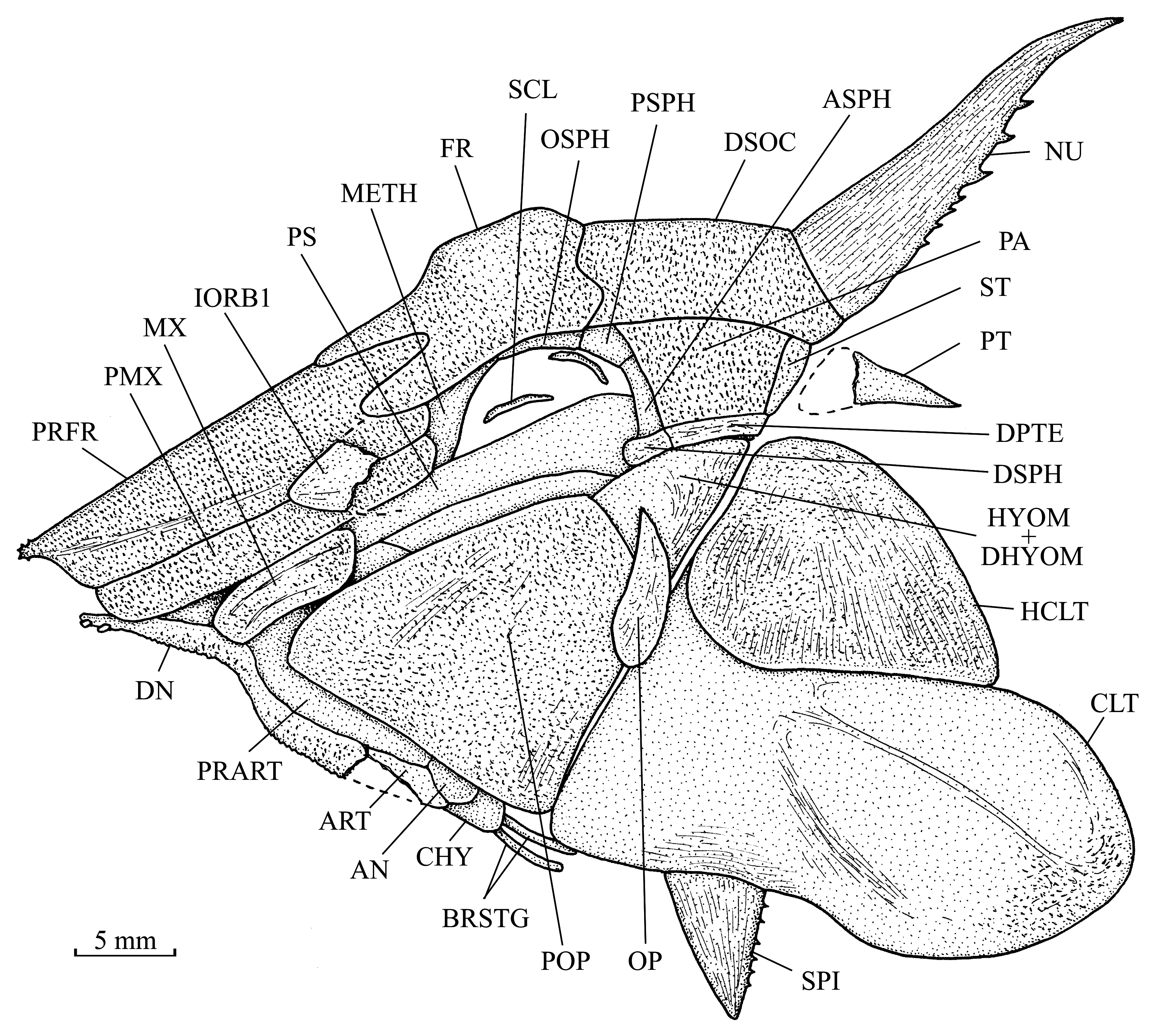
1. The skull ( Fig. 14 View Fig )
The general morphology of the skull is rather close to that of Joinvillichthys lindstroemi and the cranial dermal bones also are ornamented with small tubercles. However, there are many small differences in the head skeleton of the two fishes. Thus the description that follows will principally emphasize these differences.
The skull is shorter and deeper than in Joinvillichthys lindstroemi . Its depth, from the upper margin of the dermosupraoccipital to the lower margin of the cleithrum, is equal to 83 to 86 % of its length, from the tip of the snout to the basis of the nuchal horn.
The rostrum lengthening is less pronounced. The prefrontal is broader and has a very sinuous suture with the frontal. Its anterior tip also bears very small spines but is less outpacing of the lower jaw level. The frontal is broader but does not outpace posteriorly the level of the orbit. The bone bears a small dorsal prominence. The dermosupraoccipital is longer and is sutured to the parietal and the supratemporal but not with the dermopterotic. The parietal is considerably larger. The dermopterotic is longer but much thinner. The supratemporal is sutured to the parietal and reaches the dermopterotic at only one point. As in Joinvillichthys lindstroemi , the long nuchal horn is supported only by the dermosupraoccipital.
The orbitosphenoid and the pleurosphenoid are present in the orbit, just below the frontal, but the basisphenoid is not visible.
The toothless premaxilla is longer and narrower. The toothless maxilla also is narrower and more elongate. The lower jaw is composed with the same bones but is longer. The dentary bears two small incisiform teeth and its ventral margin is denticulated. The articulation with the quadrate is located at the level of the posterior border of the orbit.
A fragment of a large first infraorbital is preserved on the suture between the prefrontal and the premaxilla. The sclerotic ring is visible in the orbit.
The hyomandibula and the preopercle are sutured together. The exposed part of the hyomandibuladermohyomandibula is larger than in Joinvillichthys lindstroemi but still much smaller than the considerably enlarged preopercle. The opercle is broader and comma-shaped, with the sharp end dorsally located. A part of the anterior ceratohyal and two small branchiostegal rays are visible behind the lower jaw.
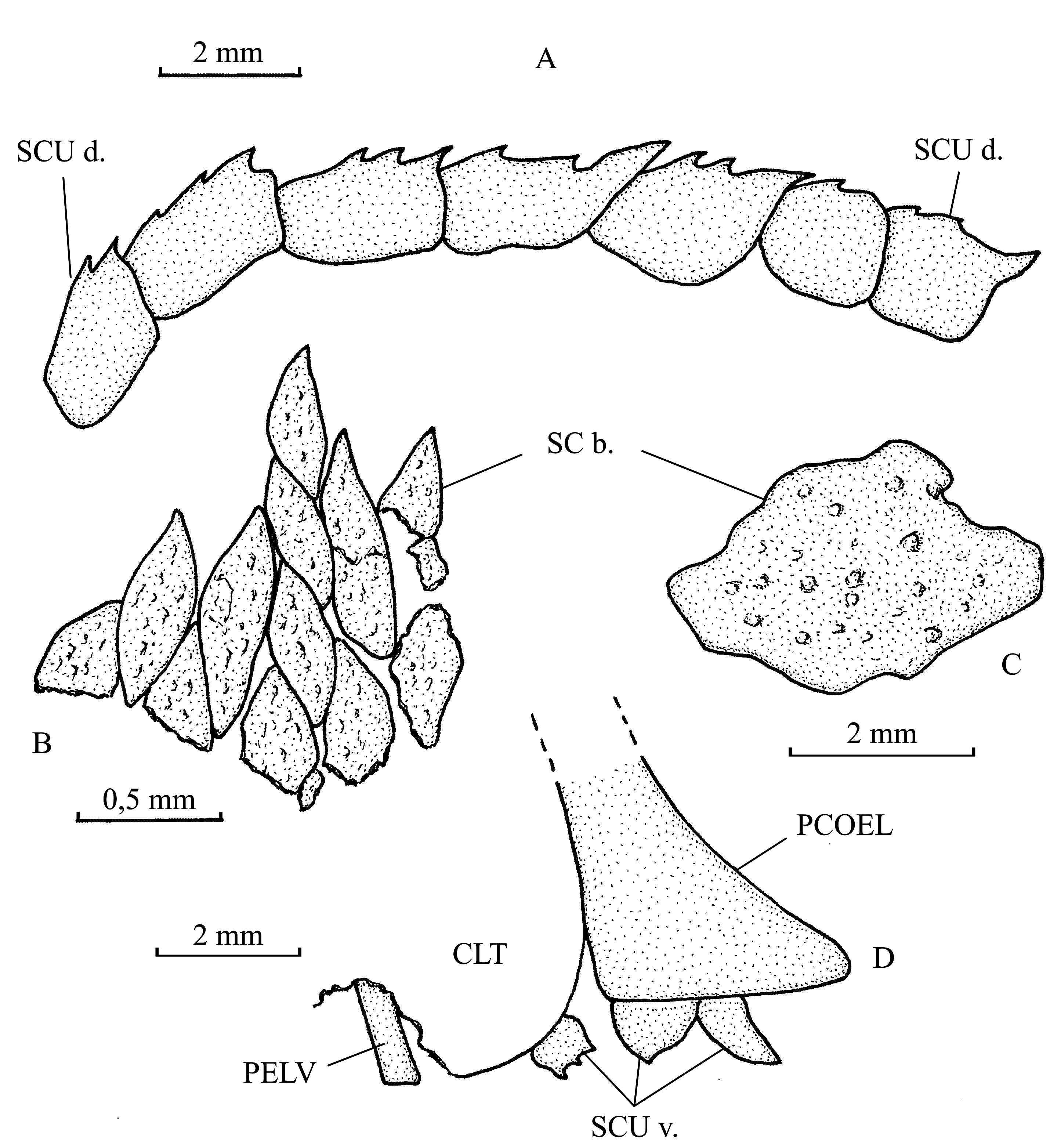
2. The girdles ( Figs 14 View Fig , 16 View Fig )
The bones of the gigantic pectoral girdle have the same size and shape as in Joinvillichthys lindstroemi . However, two important differences exist. The anterior ventral branch of the cleithrum is lost. No postcleithrum is visible, but that is perhaps due to the fossilization. The pectoral spine is shorter, much broader and is not articulated with the rear of the cleithrum but more anteriorly on its lower margin.
A small pelvic girdle is present. Indeed, a part of a vertically oriented pelvic bone is visible under a broken part of the cleithrum.
3. The axial skeleton ( Fig. 13 View Fig )
The trunk is fusiform but proportionally deeper than in Joinvillichthys lindstroemi . The axial skeleton is incomplete. Three vertebral segments are missing near the caudal peduncle. There are 16 neural spines (the three missing ones included) before the epichordal series. Only 8 haemal spines are preserved. The total number of haemal spines must be around 12 or 13. The neural and haemal spines are short but broad. The neural and haemal arches surround almost completely the notochord. No ribs are visible. The postcoelomic bone is well developed and backwardly oriented.
4. The dorsal and anal fins ( Fig. 13 View Fig )
The short dorsal and anal fins are located at the mid-length of the body. There are 9 pterygiophores and 9 rays in the dorsal fin. The anal fin contains 7 rays, but the number of anal pterygiophores is not determinable. The first dorsal and anal ray is spiny. The other rays are segmented.
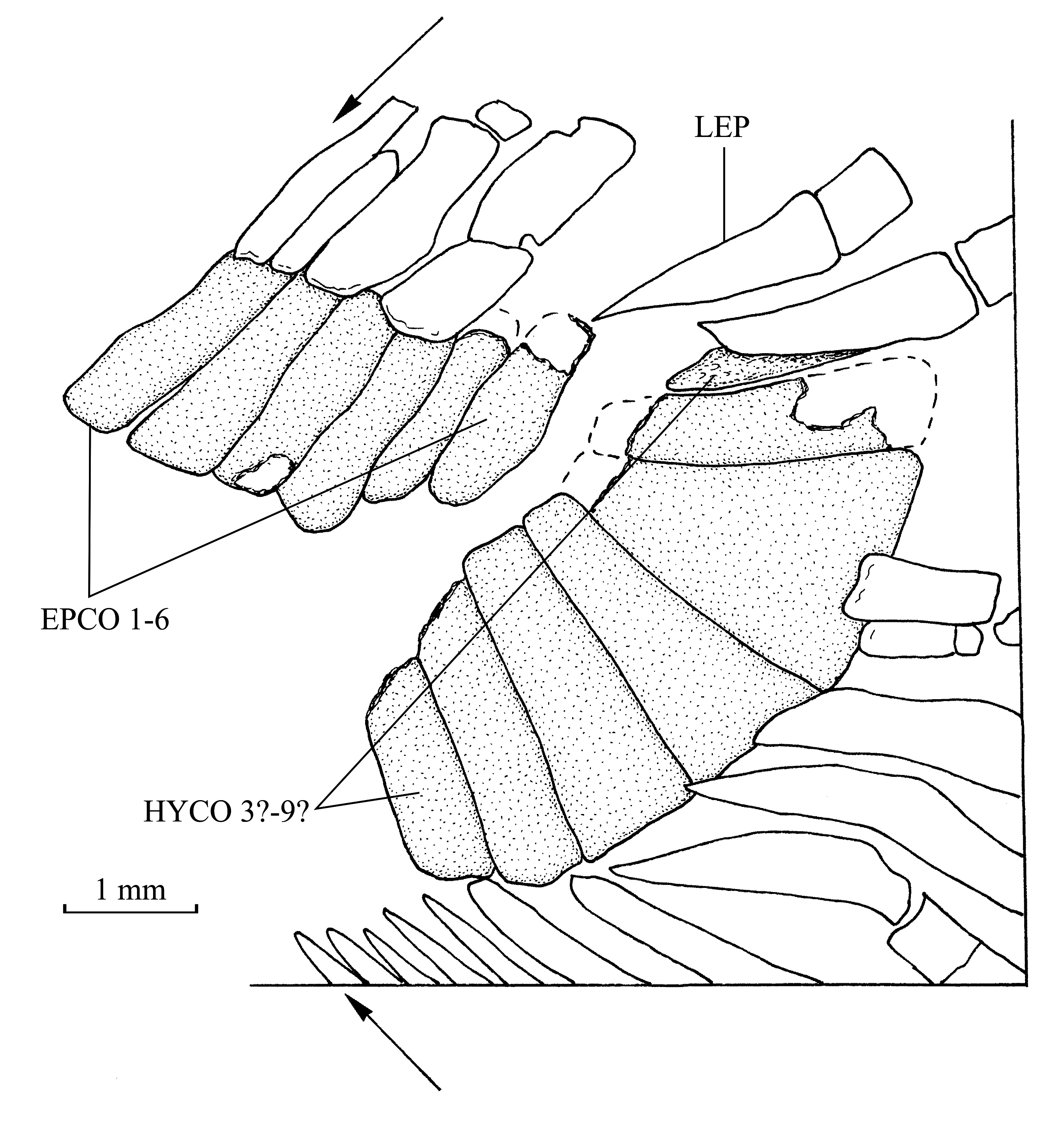
5. The caudal skeleton ( Fig. 15 View Fig )
The caudal skeleton of the holotype is partly preserved. There are 6 short and broad epichordals and 7 hypochordals. However, one or two anterior hypochordals are missing, so the complete series must be composed of 8 or 9 elements. The fifth preserved hypochordal is strongly enlarged. No urodermal is visible, but the region where theses bones are usually present is not preserved.
The caudal fin is double emarginated ( Poyato-Ariza & Wenz 2002: fig. 36E) and contains 19 principal segmented caudal rays, the 2 external being pointed and the 17 others branched. There are 6 ventral and at least 4 dorsal procurrent rays.
6. Squamation ( Fig. 16 View Fig )
The squamation is the same as in Joinvillichthys lindstroemi . There are 7 spiny scutes in the dorsal ridge and at least 3 spiny scutes in the ventral keel. The two posterior ventral scutes are associated with the ventral margin of the postcoelomic bone. The body scales are slightly ornamented with tubercles. Anteriorly, they are small and flake-like. Posteriorly, there are much larger, irregular and scute-like shaped.

| AMNH |
American Museum of Natural History |
No known copyright restrictions apply. See Agosti, D., Egloff, W., 2009. Taxonomic information exchange and copyright: the Plazi approach. BMC Research Notes 2009, 2:53 for further explanation.
|
Kingdom |
|
|
Phylum |
|
|
Class |
|
|
Order |
|
|
Family |
|
|
Genus |