
Joinvillichthys lindstroemi ( Davis, 1890 )
|
publication ID |
https://doi.org/10.5852/ejt.2014.101 |
|
publication LSID |
lsid:zoobank.org:pub:C320F2F8-E9BB-46FC-9A0C-56ECF4B1CE2B |
|
DOI |
https://doi.org/10.5281/zenodo.3852238 |
|
persistent identifier |
https://treatment.plazi.org/id/03C987FA-DB6D-FFBA-501A-F9CBFD94F9AE |
|
treatment provided by |
Tatiana |
|
scientific name |
Joinvillichthys lindstroemi ( Davis, 1890 ) |
| status |
|
Joinvillichthys lindstroemi ( Davis, 1890)
Figs 1-11 View Fig View Fig View Fig View Fig View Fig View Fig View Fig View Fig View Fig View Fig View Fig
Diagnosis
Joinvillichthys with a body depth comprising between 23.8 and 34.0 % of the standard length. No dorsal prominence on the frontal. Maxilla triangular in shape. Small parietal. Dermosupraoccipital sutured with the parietal and the dermopterotic. Dermopterotic deeper than long. Large dermosphenotic. Thin, rodlike opercle. Anterior ventral branch of the cleithrum present. Thin pectoral spine articulated on the rear of the cleithral posterior process. Caudal fin with a convex posterior margin.
Synonymy
Coccodus Lindstroemi Davis, 1890: 567 , pl. 22.
“ Coccodus ” lindstroemi – Poyato-Ariza & Wenz, 2002: 145 View Cited Treatment .
Holotype
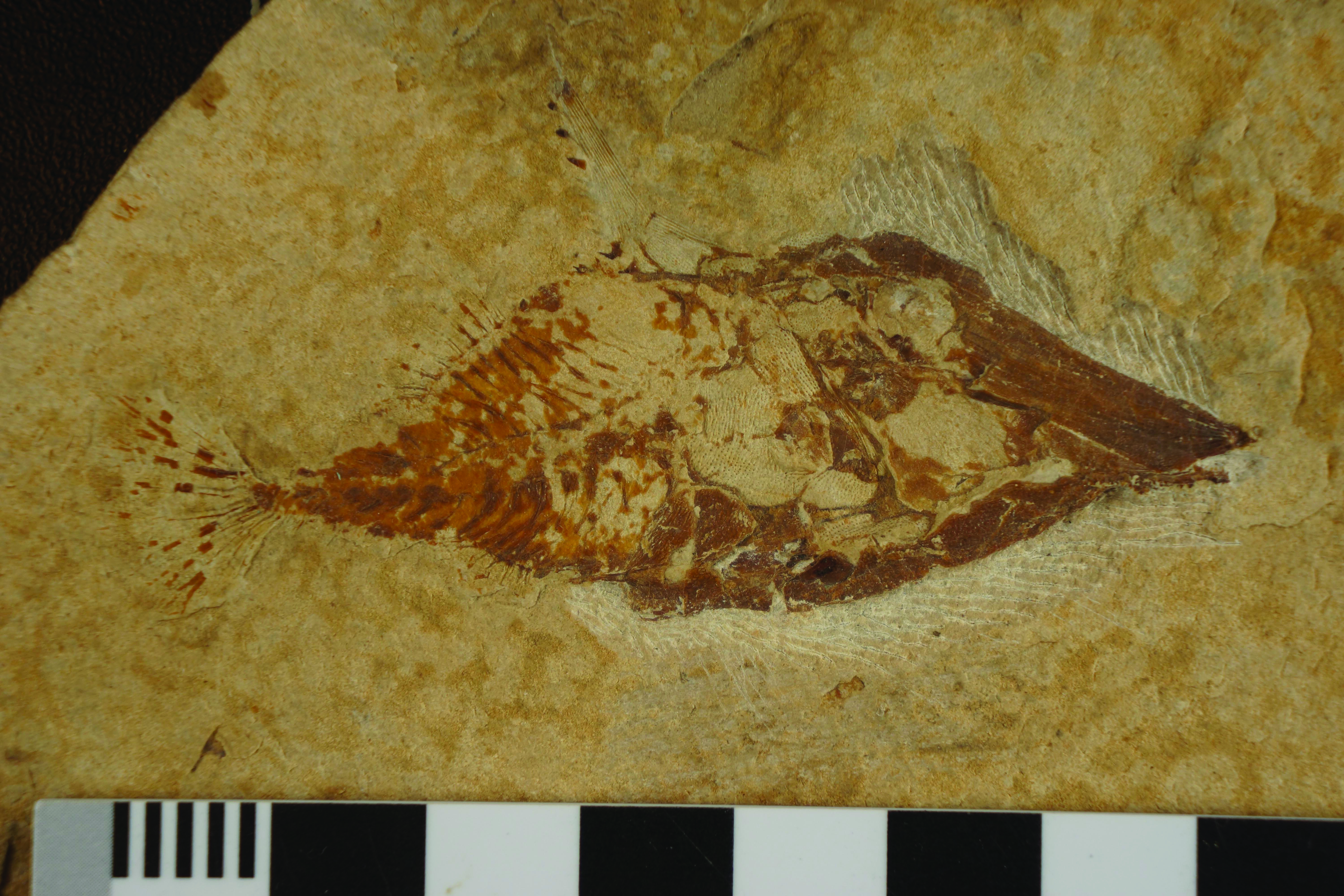
LEBANON: sample NRM PZ P. 2073, a complete specimen from Haqel ( Fig. 1 View Fig ), total length: 76 mm.
Other specimens
LEBANON: sample IRSNB N° P 9276, a nearly complete specimen (the caudal fin is missing) from Hgula ( Fig. 2 View Fig ), length: 93 mm; sample CLC S-138, a nearly complete specimen (a part of the caudal fin is missing) from Haqel ( Fig. 3 View Fig ), length: 82 mm; sample CLC S-324, a complete specimen from Haqel, total length: 76 mm.
Formation and locality
Marine Upper Cenomanian, Haqel and Hgula, Lebanon.
Morphometric data ( Fig. 4 View Fig )
The morphometric data are given in % of the standard length for the holotype NRM PZ P. 2073 ( 64 mm) and for sample IRSNB N° P 9276 ( 93 mm). These two specimens represent the two extremes of the species morphometric variation, as measured on the four studied samples
Holotype P9276
Length of the head (dermosupraoccipital included) ............................................. 55.8 % 56.3 %
Length of the cephalo-thorax (cleithrum included) .............................................. 63.9 % 68.1 % Depth of the head (without the nuchal horn) ........................................................ 35.4 % 25.0 % Length of the nuchal horn ..................................................................................... 25.2 % 25.6 % Maximum depth of the body (just behind the nuchal horn) ................................. 34.0 % 23.8 % Predorsal length .................................................................................................... 74.8 % 73.1 % Basal length of the dorsal fin ................................................................................ 9.5 % 10.6 % Preanal length ....................................................................................................... 72.1 % 73.7 % Basal length of the anal fin ................................................................................... 8.2 % 9.4 % Depth of the caudal peduncle ............................................................................... 5.4 % 5.3 %
The important individual differences in the values of the head and body depths are probably due to sexual or seasonal variations.
Osteology
1. The skull ( Figs 5–8 View Fig View Fig View Fig View Fig )
The head is very large. Its length, from the tip of the snout to the basis of the nuchal horn, is equal to the body length. According to the specimens, its depth, from the upper margin of the dermosupraoccipital to the lower margin of the cleithrum, represents from 48 to 67 % of its length. The dermal bones of the skull are ornamented with small tubercles.
The long, pointed rostrum slightly outpaces the lower jaw and is formed by two large paired bones, the prefrontal and the premaxilla. The anterior tip of the prefrontal bears two or three very small spines. Posteriorly, the bone reaches the orbit level. Only the most posterior part of the mesethmoid is visible. The vomer is completely hidden by the premaxilla. However, a small anterior fragment of the premaxilla is lost on sample CLC S-138 and a part of the vomer is visible. The bone bears small, rounded molariform teeth that are irregularly ranged.
The frontal is short, not curved and slightly broader posteriorly than anteriorly. The posterior portion of the frontal outpaces the level of the orbit. The posterior lateral part of the skull roof is formed on each side by four small bones, the parietal, the dermosphenotic, the dermopterotic and the supratemporal. The dermosphenotic partly covers the autosphenotic. The dermopterotic is deeper than long. The supratemporal is sutured to the dermopterotic and does not reach the parietal. The dermosupraoccipital occupies the median posterior part of the skull roof.This large bone is sutured with the frontal, the parietal, the dermopterotic and the supratemporal. A long pointed nuchal horn is fixed to the dermosupraoccipital. This horn is ornamented with long and thin striations and bears a series of spines on its posterior border.
Sample IRSNB N° P 9276 clearly shows the orbitosphenoid, the pleurosphenoid and the basisphenoid in the orbit. The three bones are pressed against the frontal. The orbitosphenoid reaches anteriorly the mesethmoid. The small basisphenoid is divided in a dorsal meningost and a short ventral belophragm.
.
The parasphenoid is very long, almost straight, but it does not reach the posterior border of the skull that is occupied by the basioccipital, as seen on the same specimen. Sample P 9276 also shows the very small prootic with a large foramen for the trigeminal nerve (V) in its anterior border.
The anterior margin of the metapterygoid and the entopterygoid is visible between the preopercle and the parasphenoid. The quadrate and the symplectic remain hidden by the preopercle and the cleithrum.
The premaxilla and the maxilla compose the upper jaw. As in other pycnodontomorph fishes, there is no supramaxilla. The broad, long and toothless premaxilla is located below the prefrontal to which it is sutured by its upper margin. The maxilla is large, toothless and triangle-shaped. The lower jaw comprises the dentary, the prearticular, the angular and the articular. The articulation with the quadrate is located at the level of the anterior border of the orbit. The prearticular is the largest bone of the series but is partly covered by the maxilla and the preopercle. The teeth of the prearticular are not visible. The articular and the angular are small bones. The dentary bears two incisiform teeth and is reduced to its ventral branch. Its lower margin is denticulated.
The orbit is large and longer than deep. No orbital bone is preserved, except the dermosphenotic that is sutured to the frontal, the parietal and the dermopterotic. Fragments of a sclerotic bony ring are visible on the holotype.
The hyomandibula-dermohyomandibula and the preopercle are sutured together. The exposed part of the hyomandibula-dermohyomandibula is much smaller than the greatly enlarged preopercle. The opercle is a long and very thin bone wedged between the preopercle and the cleithrum.
Small fragments of branchial bones with a few long and acuminate branchiospines are visible on sample IRSNB N° P 9276.
2. The girdles ( Figs 4–6 View Fig View Fig View Fig )
As in all Gladiopycnodontidae , the enlarged pectoral girdle is closely associated to the skull, forming a sort of cephalo-thorax. The dermal bones are ornamented with small tubercles. The postemporal rests on the large ovoid hypercleithrum (= supracleithrum) and is articulated with the dermosupraoccipital by its broad anterior border. Its posterior extremity is acuminate. The cleithrum is enormous, with a well developed anterior branch and a very wide posterior process. There is a small postcleithrum. The pectoral fin is replaced by a long and thin spine that is decorated with a few ridges and tubercles. The spine is articulated on the rear of the cleithral posterior process.
No trace of a pelvic girdle is visible. It is possible that reduced pelvic bones and fins were present but hidden by the gigantic cleithral posterior process. Such a situation exists in other gladiopycnodontid fishes ( Taverne & Capasso 2013: figs 8, 18).
3. The axial skeleton ( Fig. 4 View Fig )
As in most Gladiopycnodontidae , the trunk is fusiform and not deep-bodied. Sample IRSNB N° P 9276 has lost the scales on the body and so the well preserved axial skeleton is completely accessible. The vertebrae are constituted by the dorsal and ventral arcocentra. They surround almost completely the notochord. There are 17 neural spines before the epichordal elements and 10 haemal spines before the hypochordal series. Before the level of the dorsal fin, the neural spines are long and narrow. Posteriorly, the neural spines are much shorter but also a little broader. The haemal spines are short and broad. The number of ribs is not determinable but ribs are present under the cleithral posterior process, as seen on sample CLC S-138. The last ribs are very short. The postcoelomic bone is backwardly oriented and is articulated with the ventral arcocentrum preceding the one bearing the first haemal spine. The bone is broader ventrally than dorsally.
4. The dorsal and anal fins ( Fig. 4 View Fig )
The dorsal and anal fins are short and located in the middle of the body length. There are 8 or 9 dorsal pterygiophores and also 8 or 9 anal pterygiophores, each of them bearing a ray. The first dorsal and anal ray is spiny. The other rays are segmented.
5. The caudal skeleton ( Figs 9–10 View Fig View Fig )
Sample IRSNB N° P 9276 presents the best preserved caudal skeleton. The caudal peduncle is long and includes 5 or 6 vertebral segments. There are 6 epichordals and a least 8 hypochordals. The hypochordals are broader than the long, thin and pointed epichordals. In specimen IRSNB N° P 9276, the sixth and seventh hypochordals are moderately broadened and partly fused together. In sample CLC S-138, the sixth and seventh hypochordals are not fused and the broadening only exists on the seventh element.
The caudal fin has a convex posterior margin ( Poyato-Ariza & Wenz 2002: fig. 36B) and contains 17 or 18 principal caudal rays. There are a few procurrent rays in each lobe.
6. Squamation ( Fig. 11 View Fig )
The body is entirely covered by scales ornamented with tubercles and imbricated one into another. Anteriorly, these scales are very small, flake-like and they extend on the cleithral posterior process. Posteriorly to the median fins, these scales are a much larger, irregular and scute-like shaped.
Between the nuchal horn and the dorsal fin, the dorsal ridge is composed by 7 to 9 scutes with a spiny upper margin.
The ventral keel contains at least 4 scutes. The first three are located in the cloacal region. The posterior one of these three has a spiny lower margin. A fourth spiny scute is associated with the ventral extremity of the postcoelomic bone.


No known copyright restrictions apply. See Agosti, D., Egloff, W., 2009. Taxonomic information exchange and copyright: the Plazi approach. BMC Research Notes 2009, 2:53 for further explanation.
|
Kingdom |
|
|
Phylum |
|
|
Class |
|
|
Order |
|
|
Family |
|
|
Genus |
Joinvillichthys lindstroemi ( Davis, 1890 )
| Taverne, Louis & Capasso, Luigi 2014 |
Coccodus Lindstroemi Davis, 1890: 567
| Davis J. W. 1890: 567 |