
Pankowskichthys libanicus, Taverne & Capasso, 2014
|
publication ID |
https://doi.org/10.5852/ejt.2014.101 |
|
publication LSID |
lsid:zoobank.org:pub:C320F2F8-E9BB-46FC-9A0C-56ECF4B1CE2B |
|
DOI |
https://doi.org/10.5281/zenodo.3852240 |
|
persistent identifier |
https://treatment.plazi.org/id/F3762EEA-A774-4377-9D4D-8E9DF3E28EE7 |
|
taxon LSID |
lsid:zoobank.org:act:F3762EEA-A774-4377-9D4D-8E9DF3E28EE7 |
|
treatment provided by |
Tatiana |
|
scientific name |
Pankowskichthys libanicus |
| status |
gen. et sp. nov. |
Pankowskichthys libanicus gen. et sp. nov.
urn:lsid:zoobank.org:act:
Figs 17-21 View Fig View Fig View Fig View Fig View Fig
Diagnosis
Gladiopycnodontid with the elongate prefrontal forming a long, acuminate rostrum greatly outpacing the lower jaw level. Long frontal with a weakly developed dorsal prominence. Large parietal. Small dermopterotic. Orbitosphenoid present and separated from the mesethmoid. Premaxilla rather short, toothless, with a hook-like anterior process, and sutured by its upper margin to the prefrontal. Small, toothless triangular maxilla. Dentary curved and bearing 2 incisiform teeth. Hypertrophied trapezoid preopercle covering the cheek. Exposed part of the hyomandibula-dermohyomandibula much smaller than the preopercle. Extremely long nuchal horn, with a spiny posterior margin, and resting on both the dermosupraoccipital and the parietal. Dermosphenotic narrow. Pectoral girdle closely associated to the skull, forming a cephalo-thorax. Cleithrum hypertrophied, with a long, pointed, ventral branch. Hypercleithrum hypertrophied. Small posttemporal. Very short and broad pectoral spine articulated on the ventral margin of the cleithrum. Long dorsal fin, beginning just behind the nuchal horn. Short anal fin with 7 rays. A very broad hypural plate present in the hypochordal series. Body entirely covered by large, scute-like scales. A short, ventral spine articulated on the postcoelomic bone.
Etymology
The species name refers to Lebanon.
Holotype
LEBANON: sample IRSNB P 9278 View Materials , a complete and well preserved specimen from Hgula ( Fig. 17 View Fig ), total length: 41 mm.
Formation and locality
Marine Upper Cenomanian, Hgula, Lebanon. The species has also been found at Haqel ( Gayet et al. 2012: 87).
Morphometric data ( Fig. 18 View Fig )
The morphometric data are given in % of the standard length of the holotype ( 36 mm).
Length of the head (dermosupraoccipital and parietal included) ……………………………… 73.1 % Length of the cephalo-thorax (cleithrum included) …………………………………………… 72.5 % Depth of the head (without the nuchal horn) …………………………………………………… 42.4 % Length of the nuchal horn ……………………………………………………………………… 83.5 % Maximum depth of the body (just behind the nuchal horn) ……………………………………… 35.4 % Predorsal length ………………………………………………………………………………… 82.6 % Basal length of the dorsal fin …………………………………………………………………… 20.3 % Preanal length …………………………………………………………………………………… 75.9 % Basal length of the anal fin ………………………………………………………………………… 8.9 % Depth of the caudal peduncle ……………………………………………………………………… 8.5 %
Osteology
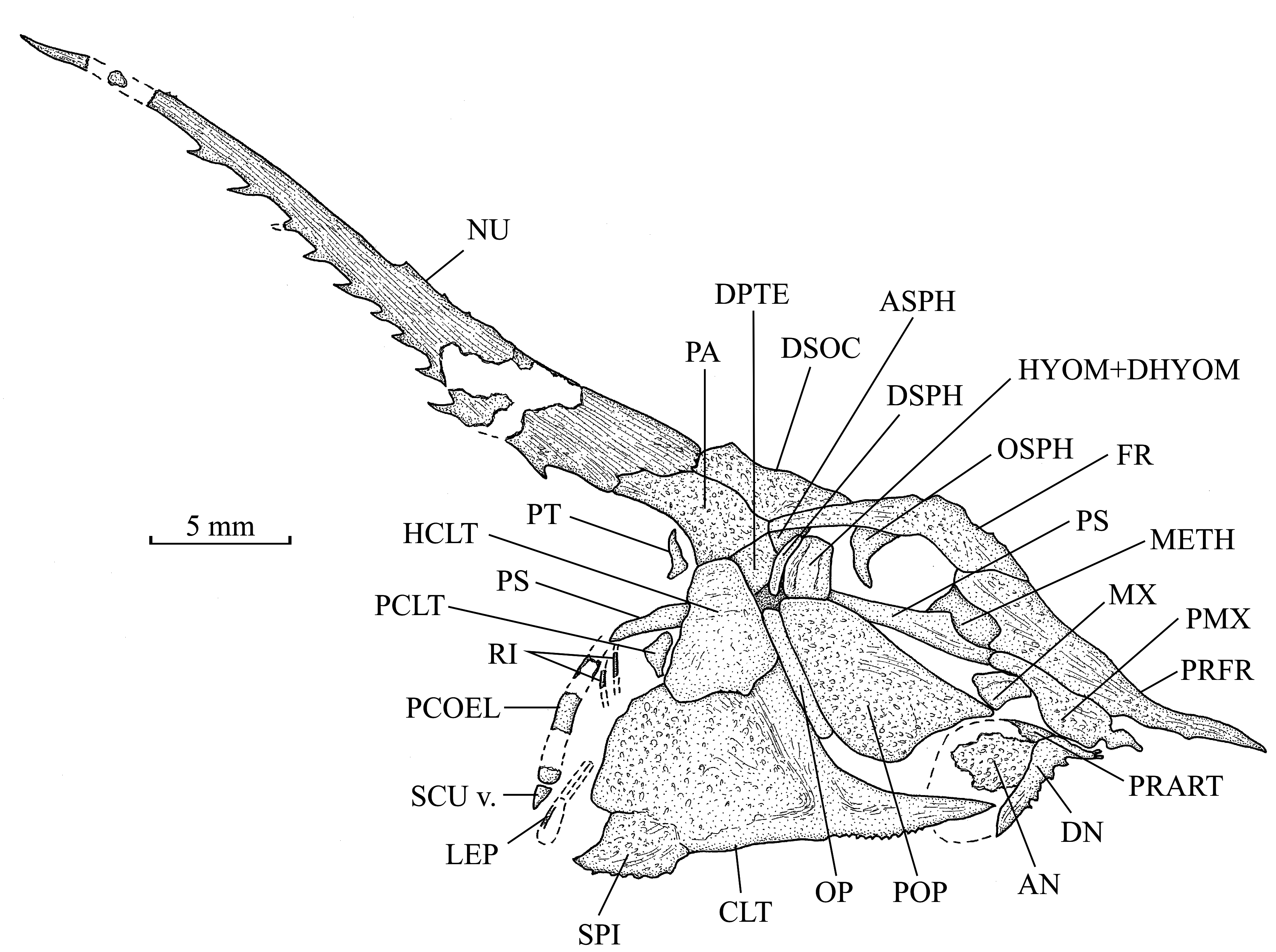
1. The skull ( Fig. 19 View Fig )
The head and the pectoral girdle are closely associated, forming a cephalo-thorax that is gigantic when compared to the body size. This character and the feeble ossification of the axial skeleton probably indicate that the concerned sample is a juvenile fish. The dermal bones are ornamented with tubercles, alveoli and thin ridges.
The rostrum is longer than in Joinvillichthys gen. nov. It is formed by the acuminate anterior extremity of the prefrontal. This rostrum greatly outpaces the lower jaw level. The preorbital length, rostrum included, represents 54.5 % of the total length of the head. The prefrontal is long, anteriorly pointed and rather broad posteriorly, hiding completely the vomer and a great part of the mesethmoid.
The frontal is narrow, rather long and exhibits a weakly developed median protuberance located just above the orbit. The elongate posterior part of the frontal extends far behind the orbit, under the dermosupraoccipital ventral margin, and reaches the parietal. The dermopterotic is small but the dermosupraoccipital and the parietal are large bones that protrude posteriorly. The small autosphenotic is located in front of the dermopterotic and just below the posterior extremity of the frontal. An extremely long nuchal horn with a spiny posterior margin is articulated on both the parietal and the dermosupraoccipital.
The parasphenoid is very long, straight and toothless. Its posterior extremity greatly outpaces the rear of the skull. The orbitosphenoid is separated from the mesethmoid. No other endocranial bone of the braincase and no bone of the palato-quadrate arch are visible.
The premaxilla and the maxilla are toothless. There is no supramaxilla. The premaxilla is long but much shorter than the prefrontal, to which it is sutured by its upper margin. This premaxilla exhibits a small, anterior hook-shaped process, a broad anterior part and a much narrower posterior region. The premaxilla does not contribute to the rostrum. The small, triangular maxilla is located below the posterior part of the premaxilla. The dentary, reduced to its ventral branch, is well developed. It bears two very small, incisiform teeth. Its posterior part forms a right angle with its anterior extremity. Its lower margin is spiny. The angular is a rather large bone. Only a very small part of the prearticular is visible. The articular is not preserved.
The orbit is wide. The long and very thin dermosphenotic is placed against the autosphenotic and the dermopterotic. No other bone of the orbital series is present.
The hyomandibula and the preopercle are sutured together. The exposed part of the hyomandibuladermohyomandibula is deeper than broad but much smaller than the hypertrophied, trapezoid-shaped preopercle that covers the cheek. The opercle is a long, rod-like bone pressed between the preopercle and the pectoral girdle.
2. The girdles ( Figs 18–19 View Fig View Fig )
The hypertrophied pectoral girdle is pressed against the skull. The bones are ornamented with tubercles, alveoli and thin ridges. The gigantic cleithrum possesses a long, narrow and pointed ventral branch, with a serrated lower margin, and a very broad posterior process. The hypercleithrum (= supracleithrum) is a large bone, broader ventrally than dorsally. A small, triangular posttemporal, with an acuminate upper extremity, is located near the parietal. A small postcleithrum is visible behind the hypercleithrum. The pectoral fin is replaced by a short but very broad spine that is articulated and partly fused to the ventral margin of the wide cleithral posterior process.
A fragment of a pelvic ray is visible a little before the postcoelomic bone. The pelvic bones are not preserved.
3. The axial skeleton ( Fig. 18 View Fig )
The axial skeleton is poorly ossified. Only the first thirteen vertebral segments are partly preserved. The complete vertebral axis probably contained a little less than twenty segments. Well developed neural arches are present but the haemal arches are not visible. Thus, the notochord is not surrounded by the arches. The first neural and haemal spines are long and the last ones very short. Fragments of ribs are visible. The postcoelomic bone is broad but rather short.
4. The dorsal and anal fins ( Fig. 18 View Fig )
The dorsal fin has a rather long basis and begins just behind the nuchal horn. The number of rays and pterygiophores is unknown. The anal fin has a shorter basis and contains 7 rays. Traces of at least 5 anal pterygiophores are visible.
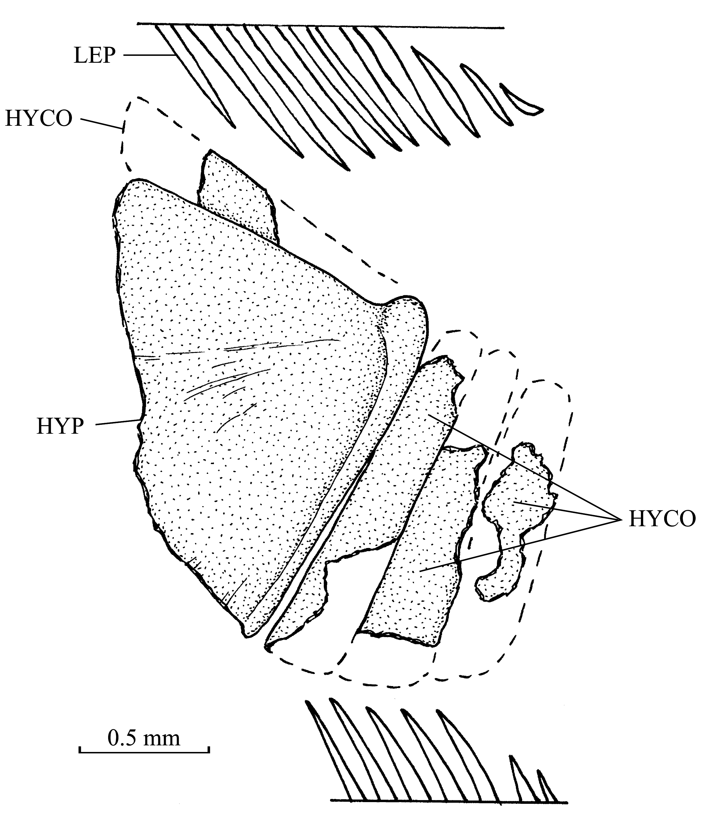
5. The caudal skeleton ( Fig. 20 View Fig )
The caudal skeleton is incompletely preserved. Only the hypochordal series is present with 5 elements. The fourth hypochordal is greatly enlarged, forming a very broad hypural plate. This plate probably results from the fusion of several hypochordals. The epichordals and the urodermals are not known.
Only a part of the caudal fin is present.
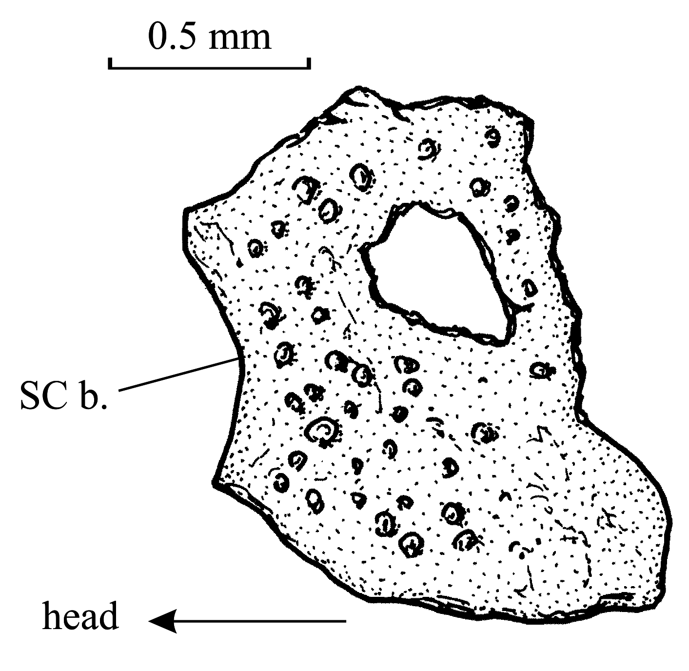
6. Squamation ( Fig. 21 View Fig )
The squamation is badly preserved. However, some fragments and prints of scales are visible on the entire body. These scales are large, scute-like, irregularly shaped and ornamented with tubercles and alveoli.
A small spine is articulated on the ventral extremity of the postcoelomic bone.


| IRSNB |
Institut Royal des Sciences Naturelles de Belgique |
No known copyright restrictions apply. See Agosti, D., Egloff, W., 2009. Taxonomic information exchange and copyright: the Plazi approach. BMC Research Notes 2009, 2:53 for further explanation.
|
Kingdom |
|
|
Phylum |
|
|
Class |
|
|
Order |
|
|
Family |
|
|
Genus |