
Pseudopyrochroa fainanensis (Pic)
|
publication ID |
https://doi.org/ 10.11646/zootaxa.4061.5.8 |
|
publication LSID |
lsid:zoobank.org:pub:FC056B8F-0ED3-4FD1-9367-9BED563DF1B8 |
|
DOI |
https://doi.org/10.5281/zenodo.6088190 |
|
persistent identifier |
https://treatment.plazi.org/id/03C9D23A-7602-B70D-FF4F-46558E52FA2A |
|
treatment provided by |
Plazi |
|
scientific name |
Pseudopyrochroa fainanensis (Pic) |
| status |
|
Pseudopyrochroa fainanensis (Pic)
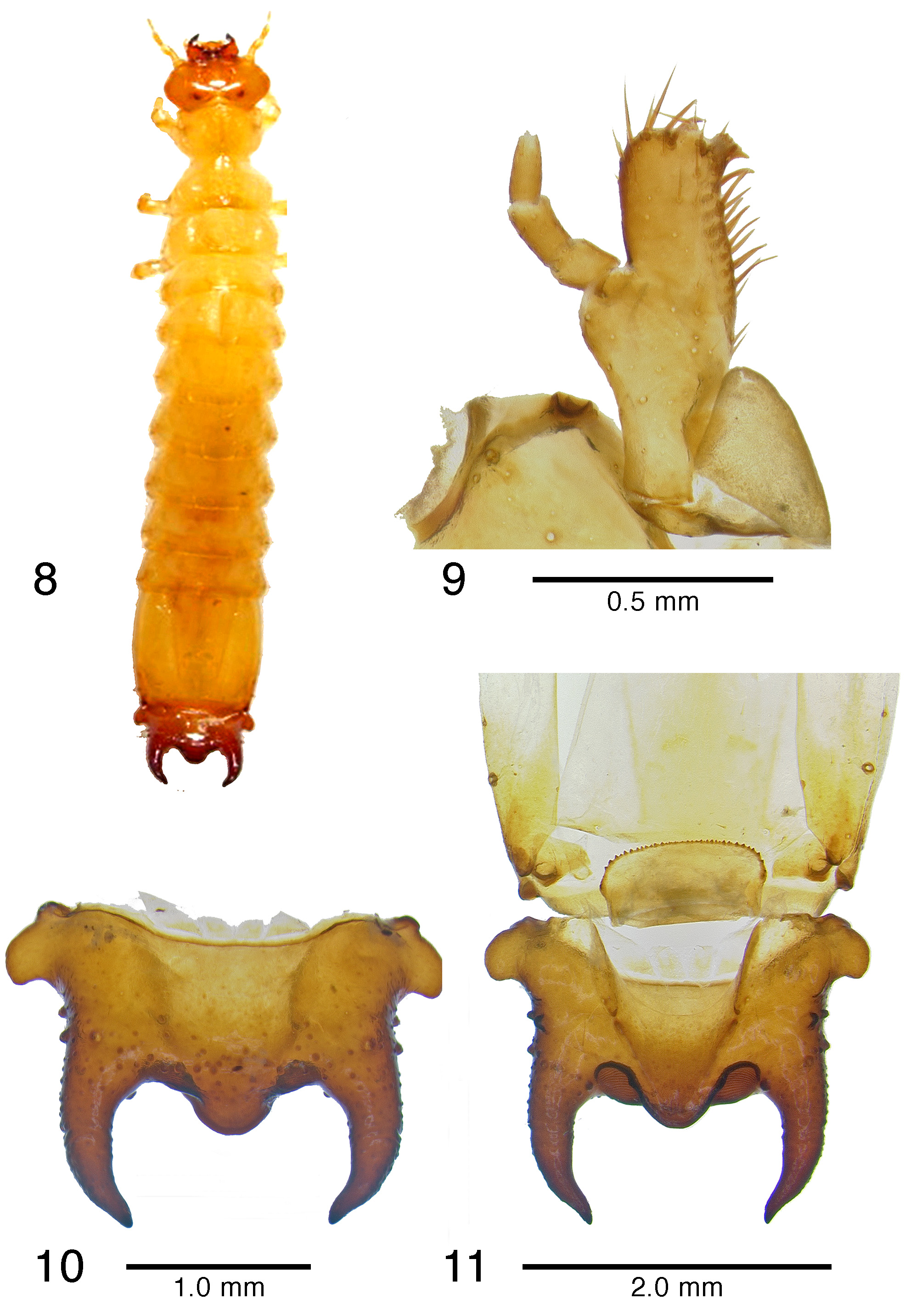
( Figures 8–11 View FIGURES 8 – 11 )
Description of mature larva. Mature larvae ( Fig. 8 View FIGURES 8 – 11 ; n = 2) attain lengths (mesal labral apex to apices of urogomphi) of 15–17 mm and widths (across widest portion of eighth abdominal segment) of 2.0– 2.4 mm. Body orthosomatic with sides subparallel; moderately sclerotized except much of cranium, mandibles, and urogomphal plate more heavily sclerotized; body vestiture consisting of short to moderately elongate, scattered setae. Thoracic and abdominal terga lacking parabasal ridges. Head and body creamy-yellowish to amber, melanization much darker in areas of heavy sclerotization such as tips of mandibles, urogomphi, urogomphal lip and urogomphal pits.
Head: Prognathous, flattened, exserted from prothorax. Epicranial suture lyriform with stem short, frontal arms complete nearly to antennal insertions; endocarinae absent. Free, symmetrical labrum anterad fused frontoclypeal region. Stemmata on each side 4, in 2 groups of 2, lower pair parallel with antennal insertions. Antennal insertions fully exposed, antennae moderately elongate, filiform, 3-segmented, 1st antennomere 0.1–0.2 longer than antennomeres 2 and 3, sensorium of segment 2 small, conical, 3rd antennomere narrower than antennomeres 1–2, acutely rounded apically. Mouthparts retracted. Mandibles heavily sclerotized, movable, asymmetrical, molar area of mandibles well developed, left mandible bearing a prominent molar tooth; apex of right mandible distinctly tridentate, that of left mandible bidentate; dorsal mandibular surface basad the molae with microtrichial complex well developed. Maxillae ( Fig. 9 View FIGURES 8 – 11 ) each with transverse, 1-segmented cardo which is diagonally twisted upward upon itself toward the stipes and thus appearing 2-segmented, a well developed, undivided, pad-like maxillary articulating area; ventral surface of stipes relatively glabrous, bearing fewer than 20, scattered fine to stout setae; galea and lacinia fused to form maxillary mala; mala bearing stout apical and adoral setae and spinose-dentiform uncus at apico-adoral margin; 3-segmented, stoutly filiform maxillary palpus, 2nd palpomere slightly less than 2X length of first, 3rd palpomere subequal in length to that of 2nd, 3rd palpomere slightly tapering distally, obtusely rounded apically. Labium with mentum ovate-subquadrate, submentum elongate with sides shallowly sinuate basally, convexly rounded; ligula elongate, well developed; each labial palpus short, 2-segmented. Hypopharyngeal sclerome well sclerotized, molar-like; proximal region of hypopharynx with complex setal brushes. Hypostomal rods well developed, divergent; gular sutures separate.
Thorax and Abdomen: Thoracic segmentation well developed, sides parallel; cervicosternum divided into three plates. Legs well developed, moderately short, 5-segmented including tarsungulus, vestiture consisting of sparse, short setae; coxae large, separated by 2–3 coxal diameters. Abdomen flattened, moderately sclerotized, tergites 1–7 subequal in length and width; 8th tergite approximately 2X length of 7th. Sternite 8 emarginate apically. Tergite 9 ( Fig. 10 View FIGURES 8 – 11 ) hinged, capable of considerable dorso-longitudinal movement, extending ventrally, thus forming the urogomphal plate ( Figs. 10–11 View FIGURES 8 – 11 ), widest basally where it forms well developed, subquadrate to slightly acuminately rounded lateral lobes; surface of urogomphal plate bearing numerous, well-developed callosities and a few larger, dorsal and lateral setigerous calli on the dorsal and lateral surfaces of the urogomphi; urogomphi heavily sclerotized, relatively short, stout, strongly incurved distally, tapering, slightly recurved and acuminate apically; urogomphal plate strongly produced apico-mesally, forming broad, obtusely rounded mesal lobe between urogomphi; ventral surface of urogomphal plate shallowly, but rather abruptly excavate basally at articulation with 9th and 10th sternites, excavation narrowing distally to bases of urogomphi and becoming obsolete along ventral face of urogomphal lip. Urogomphal plate possessing a well developed, heavily sclerotized, apico-mesally rounded urogomphal lip; paired, well defined, subovate, heavily sclerotized, recessed urogomphal pits ( Fig. 11 View FIGURES 8 – 11 ) present apically, one each between the heavily sclerotized, fixed urogomphi, and the apico-mesal lobe of the urogomphal lip; urogomphal pits bearing characteristic fingerprint-like rugulae ( Fig. 11 View FIGURES 8 – 11 ). Sternite 9 ( Fig. 11 View FIGURES 8 – 11 ) transverse, partially recessed into shallow emargination of 8th sternite, possessing continuous semicircular arch of approximately 36–40 fine asperites along anterior margin. Tenth segment strongly reduced, transverse, visible ventrally.
Spiracles: One pair of well-developed, ovate thoracic spiracles, situated ventro-laterally on laterotergite along anterior end of mesothorax. Paired, annular abdominal spiracles, subequal in size, located on dorsolateral margins of abdominal tergites and ventrolateral margins 1–7; paired spiracles of abdominal laterotergite 8 ( Fig 11 View FIGURES 8 – 11 ) larger, annular, located laterally at distal 2/3 of its length.
Material examined. [1 ♀ with exuvium]: TAIWAN: Heping District, // Taichung City, Tahsuehshan, // Dasyueshan trail 18.8k, // 24.250448°N / 120.917915°E; // elevation 1240m (larva D) // 25 November 2012 // W.-R. Liang; [2nd label]: larva collected beneath bark of // decaying hardwood log; // 29.I.2013, pupated // 5.II.2013, eclosed; [3rd label]: Pseudopyrochroa // fainanensis ♂ // (Pic) // det. D. K. Young]. [1 partial exuvium]; TAIWAN: Nantou Hsien; 2100m // Meifeng; National Taiwan // University Highland Experimental Farm // 24.088607°N / 121.176065°E // 24.XI.2013 // Yun Hsiao, leg // collected from beneath bark // and in moist, decaying wood of // dead hardwood tree; [2nd label]: Pseudopyrochroa // fainanensis // (Pic) // det. D. K. Young. [1 mature larva]; TAIWAN: Nantou Hsien; 2068m // Meifeng; National Taiwan // University Highland Experimental Farm // 24.091319°N / 121.176258°E // 2.V.2014 // Yun Hsiao, leg // collected from beneath bark // and in moist, decaying wood of // dead hardwood tree; [2nd label]: Pseudopyrochroa // fainanensis // (Pic) // det. D. K. Young.
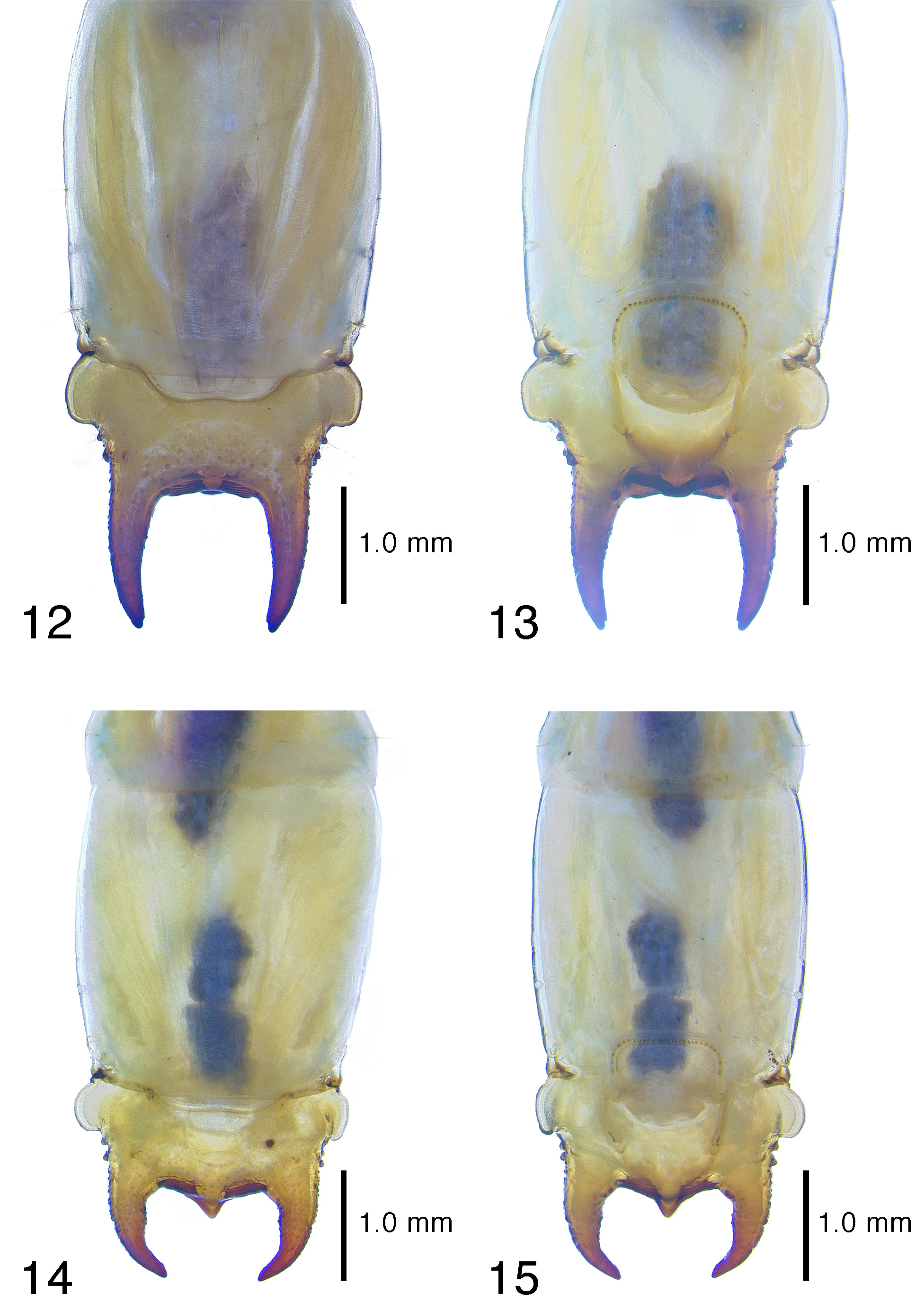
Diagnosis. The most diagnostically useful characters of P. f a i n an e n s i s larvae are the short, stout, incurvate urogomphi ( Figs. 10–11 View FIGURES 8 – 11 ), the strongly, apico-mesally produced urogomphal plate and fused, heavily sclerotized urogomphal lip, and the well defined urogomphal pits with characteristic fingerprint-like rugulae ( Fig. 11 View FIGURES 8 – 11 ). The only presently known pyrochroid larva with a urogomphal configuration approaching that of P. fainanensis is the southeast Asian Pseudopyrochroa basalis (Pic) ( Figs. 14–15 View FIGURES 14 – 15 ). Like P. fainanensis ( Figs. 10–11 View FIGURES 8 – 11 ), the urogomphi of P. basalis are short, stout, and conspicuously incurvate. Larvae of P. basalis have the apico-mesal extension of the urogomphal plate far more acuminately produced ( Figs. 14–15 View FIGURES 14 – 15 ) and the urgomphal pits are considerably less exposed in ventral view ( Fig. 15 View FIGURES 14 – 15 ).
No known copyright restrictions apply. See Agosti, D., Egloff, W., 2009. Taxonomic information exchange and copyright: the Plazi approach. BMC Research Notes 2009, 2:53 for further explanation.