
Paulasterias undetermined
|
publication ID |
https://doi.org/ 10.1111/zoj.12229 |
|
persistent identifier |
https://treatment.plazi.org/id/03CA878C-FF8B-226E-56C3-FB94FEEFEABB |
|
treatment provided by |
Carolina |
|
scientific name |
Paulasterias undetermined |
| status |
|
PAULASTERIAS TYLERI GEN. ET SP. NOV.
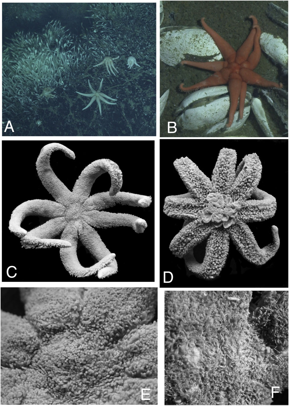
FIGS 3A–F View Figure 3 , 4A–E View Figure 4 , 5A–D View Figure 5
Stichasteridae View in CoL sp. (or ‘stichasterid asteroid’) Rogers et al. 2012: 7, table 2, figure 3E; Marsh et al., 2012: 1, 2, 6, 8, 15, figures 4, 5, table 1; Reid et al. 2013: 1, 6, 8, 9, tables 3 and 5.
Etymology
The genus and species are named for Professor Paul Tyler, Member of the Most Excellent Order of the British Empire (MBE), National Oceanography Centre, at the University of Southampton, in honor of his voluminous contributions to deep-sea biology.
Ecological observations
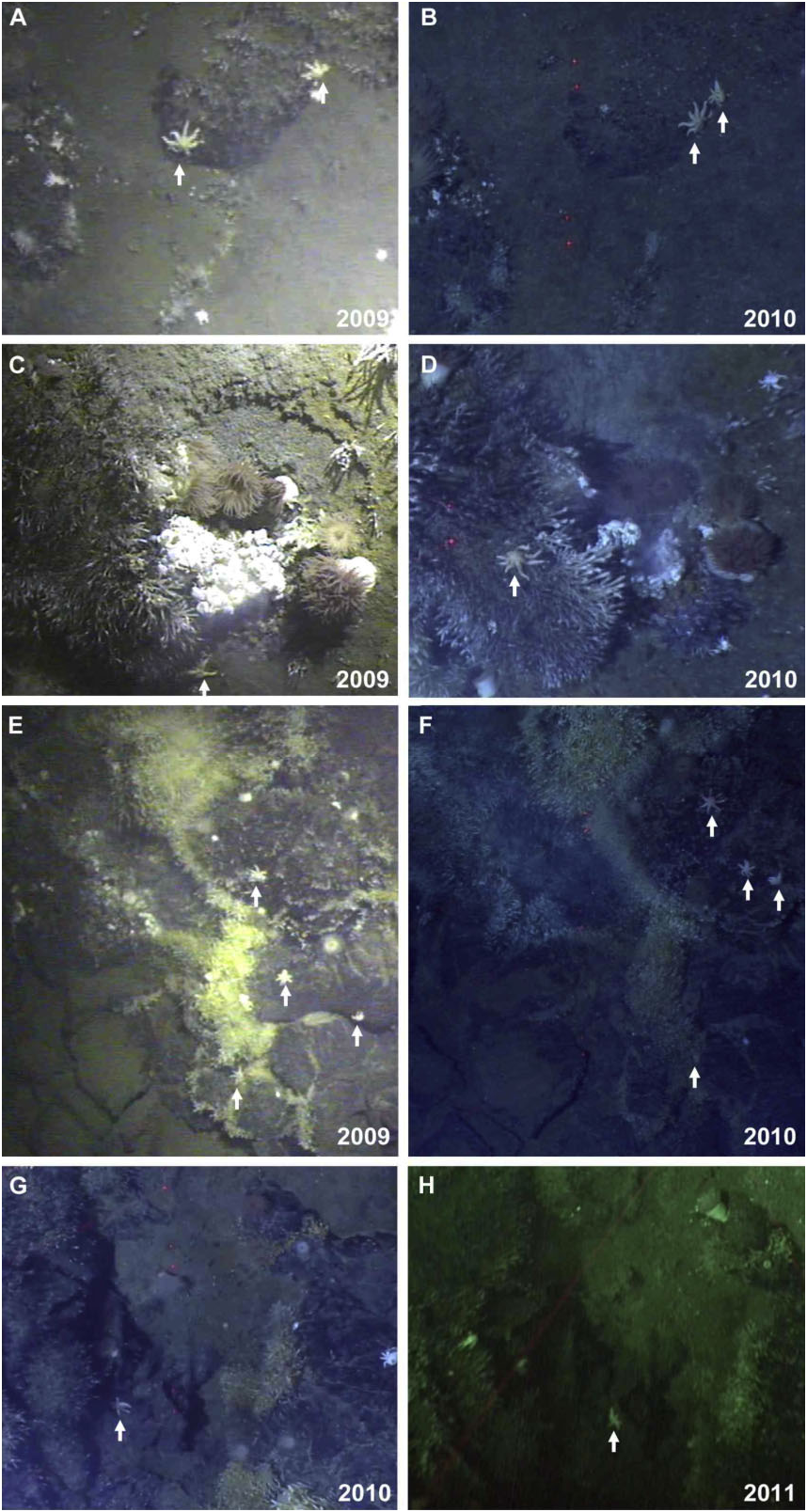
This species was observed towards the base of vent chimneys and peripheral to areas of low-temperature diffuse venting ( Fig. 3A,B View Figure 3 ), and was classified as part of the ‘peripheral assemblage’ as described by Marsh et al. (2012). This species was observed individually as well as in small aggregations of between two and five individuals (with 0.11 m being the closest proximity between the centre points of the discs). Throughout the three ChEsSo cruises to the East Scotia Ridge (2009 JR224 RRS James Clark Ross; 2010 JC42 RRS
FS, furrow spine.
James Cook; 2011 JC55 RRS James Cook), four areas of low-lying diffuse flow seabed at the E9 vent field were imaged in subsequent years [ Fig. 6 View Figure 6 ; 2009 and 2011, Seabed High Resolution Imaging Platform SHRIMP; 2010, remotely operated vehicle (ROV) Isis ]. Within these areas of observed seabed, absolute numbers of P. tyleri gen. et sp. nov. remain constant, and using seabed geological features for reference, the positions of individuals on the seafloor have changed by less than 1 m from the locations where they were first observed ( Fig. 6 View Figure 6 ).
Occurrence
East Scotia Ridge segments E2 and E9, South Sandwich Islands, Kemp Caldera to Ross Sea, 1420– 2609 m.
Taxonomic comments
Paulasterias tyleri gen. et sp. nov. displays a body form similar to that of several different multi-armed Antarctic asteriid taxa, all of which can be confused with P. tyleri gen. et sp. nov., including species within Psalidaster , Saliasterias , Lysasterias , and Diplasterias . Differences between P. tyleri gen. et sp. nov. and these species are outlined in Table 2.
Description
Between six and eight arms, but most specimens observed with seven; disc small (R / r = 3.78–9.00, but most with ∼5–6, where R is the length from disk center to end of arm and r is radius of the disc). One smaller specimen (NHMUK 2014.23, R = 2.2 cm) has six arms, with an incipient seventh growing between two other arms.
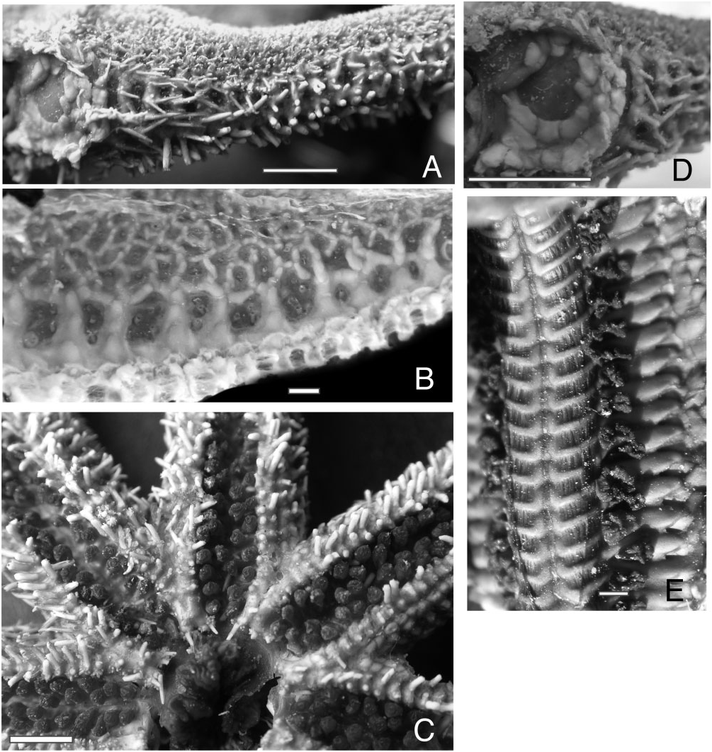
Based on measurements of the disc size compared with the R / r ratio, individuals with smaller discs show a more strongly stellate body form, whereas arm length varies between more strongly and weakly stellate individuals. Arms proximally swollen, with distinct septa present inter-radially at contact points between arms ( Fig. 4D View Figure 4 ). Spination absent from surface in this region. Endoskeleton weakly calcified, with widely spaced reticulation (observable when tissue is denuded; Fig. 4B View Figure 4 ). Two types of pedicellariae present: straight pedicellariae on arm surface and adambulacral plate in tube foot furrow, with crossed-type pedicellariae covering the majority of the abactinal/lateral surface ( Fig. 5 View Figure 5 ) .
Abactinal plates forming widely spaced reticulate pattern. Surface covered by an abundance of dense spinelets ( Fig. 3E, F View Figure 3 ), fleshy tissue, and pedicellariae. All abactinal plates (i.e. the reticulate patterns in Fig. 4B View Figure 4 ) obscured. Spination forms a fur-like appearance to the surface ( Fig. 3E, F View Figure 3 ). Spinelets, slender, covered in a sheath of tissue (best observed in dry specimens), range in height from 0.5 to 1.0 mm (base to tip) at densities of 20–30 along a 1.0-cm line (at R = 4.0– 6.0 cm, respectively). Individual plates with approximately between two and six spinelets, each arranged in a crescentic or irregular series. Some individual spinelets on single plates. Spinelet density heaviest on abactinal and upper lateral surfaces ( Fig. 3C, E, F View Figure 3 ). Abactinal plates are smaller (50–75% of the size) relative to the elongate and larger plates present in the marginal plate/lateral arm region. Plates tightly reticulate, with very close articulation becoming almost imbricate. Abactinal plates more irregularly arranged than those on marginal/lateral arm regions, forming a continuum with the more regularly arranged plates present along the marginal/lateral arm series ( Fig. 4A, B View Figure 4 ). Abactinal plates reticulate, widely arranged on the body surface, becoming more densely arranged, almost imbricate, along marginal series. Gaps between tightly articulated abactinal plates filled with tissue often have between one and three (mostly single) papula present between plates. Discrete carinal series not observed.
Papulae are large and single but obscured among spinelet-covered surface. Madreporite with deep sulci, partially to completely covered and encircled by between two and eight spinelets, arranged in a single series. On some specimens, madreporite surface free of spination.
Two types of pedicellariae observable on the abactinal surface. Straight pedicellariae less abundant and not observed on some individuals, present interadially on the lateral arm surface but not observed on the disc surface, similar in form but smaller (∼ 0.5 mm in length) than those observed inter-radially in P. mcclaini gen. et sp. nov. These present interadially on the lateral arm surface but not observed on the disc surface.
Most abundant crossed pedicellariae with rounded valve edges ( Fig. 5A–D View Figure 5 ). Pedicellariae width approximately <0.5 mm, present widely on body surface, interspersed among spines and papulae. Valves rounded with multiple jagged edges. Pedicellariae on abactinal/ lateral surface crossed, and similar in form to those observed in other forcipulataceans. Valves of these pedicellariae articulate at their bases ( Fig. 5A, B View Figure 5 ). The distal part of the valve containing 25–40 sharp, conical teeth, with smaller teeth at the distalmost edge and larger teeth present more proximally on the roof. The roof (i.e. present nearer to the base of the valve) of the pedicellariae with 20–40 sharp conical teeth ( Fig. 5C, D View Figure 5 ). Numerous circular pores present where muscle and tissue connect along the surface of each valve. Attachment area at proximal end of each valve with numerous fenestrae.
Marginal plate series partially obscured by spinelets and fleshy tissue ( Fig. 4A View Figure 4 ). Abactinal plates articulate with superomarginals elongate and forming ‘top edge’ of window-like spaces between each superomarginal–inferomarginal pairing, with each containing papulae ( Fig. 4B View Figure 4 ). Each of these large spaces flanked by approximately between eight and 12 elongate, round- to rectangular-shaped, spine-bearing plates forming a quadrate frame articulating with the superomarginal series. Superomarginal plate series, imbricate, trilobate in shape, smaller than inferomarginals (<50% of size), flanked by other imbricate plates articulating with adjacent superomarginals. Superomarginals + intermarginals with between one and four large spines ( Fig. 4A View Figure 4 ). Larger spines present proximally, becoming smaller distally. Intermarginal plates present between superomarginals and inferomarginals, with between two and four (mostly three) forming borders around papular regions. Inferomarginals multilobate, larger than superomarginals. Single, large spine present on inferomarginals, decreasing in size proximally to distally. Papulae, between two and four, mostly three, present between spaces formed by lobes between superomarginal plates.
Inter-radial septa present between arms ( Fig. 4D View Figure 4 ), flanking large membrane-like window at proximal articulation between arms. Plates that form the septa are large, scalar, and articulate with actinolateral and adambulacral plates. Lower end of inter-radial septa articulates via actinal plating with adjacent arm septa. Actinal plates, individually diamond to polygonal in shape. Present in series adjacent to adambulacrals along arm. Actinal plates form irregular imbricate pavement inter-radially on disc. Adoral carina composed of one or two abutted adambulacrals. Tissue present between abutted contact along plates of adjacent adambulacrals (implying no direct skeletal fusion). Adoral carina with two abutting adambulacrals observed in slightly larger individuals (e.g. R = 6.1 cm, USNM 1087711).
Each paired oral plate articulated with tissue, shieldshaped, with two spines present on surface ( Fig. 4C View Figure 4 ). In addition to these surficial oral spines, two more elongate spines present on each oral plate project into mouth, each with approximately a dozen straight pedicellariae clustered around the base. These two elongate oral spines are consistent in numeration and morphology, with furrow spines present on each adambulacral plate. Furrow spines, two per adambulacral plate, are tissue-covered, thick, and round in cross section. Approximately between four and a dozen pedicellariae, beak-like, differing from those on the abactinal surface, occur on the furrow-side of each adambulacral plate at the base of each set of furrow spines. Adambulacral plates quadrate in outline, blockish, punctuated by tissue present between plates ( Fig. 4B View Figure 4 ).
Ambulacral ossicles compressed and wing-shaped ( Fig. 4E View Figure 4 ), but with relatively broad keel and wide wing relative to Asteriidae . Tissue present between ossicles.
A well-developed cardiac stomach observed ( Fig. 3D View Figure 3 ) extruded in all Scotia Arc specimens.
Living colour of animal is white to light reddish orange.
Material examined
See Table 1.

| R |
Departamento de Geologia, Universidad de Chile |
No known copyright restrictions apply. See Agosti, D., Egloff, W., 2009. Taxonomic information exchange and copyright: the Plazi approach. BMC Research Notes 2009, 2:53 for further explanation.
|
Kingdom |
|
|
Phylum |
|
|
Class |
|
|
Family |
|
|
Genus |
Paulasterias undetermined
| Mah, Christopher, Linse, Katrin, Copley, Jon, Marsh, Leigh, Rogers, Alex, Clague, David & Foltz, David 2015 |
Stichasteridae
| Reid WDK & Sweeting CJ & Wigham BD & Zwirglmaier K & Hawkes JA & McGill RAR & Linse K & Polunin NVC 2013: 1 |
| Rogers AD & Tyler PA & Connelly DP & Copley JT & James R & Larter RD & Linse K & Mills RA & Naveira-Garabato A & Pancost RD & Pearce DA & Polunin NVC & German CR & Shank T & Boersch-Supan PH & Alker BJ & Aquilina A & Bennett SA & Clarke A & Dinley RJJ & Graham AGC & Green DRH & Hawkes JA & Hepburn L & Hilario A & Huvenne VAI & Marsh L & Ramirez-Llodra E & Reid WDK & Roterman CN & Sweeting CJ & Thatje S & Zwirglmaier K 2012: 7 |
| Marsh L & Copley JT & Huvenne VAI & Linse K & Reid WDK & Rogers AD & Sweting CJ & Tyler PA 2012: 1 |