
Metriogryllacris ( Metriogryllacris ) hamata, Zhang & Pang & Bian, 2022
|
publication ID |
https://doi.org/10.11646/zootaxa.5213.1.2 |
|
publication LSID |
lsid:zoobank.org:pub:77564E5C-0F0D-4AC3-A28F-78B5DA132C50 |
|
DOI |
https://doi.org/10.5281/zenodo.7360674 |
|
persistent identifier |
https://treatment.plazi.org/id/03CA879A-0251-FFCE-FF07-3308BDA3CA43 |
|
treatment provided by |
Plazi |
|
scientific name |
Metriogryllacris ( Metriogryllacris ) hamata |
| status |
sp. nov. |
Metriogryllacris ( Metriogryllacris) hamata sp. nov.
小Ẇaeḋỡ
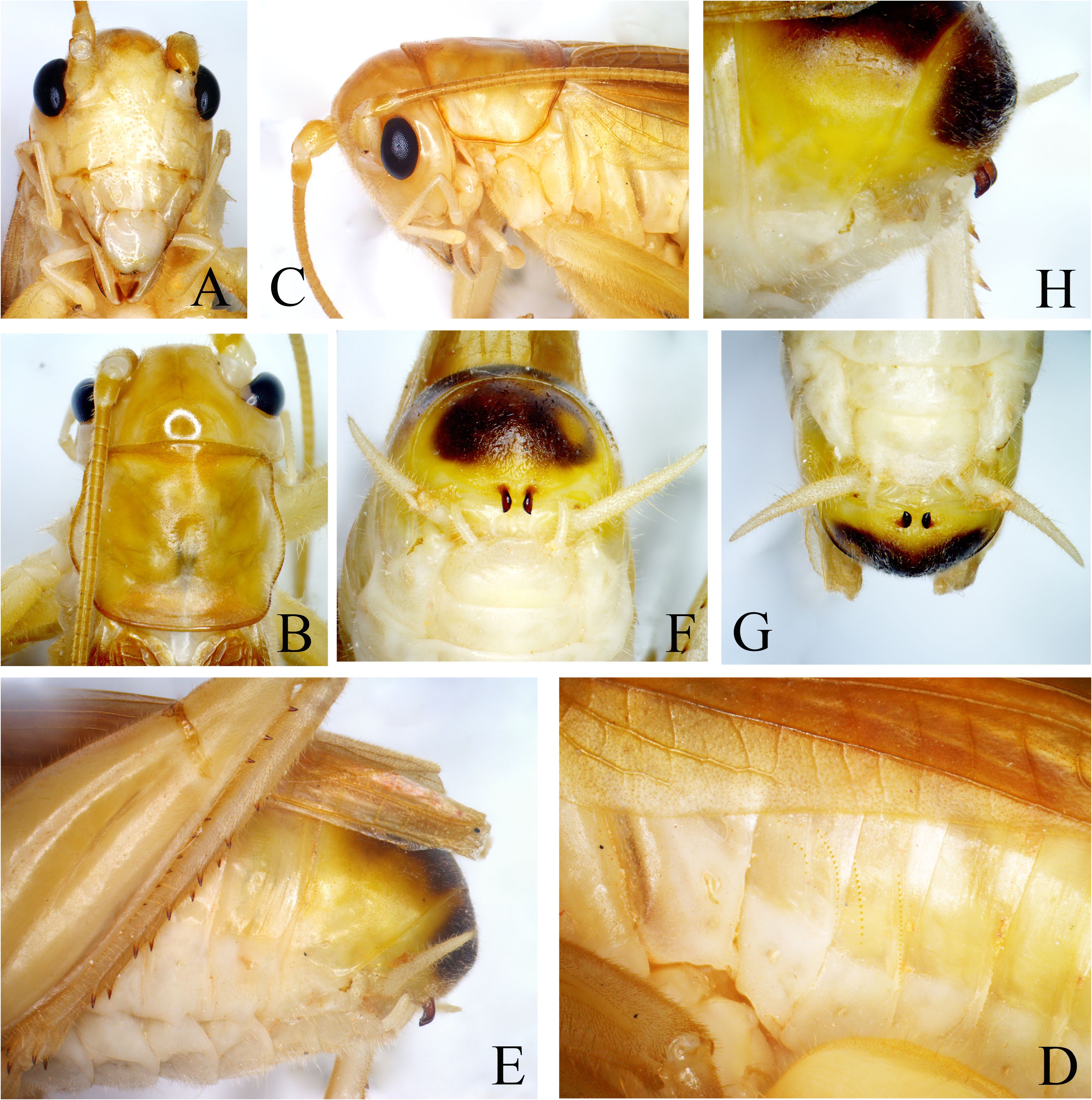
Figures 6–7 View FIGURE 6 View FIGURE 7 , 8A View FIGURE 8 , 9A–D View FIGURE 9
Diagnosis. The new species differs from Metriogryllacris ( Metriogryllacris) permodesta (Griffini, 1914) ( Liu et al., 2021: Figs. 3–6 View FIGURE 3 View FIGURE 4 View FIGURE 5 View FIGURE 6 ) in: the hooks of male ninth abdominal tergite small and widely separated from each other, basal area of the hook narrow in lateral view ( Fig. 6H View FIGURE 6 ); the sclerotized areas of copulatory depressions of female seventh sternite shorter, widely separated from each other with median furrow ( Fig. 7F View FIGURE 7 ).
Description. Male. Face nearly smooth with scattered impressed dots; fastigium verticis distinctly wider than scapus; ocelli indistinct ( Fig. 7A View FIGURE 7 ). Wings reaching eighth abdominal tergite. Tegmina: In left tegmen ( Fig. 9A View FIGURE 9 ), radius forks into two branches before apical third of tegmen, R forked near apical area; MA free from base, then fused with R in short distance and then again divided; media posterior absent; cubitus undivided, free throughout. While right tegmen ( Fig. 9B View FIGURE 9 ), radius releases RS about seven tenths of tegmen; media anterior leaning on radius; media anterior free from base, which divides into two branches about basal third of tegmen, the first branch receives a short connection branch from MA, but remains single-branched as MP+CuA1. Both tegmina: Cubitus posterior undivided, free throughout; with 3 anal veins. Fore coxae with 1 small spine. Fore and middle tibiae ventrally with 4 pairs of spines and 1 pair of apical spines; middle tibiae with 1 internal spine on dorsal surface. Hind femora with 10–13 internal and 9–11 external spines on ventral surface; tibiae with 4 internal and 6 external spines on dorsal surface, ventral surface with 1 pair of subapical spines, apices with 1 pair of dorsal spines and 2 pairs of ventral spines. Second and third abdominal tergites each with 2 rows of stridulatory pegs ( Fig. 6D View FIGURE 6 ). Eighth abdominal tergite prolonged to backward and ventrad. Ninth abdominal tergite semicircular, ventral margin with 1 pair of small hooks ( Fig. 6E, H View FIGURE 6 ) and separated from each other ( Fig. 6F–G View FIGURE 6 ), with narrow bases ( Fig. 6F, H View FIGURE 6 ). Subgenital plate wider than long, the lateral margins convex; apical area projected, subtrapezoidal, posterior margin almost straight with faintly concave in middle; styli inserted at the lateral constriction ( Fig. 6G View FIGURE 6 ).
Female. Tegmina: Radius with two branches, both forked near tip ( Fig. 9C View FIGURE 9 ). In left tegmen ( Fig. 9A View FIGURE 9 ), media free from base, cubitus anterior forks into two veins before middle of tegmen ( Fig. 9C View FIGURE 9 ). In right tegmen, media divides into two branches behind middle of tegmen, cubitus anterior single-branched ( Fig. 9D View FIGURE 9 ). Both tegmina: Cubitus posterior undivided, free throughout; with 3 anal veins. Hind femora ventrally with 10–13 internal and 11–12 external spines; tibiae with 6 pairs of dorsal spines. Ventral surface of seventh abdominal sternite concave with convex converging lateral margins, posterior margin of the concave widely separated by a wider furrow along the midline and the lateral margins obviously sclerotized, at tips with 1 copulatory depression on each side ( Fig. 7F View FIGURE 7 ); it follows a membranous zone which connected with subgenital plate. Subgenital plate wider than long, posterior margin widely rounded with faintly concave in middle. Ovipositor short, apart from subbasal curvature straight, apices obtuse; basal areas of ventral valvulae with 1 pair of long spine-shaped lateral lobes which directing downward and 1 pair of short processes ( Fig. 7E–F View FIGURE 7 ).
Coloration. Body yellow green ( Fig. 8A View FIGURE 8 ). Eyes black. Margins of pronotum and tegmina brown. Dorsal surfaces of eighth and ninth abdominal tergites black. Hooks of male ninth abdominal tergite and sclerotized areas of female seventh abdominal sternite black.
Material examined. Holotype: male, Wanggangshan, Qinzhou , Guangxi, August 15, 2022, coll. by Qianwen Zhang and Shan Li . Paratype: 1 female, Wanggangshan, Qinzhou, Guangxi, August 14, 2022, coll. by Qianwen Zhang and Shan Li .
Measurements (mm). Male: BL 17.49, PL 4.19, TL 12.57, HFL 9.17; Female: BL 17.47, PL 5.05, TL 12.94, HFL 9.82, OvL 8.51.
Distribution. Guangxi (Qinzhou).
Etymology. The new species name derives from the male ninth abdominal tergite with 1 pair of small processes which hooked in lateral view.


No known copyright restrictions apply. See Agosti, D., Egloff, W., 2009. Taxonomic information exchange and copyright: the Plazi approach. BMC Research Notes 2009, 2:53 for further explanation.
|
Kingdom |
|
|
Phylum |
|
|
Class |
|
|
Order |
|
|
Family |
|
|
Genus |