
Stentor tartari Murthy & Bai, 1974
|
publication ID |
https://doi.org/ 10.11646/zootaxa.4732.3.6 |
|
publication LSID |
lsid:zoobank.org:pub:76254E06-3555-4C8D-9B25-1DF8BD9C55ED |
|
DOI |
https://doi.org/10.5281/zenodo.3671746 |
|
persistent identifier |
https://treatment.plazi.org/id/03CA9569-FFB4-FF98-65EC-FA6910F19025 |
|
treatment provided by |
Plazi |
|
scientific name |
Stentor tartari Murthy & Bai, 1974 |
| status |
|
Stentor tartari Murthy & Bai, 1974 View in CoL
( Figs. 6–8 View FIGURE 6 View FIGURE 7 View FIGURE 8 , Tables 1 View TABLE 1 , 2 View TABLE 2 )
Improved diagnosis. Body size 200–355 × 85–135 µm in vivo (on average 300 × 105 µm), 250–700 × 70–135 µm when extended, about 200 × 160 µm after protargol impregnation. Body slender trumpet-shaped to conical when extended, irregular pinkish and colorless cortical granules scattered throughout whole body, symbiotic green algae present, contractile vacuole located in anterior 1/3 of body near left margin with a collecting canal, 8–13 peristomial kineties, 62–106 somatic ciliary rows, 1–4 (average 2) macronuclear nodules located at mid-body. 5–18 micronuclei located around macronuclear nodules.
Locality and habitat. Small freshwater pond (0 psu), Cheongnyang-myeon, Ulju-gun, Ulsan, Korea (35°31′ 47°N, 129°13′ 41°E).
Voucher material. A protargol-stained slide with fixed specimens was deposited at the National Institute of Biological Resources ( NIBR), Incheon, Korea, with registration number NIBRPR0000107178. The voucher specimens are marked by a black ink circles on the slide.
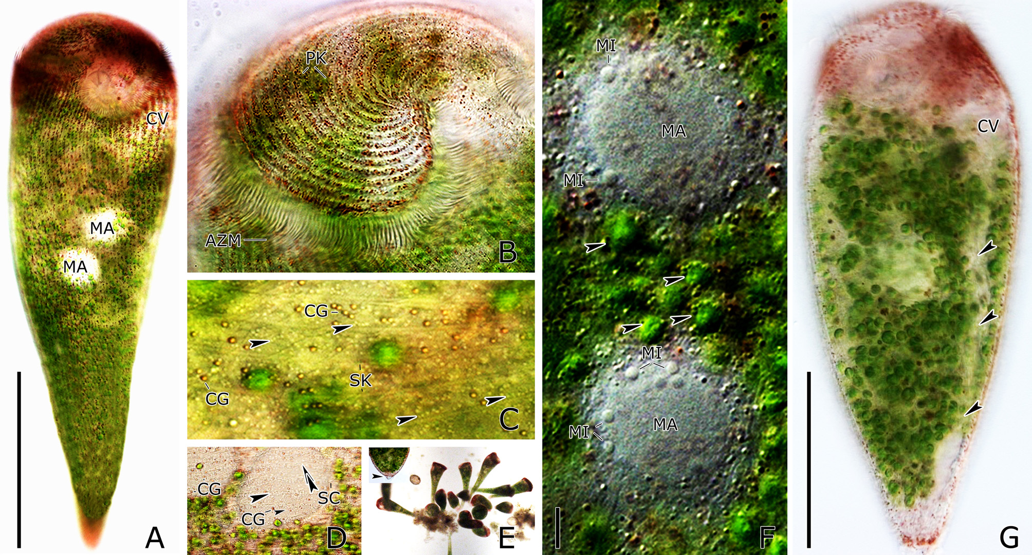
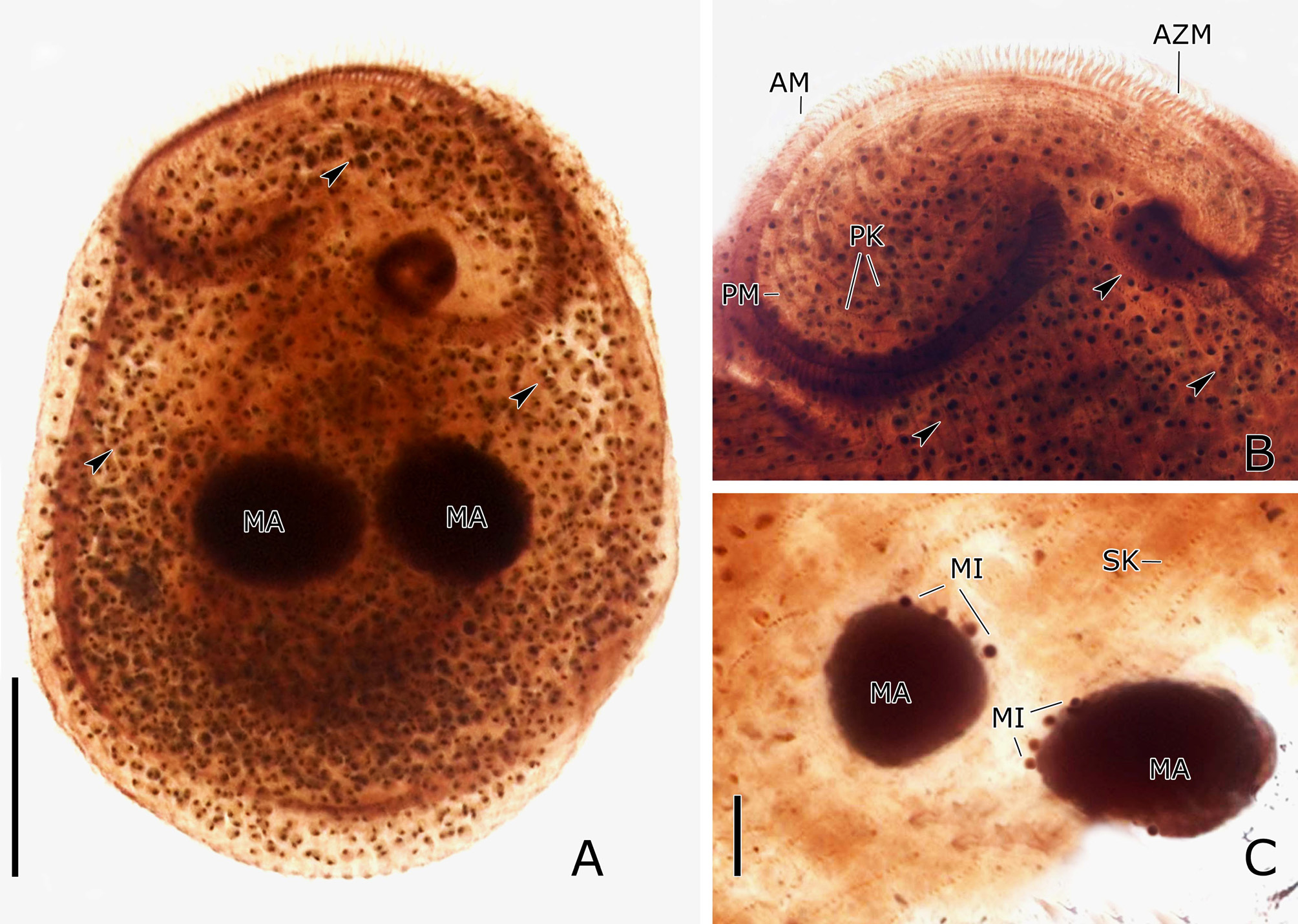
Description. The freely motile cell is inverted pear-shaped and size ranges, 200–355 × 85–135 µm (on average 300 × 105 µm) in vivo ( Fig. 7A View FIGURE 7 ). After extension, when cell relaxed, the body looks like trumpet-shaped and size ranges, 250–700 µm in length and 70–135 µm in width in the peristomial area. Specimens shrink considerably after protargol impregnation, i.e., 95–310 × 75–230 µm (on average 200 × 160 µm) ( Fig. 8A View FIGURE 8 ). Two types of cortical granules, colored and colorless, irregularly arranged between somatic kineties ( Fig. 7C, D View FIGURE 7 ). The colored cortical granules appeared pinkish red, reddish, or brown red, round or oval-shaped 0.5–1 µm in diameter ( Fig. 7C, D View FIGURE 7 ). Density of the colored cortical granules is higher in anterior than in mid to posterior parts of body ( Fig. 7A View FIGURE 7 ). Colorless cortical granules are smaller than reddish granules; shape of colorless cortical granules is round to oval, 0.3–0.5 µm in diameter ( Fig. 7C View FIGURE 7 ). Transparent cortex makes cytoplasmic organelles easily identifiable ( Fig. 7A, C, F View FIGURE 7 ). Sixtytwo to 106 parallel somatic kineties composed of dikinetids longitudinally arranged parallel to antero-posterior axis of cell ( Fig. 6E, F View FIGURE 6 , 7C View FIGURE 7 , 8B View FIGURE 8 ). Each somatic cilium is 6–12 µm long ( Fig. 7D View FIGURE 7 ). Average gap between adjacent somatic kineties is 7 µm in vivo. Mitochondria (?)-like organelles distributed on pellicle slightly more densely packed near somatic kineties ( Fig. 7D View FIGURE 7 , arrowhead). One contractile vacuole with a long collecting canal located left of the buccal cavity about 35 µm in diameter during diastole ( Fig. 6A View FIGURE 6 , 7G View FIGURE 7 ). Eight to 17 peristomial kineties were observed in peristomial region ( Fig. 6E View FIGURE 6 , 7B View FIGURE 7 , 8B View FIGURE 8 ) and size of peristomial cilia was similar to the size of somatic cilia. Buccal pouch absent. Adoral zone of membranelles conspicuous consist of 110–180 membranelles ( Fig. 6E View FIGURE 6 , 7B View FIGURE 7 , 8B View FIGURE 8 ). Adoral zone of membranelles surrounds the peristome and finally ends at the buccal cavity ( Fig. 7A View FIGURE 7 , 8A View FIGURE 8 ). The longest adoral membranelle, 12 µm long. The Paroral membrane is typical of the genus, consisted of a single and continuous row of thin cilia, 20–35 µm long ( Fig. 6E View FIGURE 6 , 8B View FIGURE 8 ). Nuclear apparatus comprised of 1–5 spherical macronuclear nodules (on average 2), about 20–35 µm in diameter in vivo, usually located at the mid-body, however the position of macronuclear nodules is not fixed ( Fig. 7A, F View FIGURE 7 , 8A, C View FIGURE 8 ). Five to 18 spherical micronuclei with a diameter of 1–2 µm in vivo adjacent to macronuclear nodules ( Fig. 7F View FIGURE 7 , 8C View FIGURE 8 ). Symbiotic algae, 2–4 µm in diameter in vivo, present throughout body giving it a dark to light green appearance ( Fig. 7A, E View FIGURE 7 , 8A View FIGURE 8 ). Food vacuoles are 5–30 µm in diameter; contain bacteria, diatoms as food ( Fig. 6A View FIGURE 6 ). Lipid droplets 2–5 µm in diameter scattered throughout body. Holdfast organelle present at posterior end of cell ( Fig. 7E View FIGURE 7 , inset: arrowhead); colored cortical granules densely packed in this region exhibiting a red appearance ( Fig. 7A View FIGURE 7 ).

| NIBR |
National Institute of Biological Resources |
No known copyright restrictions apply. See Agosti, D., Egloff, W., 2009. Taxonomic information exchange and copyright: the Plazi approach. BMC Research Notes 2009, 2:53 for further explanation.
|
Kingdom |
|
|
Phylum |
|
|
Class |
|
|
Order |
|
|
Family |
|
|
Genus |