
Enochrus (Hugoscottia) variegatus (Steinheil, 1869)
|
publication ID |
https://doi.org/ 10.5281/zenodo.188528 |
|
DOI |
https://doi.org/10.5281/zenodo.5630372 |
|
persistent identifier |
https://treatment.plazi.org/id/03CB0A3E-FF9F-FFEE-60C5-DF70A2640D9C |
|
treatment provided by |
Plazi |
|
scientific name |
Enochrus (Hugoscottia) variegatus (Steinheil, 1869) |
| status |
|
Enochrus (Hugoscottia) variegatus (Steinheil, 1869) View in CoL
( Figs. 1 View FIGURES 1 – 2 , 3–6 View FIGURES 3 – 6 , 11–17 View FIGURES 11 – 17 , 22–23 View FIGURES 22 – 25 , 26–27 View FIGURES 26 – 29 , 30 View FIGURES 30 – 31 , 32–34 View FIGURES 32 – 34 )
Material examined. ARGENTINA: Buenos Aires City: “Paseo de las Américas” Park, 6 breeding adults collected on 02.xi.2006 by B. Byttebier (7 egg cases, 30 instar I, 6 instar II and 9 instar III larvae, 2 pupae, 12 emerged adults).
Diagnosis. Larvae. Coronal sulcus absent (instars I–II) or very short, in some cases difficult to observe (instar III); left mandible with two inner teeth; AN10 and AN11 situated apically; MX 4 and MX 6 located distally on outer margin, MX 5 situated somewhat mesally; PA26 situated far from PA27 and closer to PA17; sterna III–VII with a pair of prolegs, each one bearing a group of cuticular spines.
Egg case ( Fig. 1 View FIGURES 1 – 2 ). Whitish, suboval and flat in shape (total length = 3.97–5.95 mm, width = 1.31–2.46 mm; length without mast = 1.91–3.15 mm); generally constructed on aquatic vegetation, made of two layers of silk, second one covering the eggs. Mast ribbon-like, of variable length and width, attached to the substrate (length = 1.46–3.47 mm).
Eggs whitish or dark brown in color, laid out in two rows within the egg case (length = 0.52–0.60 mm, width = 0.20–0.30 mm). Number of eggs per case variable, between 11 and 14 (N = 7).
First-instar larva ( Figs. 3–6 View FIGURES 3 – 6 , 11–17 View FIGURES 11 – 17 , 26–27 View FIGURES 26 – 29 , 32 View FIGURES 32 – 34 ). Morphometric measures detailed in Table 2 View TABLE 2 .
Color. Head capsule light brown, without color pattern; ocular area lighter. Head appendages light brown. Thoracic tergites and thoracic appendages light brown; membranous areas of the thorax testaceous. Abdominal sclerites, urogomphi and lateral appendages of abdominal segment VIII light brown; membranous areas of the abdomen testaceous. Non-sclerotized integument covered by fine pubescence.
Body ( Fig. 32 View FIGURES 32 – 34 ). Subcylindrical, slightly narrowing towards abdominal apex.
Head ( Figs. 3–6 View FIGURES 3 – 6 , 11–17 View FIGURES 11 – 17 ). HL = 0.19–0.22 mm; HW = 0.26–0.29 mm. Head capsule subquadrate ( Fig. 4 View FIGURES 3 – 6 ), slightly broader than long, somewhat retracted within prothorax; occipital foramen wide, dorsal part of cervix with two small suboval cervical sclerites. Frontoclypeal suture absent, frontal sulci V-shaped, coming together close to base of head capsule; coronal sulcus absent. Ocular areas with six stemmata on each side, arranged in 2 rows of 3, one of the inferior row laterally separate from the other. Labroclypeus asymmetrical ( Fig. 5 View FIGURES 3 – 6 ). Nasale projecting obliquely, with six or seven irregular teeth, right teeth projecting further than left ones. Right epistomal lobe projecting further than nasale teeth, left one more rounded and short. Antenna ( Fig. 13 View FIGURES 11 – 17 ). Three-segmented. A1 subequal or shorter than A2; A2 slightly slender than A1, with sensorium located on outer apical margin; sensorium about one half the length of A3. A3 the shortest. Mandible ( Figs. 11–12 View FIGURES 11 – 17 ).
Enochrus variegatus Enochrus vulgaris Asymmetrical, sharply pointed. Left and right mandible with two inner teeth. Inner subapical margin of mandible and distal inner tooth serrated. Maxilla ( Figs. 16–17 View FIGURES 11 – 17 ). Six-segmented, longer than antenna. Cardo small, subtriangular. Stipes somewhat longer than maxillary palpus. MP composed of four palpomeres. MP1 the widest, bearing a short process on inner distal margin. MP1, MP2, MP3 similar in length; MP4 subconic, small. Labium ( Figs. 14–15 View FIGURES 11 – 17 ). Stout, well developed. Submentum subpentagonal, wider than mentum ( Fig. 6 View FIGURES 3 – 6 ). Mentum subquadrate, with strong spines on basodorsal surface. Prementum slightly narrower than mentum, subquadrate, 1.00–1.22 times as long as broad. Labial palpi two-segmented; LP1 short; LP2 1.50–2.15 times the length of LP1. Ligula short, subequal in length to LP1.
Thorax. Prothorax slightly wider than head capsule. Pronotal plate large and subrectangular, covering most of prothorax, formed by two plates divided by a fine sagittal line and two posterotransverse lines. Prosternum subtrapezoidal; prosternal plate almost completely divided by a sagittal line. Meso- and metathorax shorter than prothorax. Mesonotum with two pairs of sclerites, anterior pair transverse and narrow, posterior pair wider, irregular in shape; metanotum with two pairs of small sclerites. Mesosternum and metasternum membranous. Mesothoracic spiracles non-functional. Pleural areas with minute sclerites. Legs five-segmented ( Figs. 26–27 View FIGURES 26 – 29 ), visible in dorsal view. Coxa subtriangular; trochanter short; femur slender, longer than coxa; tibiotarsus slender than femur; pretarsal claw about 3/4 times the length of tibiotarsus.
Abdomen. Ten-segmented, segments IX and X reduced. Segments I–VII similar in shape, subdivided by two or three transverse folds. Segments II–VII with two pairs of small tergites (the anterior ones bearing a short seta and a pore, the posterior ones with a long seta). Segment VIII with a large, suboval, dorsal plate and a pair of short, one-segmented lateral appendages. Segment IX trilobed with a pair of one-segmented urogomphi. Sterna III–VII with a pair of prolegs, each one bearing a group of cuticular spines ( Fig. 30 View FIGURES 30 – 31 ). Spiracles on segments I–VII biforous, non-functional; spiracles on segment VIII annular, opening within the spiracular atrium.
Primary chaetotaxy ( Figs. 3–6 View FIGURES 3 – 6 , 11–17 View FIGURES 11 – 17 , 26–27 View FIGURES 26 – 29 ). Head capsule ( Figs. 3–6 View FIGURES 3 – 6 , 11–17 View FIGURES 11 – 17 ). Frontale with 46 sensilla: 2 long setae (FR1), 2 minute seta-like sensilla (FR3) and 2 pores (FR2) near frontal sulci; 5 setae (FR5, FR6, FR7, FR9, FR10) and 2 pores (FR4, FR14) near the base of each antenna. Nasale ( Fig. 5 View FIGURES 3 – 6 ) with 6 dorsal stout, short setae (gFR1) on anterior margin; 2 minute ventral setae difficult to visualize; 2 pores (FR15) and 2 long setae (FR8) placed posteriorly to median setae of nasale. Each epistomal lobe with 2 poreslike and 2 small setae difficult to distinguish in some specimens. Two pores (FR11, FR13) and 1 very short seta (FR12) placed posteriorly to the cleft between epistomal lobe and nasale. Each parietale ( Figs. 3–4, 6 View FIGURES 3 – 6 ) with 30 sensilla. Dorsal surface ( Fig. 4 View FIGURES 3 – 6 ): a group of 4 minute setae (PA1, PA2, PA4, PA5) and 1 pore (PA3) placed posteriorly in a longitudinal row; 1 pore (PA6) located close to the joint of frontal sulci; 3 long hair-like setae (PA7, PA13, PA14), 1 short (PA12) and a lateral pore (PA15) situated in a transversal row posterior to the stemmata; 4 long setae (PA8, PA9, PA20, PA21), 1 short setae (PA11) and 2 pores (PA10, PA19) in the area surrounding stemmata. Ventral surface ( Fig. 6 View FIGURES 3 – 6 ): each parietale with 3 pores (PA23–25) and 1 long seta (PA22) near mandibular acetabulum; 2 long hair-like setae (PA16, PA18) and 2 pores (PA17, PA30) on the outer margin; 2 elongated setae (PA26, PA28) and 2 pores (PA27, PA29) close to midline. Sensilla PA26 positioned far from PA27 and more distally at level of PA16–PA17. Antenna ( Fig. 13 View FIGURES 11 – 17 ). A1 without setae, with 1 dorsal pore (AN1) at mid-length, 1 ventral pore (AN5) subapically and 3 dorsal pores (AN2–4) at the tip. A2 with cuticular spines on ventral surface, 1 dorsal pore (AN6) on distal third, 2 setae on inner margin situated apically (AN11 short and AN10 long), and 3 minute, stout setae (AN7–9) apically on outer margin. A3 with a group of at least 4 short setae and 2 long apical setae (gAN). Mandible ( Figs. 11–12 View FIGURES 11 – 17 ). With 6 sensilla: 1 long seta (MN1) at mid-length, 1 minute seta (MN5) subapically and 3 pores (MN2–4) at level of the second inner teeth. MN6 on the inner margin of the apical tooth. Maxilla ( Figs. 16–17 View FIGURES 11 – 17 ). Cardo with 1 long seta ( MX 1).
Stipes with 10 sensilla: 5 stout and short setae on inner margin ( MX 7–11), apically bifid except MX 7; 2 long apical setae ( MX 5–6) and 1 pore ( MX 4) on outer margin. MX 4 and MX 6 located distally on outer margin, and MX 5 more mesally; 2 ventral pores ( MX 2–3) at about mid-length; a small spinula distal to MX 11. MP1 with 1 dorsal, spiniform seta ( MX 16) at base, 2 hair-like setae ( MX 13–14) and 1 pore ( MX 12) ventrally on distal portion; 2 pores ( MX 15 ventral, MX 17 dorsal) on membrane at base of inner process of MP1, and at least 3 setae (gAPP) at the tip of inner process. MP2 with 1 minute basal seta ( MX 27) on the outer margin and 2 pores: MX 18 ventral, MX 19 dorsal and located in membranous area. MP3 with 2 setae ( MX 21 ventral, on outer margin, MX 23 dorsal) and 2 ventral pores ( MX 20, MX 22). MP4 with 1 long basal seta ( MX 24) on inner margin, 2 pores subapically on outer margin ( MX 25 digitiform and dorsal, MX 26 ventral) and several short sensilla at the apex (gMX). Labium ( Figs. 6 View FIGURES 3 – 6 , 14–15 View FIGURES 11 – 17 ). Submentum with 2 pairs of setae, LA1 long and hair-like on anterolateral angle, LA2 minute on anterior margin ( Fig. 6 View FIGURES 3 – 6 ). Mentum with 2 short setae (LA3) and 2 ventral pores (LA4) at mid-length. Ventral surface of prementum with 2 ventral minute setae (LA5) at the laterobasal angles, 2 long dorsal setae (LA6) at mid-length and 2 pores (LA7) distally. Dorsal surface with 2 pores (LA8), 2 minute seta-like sensilla (LA9) on membrane between prementum and palpi, and several cuticular spines at the base of palpi. Ligula with 2 ventral pore-like sensilla (LA11), 2 long basodorsal setae (LA10) and 2 minute dorsodistal setae (LA12). LP1 with a minute ventral pore-like sensilla (LA13) at the base, 1 dorsodistal pore (LA14) and several cuticular spines on membranous area. LP2 with 1 dorsal pore (LA15) on outer margin near the tip, cuticular spines at dorsal mid-length and several apical setae (gLA). Legs ( Figs. 26–27 View FIGURES 26 – 29 , Table 3). The number and position of pores are the same on pro-, meso- and metathoracic legs. Coxa with 18 setae and 2 pores on posterior surface. Trochanter with 4–7 setae and 8 pores on distal portion (6 on anterior surface and 2 on posterior). Femur with 9–10 setae and 2 pores (one basal on anterior surface and one distal on posterior surface). Tibiotarsus with 17 setae and 2 anterodistal pores. Pretarsus with 2 ventral setae.
Second-instar larva
Similar to first-instar larva except for the following features.
Morphometric measures detailed in Table 2 View TABLE 2 .
Color. Head capsule and head appendages brown. Thoracic tergites and thoracic appendages brown.
Head. HL = 0.27–0.33 mm; HW = 0.34–0.38 mm.
Thorax. Prosternal plate partially divided by a sagittal line on basal 1/3.
Abdomen. Segments I with two pairs of dorsal sclerites. Segments II–VII with a row of small tubercles close to mid-line, each bearing a short seta. Segment VIII with one-segmented lateral appendages well developed.
Chaetotaxy. Head capsule. Each parietale with 9–10 secondary sensilla: 3 setae and 1 pore close to PA8–PA9; 3 setae on each lateral margin; a row of 2–3 setae between PA6 and PA7. Mandible. With 5–8 secondary sensilla. Maxilla. Stipes with 2 secondary ventral setae. Labium. Mentum with 10–12 secondary setae: 2 setae (1 minute, 1 stout) on each anterolateral angle, 2 (occasionally 3) stout setae on each lateral margin, and 2 stout setae on anterior portion of dorsal surface. Ligula with 2 minute apical processes ( Fig. 22 View FIGURES 22 – 25 ). Legs. Procoxa. 4 P setae. Meso-, metacoxa. 8 AD setae.
Enochrus variegatus Enochrus vulgaris
Segment Position Instar I Instar II Instar III Instar I Instar II Instar III Coxa A 1 / 1 / 1 1 / 1 / 1 1 / 1 / 1 1 / 1 / 1 1 / 1 / 1 1 / 1 / 1
AD 7 / 7 / 7 7 / 8 / 8 7 / 8 / 8 7 / 7 / 7 7 / 8 / 8 7 / 8 / 8
APr 2 / 2 / 2 2 / 2 / 2 2 / 2 / 2 2 / 2 / 2 2 / 2 / 2 2 / 2 / 2
DPr 3 / 3 / 3 3 / 3 / 3 3 / 3 / 3 3 / 3 / 3 3 / 3 / 3 3 / 3 / 3
P 3 / 3 / 3 4 / 3 / 3 4 / 3 / 3 3 / 3 / 3 4 / 3 / 3 4 / 3 / 3
PPr 2 / 2 / 2 2 / 1–2 / 2 2 / 2 / 2 2 / 2 / 2 2 / 2 / 2 2 / 2 / 2 Trochanter Pr 7 / 7 / 7 7 / 7 / 6–7 7 / 7 / 7 5 / 4–5 / 4–5 5 / 5 / 5 5 / 5 / 5 Femur AV 4 / 4–5 / 4 4 / 4 / 4 4 / 4 / 4 4 / 4 / 4 4 / 4 / 4 4 / 4 / 4
D 3 / 3 / 3 3 / 3 / 3 3 / 3 / 3 3 / 3 / 3 3 / 3 / 3 3 / 3 / 3
PV 2 / 2 / 2 2 / 2 / 2 2 / 2 / 2 2 / 2 / 2 2 / 2 / 2 2 / 2 / 2 Tibiotarsus ADi 6 / 6 / 6 6 / 6 / 6 6 / 6 / 6 6 / 6 / 6 6 / 6 / 6 6 / 6 / 6
APr 3 / 3 / 3 3 / 3 / 3 3 / 3 / 3 3 / 3 / 3 3 / 3 / 3 3 / 3 / 3
PDi 5 / 5 / 5 5 / 5 / 5 5 / 5 / 5 5 / 5 / 5 5 / 5 / 5 5 / 5 / 5
PPr 3 / 3 / 3 3 / 3 / 3 3 / 3 / 3 3 / 3 / 3 3 / 3 / 3 3 / 3 / 3 Pretarsus V 2 / 2 / 2 2 / 2 / 2 2 / 2 / 2 2 / 2 / 2 2 / 2 / 2 2 / 2 / 2 Third-instar larva ( Figs. 22–23 View FIGURES 22 – 25 , 30 View FIGURES 30 – 31 ). Similar to second-instar larva except for the following features.
Morphometric measures detailed in Table 2 View TABLE 2 .
Head. HL = 0.40–0.43 mm; HW = 0.47–0.51 mm. Head capsule with coronal sulcus very short.
Thorax. Prosternal plate partially divided by a sagittal line on basal 1/3.
Chaetotaxy ( Figs. 22–23 View FIGURES 22 – 25 ). Head capsule. Each parietale with 9–14 secondary sensilla: 3 setae and 1 pore close to PA8–PA9; 3–4 setae on the margin; a row of 2–6 setae between PA6 and PA7. Mandible. MN6 present but barely recognizable. Maxilla. Stipes with 2–3 secondary, ventral sensilla: 1 setae and occasionally 1 pore. Labium ( Figs. 22–23 View FIGURES 22 – 25 ). Each lateral margin of mentum generally with 3 (occasionally 2) stout secondary setae on each lateral margin of the mentum. Legs. Tibiotarsus. 1 APr secondary pore. Chaetotaxy summarized in Table 3.
Pupa ( Figs. 33–34 View FIGURES 32 – 34 ). Color. Whitish, eyes of pharate adult red to brown.
Body. TL (excluding pronotal styli and cerci) = 2.80–3.30 mm; MW = 1.26–1.30 mm.
Head. Partially hidden by pronotum. Antennae. Partially covered by head. Head appendages visible on ventral view; maxillary palpi extending to base of mesotarsi. Four supraorbital styli (2 short on inner margin of each eye and 2 longer posterior to the shortest styli).
Thorax. Pro-, meso- and metathoracic legs visible on ventral view, metathoracic appendages partially hidden by wingpads. Pronotum with 24 styli distributed as follows: 5 pairs on anterior margin, 5 pairs on posterior margin and 2 on pronotal disc. Meso- and metanotum with 1 pair of styli close to midline.
Abdomen. Narrowing towards abdominal apex. Segment I with 3 pairs of styli located on a transversal row; segments II–VII with a row of 8 styli: 3 pairs located on each side of midline of terga and the outer pair on pleural areas; segment VIII with a pair of distal styli; segment IX with a pair of cerci moderately long, wider at base.

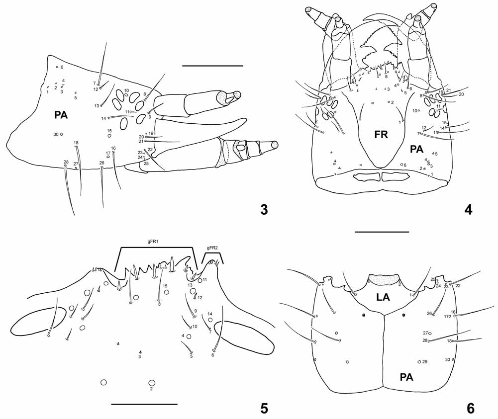
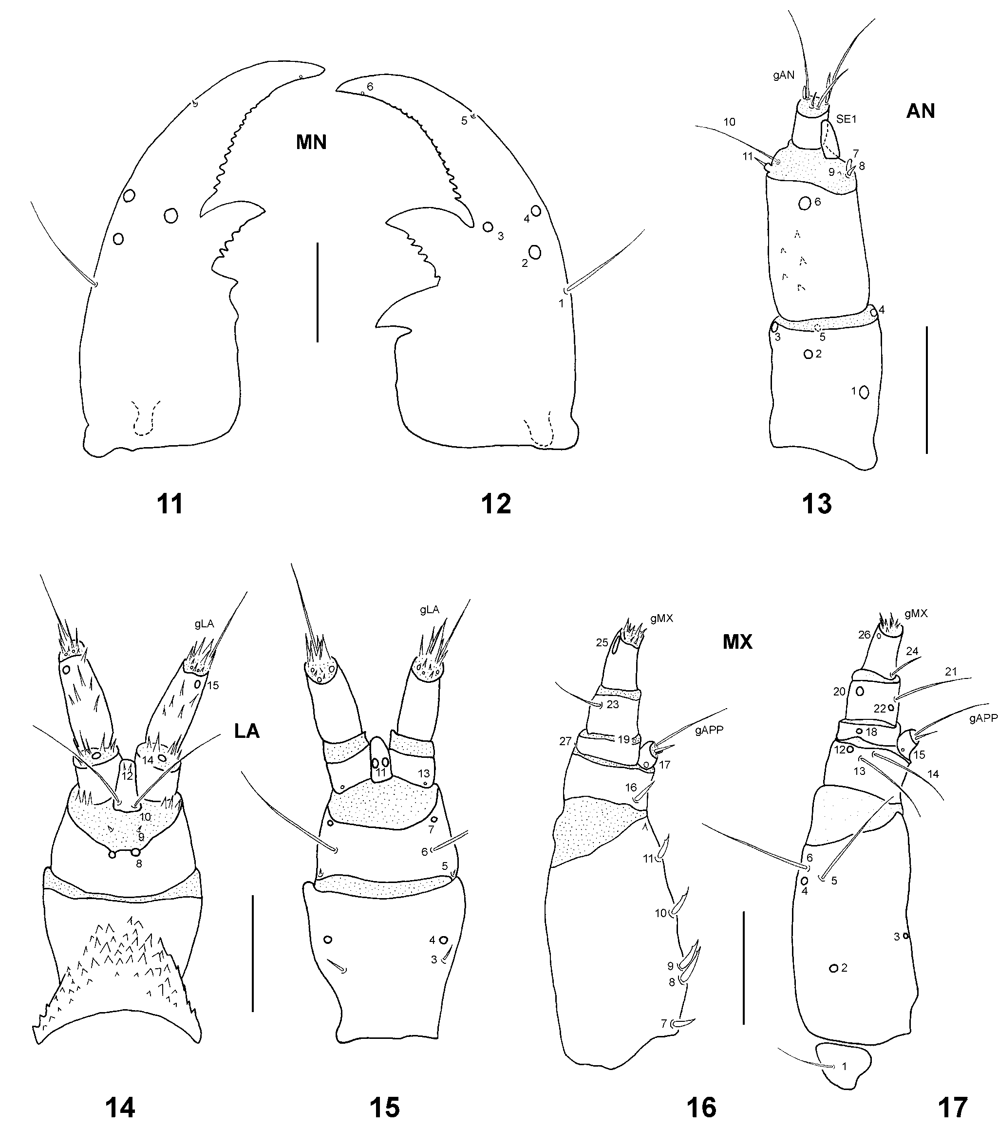
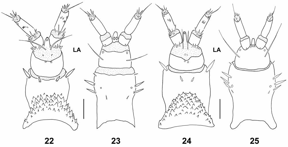
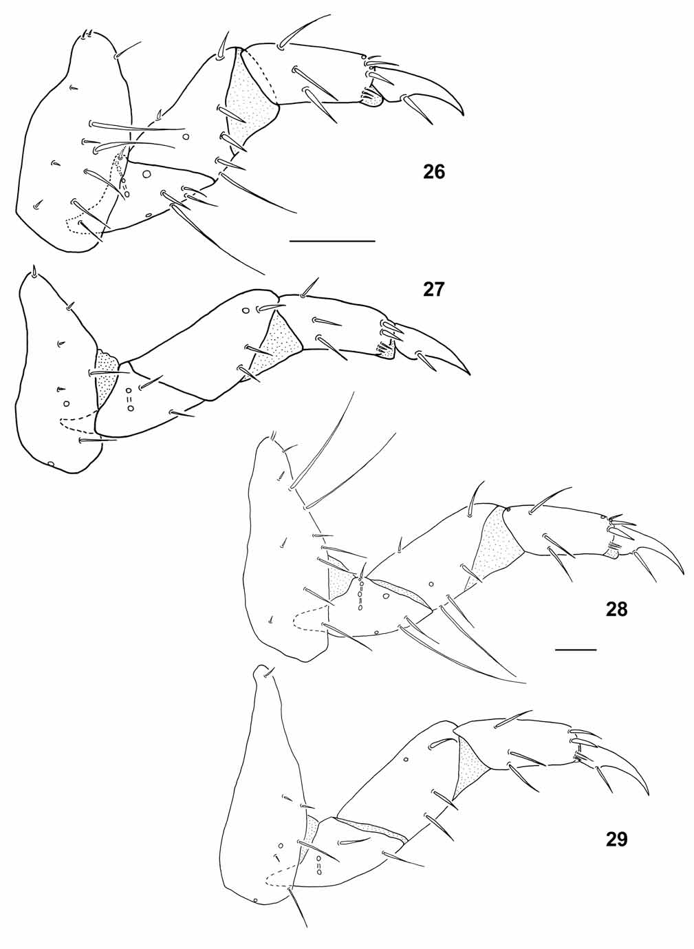


TABLE 2. (continued)
| Measure | Instar I | Instar II | Instar III | Instar I | Instar II | Instar III |
|---|---|---|---|---|---|---|
| Leg 3/Leg 1 | 0.98–1.05 | 0.95–1.04 | 0.98–1.07 | 0.91–0.96 | 0.96–0.98 | 0.62–1.03 |
| Leg 3/Leg 2 | 0.95–1.12 | 0.93–1.05 | 0.96–1.05 | 0.89–0.99 | 1.00–1.05 | 0.71–1.04 |
| Leg 1 (CO/FE) | 1.21–1.55 | 1.24–1.30 | 1.19–1.37 | 1.13–1.25 | 1.12–1.24 | 1.06–1.22 |
| Leg 2 (CO/FE) | 1.21–1.68 | 1.56–1.74 | 1.33–1.73 | 1.29–1.44 | 1.38–1.54 | 1.46–1.55 |
| Leg 3 (CO/FE) | 1.41–1.70 | 1.44–1.63 | 1.54–1.76 | 1.17–1.32 | 1.44–1.54 | 1.41–1.42 |
| Leg 1 (TITA/FE) | 0.79–0.91 | 0.69–0.81 | 0.63–0.80 | 0.77–0.83 | 0.68–0.76 | 0.65–0.75 |
| Leg 2 (TITA/FE) | 0.85–1.00 | 0.86–0.97 | 0.74–0.84 | 0.78–0.92 | 0.71–0.82 | 0.78–0.86 |
| Leg 3 (TITA/FE) | 0.86–1.03 | 0.84–0.98 | 0.78–0.88 | 0.83–0.97 | 0.82–0.92 | 0.74–0.83 |
| Leg 1 (CL/TITA) | 0.67–0.81 | 0.59–0.75 | 0.60–0.65 | 0.76–0.88 | 0.67–0.76 | 0.47–0.58 |
| Leg 2 (CL/TITA) | 0.74–0.91 | 0.74–0.82 | 0.62–0.73 | 0.91–1.10 | 0.79–0.98 | 0.60–0.75 |
| Leg 3 (CL/TITA) | 0.81–0.89 | 0.42–0.78 | 0.69–0.74 | 0.85–0.97 | 0.78–0.96 | 0.67–0.72 |
No known copyright restrictions apply. See Agosti, D., Egloff, W., 2009. Taxonomic information exchange and copyright: the Plazi approach. BMC Research Notes 2009, 2:53 for further explanation.