
Heterostomus curvipalpis Bigot, 1857
|
publication ID |
https://doi.org/10.11646/zootaxa.3616.3.4 |
|
publication LSID |
lsid:zoobank.org:pub:A4C28780-EC2C-4A86-B0C2-34A086D43FAF |
|
DOI |
https://doi.org/10.5281/zenodo.6153210 |
|
persistent identifier |
https://treatment.plazi.org/id/03CB87AC-9B18-5F1A-77CA-FDF45A6A57C6 |
|
treatment provided by |
Plazi |
|
scientific name |
Heterostomus curvipalpis Bigot, 1857 |
| status |
|
Heterostomus curvipalpis Bigot, 1857
( Figs. 1–23 View FIGURES 1 – 4 View FIGURES 5 – 8 View FIGURES 9 – 14 View FIGURES 15 – 23 )
Heterostomus curvipalpis Bigot, 1857: 285 ; Malloch, 1932: 204; Nagatomi, 1984:115; 1985: 699.
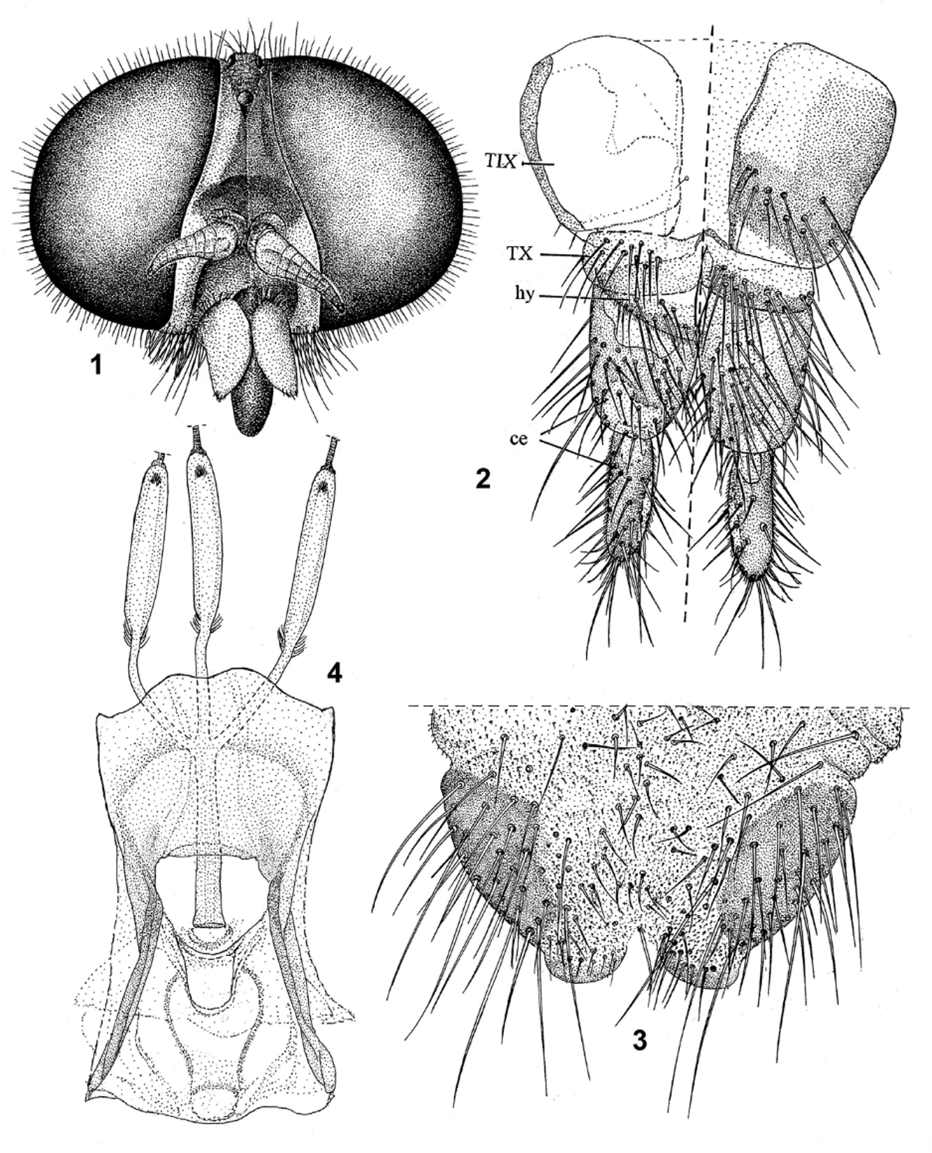
Adult ( Figs. 1–14 View FIGURES 1 – 4 View FIGURES 5 – 8 View FIGURES 9 – 14 ): Large species, wing length 13.0–18.0 mm, eyes bare; face with upper and mid lower part scarcely produced anteriorly; conspicuous ocelli; antennal flagellum with eight flagellomeres, palpus with two flattened articles, proboscis fleshy and surpassing palpus length ( Figs. 1 View FIGURES 1 – 4 , 5 View FIGURES 5 – 8 , 12, 14 View FIGURES 9 – 14 ); male holoptic. Scutum flat ( Figs. 9, 10 View FIGURES 9 – 14 ); postmesonotum and postscutellum present; metathorax with postspiracular plate. Wing elongate ( Figs. 8 View FIGURES 5 – 8 , 13 View FIGURES 9 – 14 ), vein C reaching wing apex, vein R1 setose, R2+3 anteriorly curved from medial portion and ending very close to R1 apex; basal radial cell elongate, its apex surpassing the middle of discal cell; R4 arising from anterior third of R5, which ends just at the wing apex; M1 ending posterior to the wing apex, vein M3 curved but not fusing with Cu–A1, thus cell m3 open at wing margin, CuA1 arises from the apex of basal medial cell, posterior cubital cell closed; alula scarcely developed and lower calypter well developed, with a tuft of long pubescence. Tibial spurs 1:2:2. Abdomen elongate and flattened, gradually narrowed beyond VII segment ( Fig. 11 View FIGURES 9 – 14 ).
Female genitalia: tergite IX membranous at middle, tergite X divided; cerci with two segments ( Fig. 2 View FIGURES 1 – 4 ), with a few developed subapical sensorial pits on internal side; genital fork wide basally, with thick spermathecal ducts at base ( Fig. 4 View FIGURES 1 – 4 ).
Male genitalia ( Fig. 6 View FIGURES 5 – 8 ): gonocoxites fused dorsally, gonostylus subtriangular, short and externally curved; parameral sheath surrounding the aedeagus; tip of the aedeagus well developed; ejaculatory apodeme extended posteriorly by endoaedeagal guide, lateral aedeagal apodeme large; spermatic pump subcylindric and enlarged; gonocoxal apodemes relatively thick and greatly projected anteriorly. Tergites IX and X fused ( Fig.7 View FIGURES 5 – 8 ) (= epandrium), cerci subquadrangular; sternite IX expanded (= hypoproct).
Pupa (female, emerged in the laboratory) ( Figs. 15–23 View FIGURES 15 – 23 ): Exuvial coloration reddish brown, uniformly glossy, slightly darker on thoracic and abdominal spiracles, and with blackish spines forming abdominal rings on each segment. Length 30.0 mm.
Frontal and cephalic sclerites unknown (lost during the emergence). Thorax dorsally with transverse sulcus ( Fig. 16, 17 View FIGURES 15 – 23 ) on anterior 3/4 and small lateral rugosities (wrinkled areas). Mesothoracic spiracle prominences slightly elevated (0.8 mm) ( Figs. 16, 17, 18, 19 View FIGURES 15 – 23 ). Spiracle slit placed transversely on the apex of the prominence, is arched and elongated, showing curved like a semicircle on both extremes and without projection. Wing and leg sheaths of equal length, extended surpassing a little the first abdominal segment border. Mesonotum with three separated spines on each side: one anterolateral, one mediodorsal and one submediandorsal. Abdominal spines appear seta-like,stout at base and thinly prolonged distally. First abdominal tergum with 9+9 stout spines approximately equidistant, five of them more dorsal in position; the following four spines on pleura are closer to each other. Subsequent abdominal segments II–VII with two uniseriate and continuous rows of spines and without setae, and delimiting, in combination with the intersegmental sutures, three annular stripes, all around each segment and of approximately same length ( Figs. 15, 16, 20, 21 View FIGURES 15 – 23 ). Middle stripes between spine rings smoother and glossier. First rows of spines smaller than the second, and absent ventrally between the respiratory spiracles area. The second rows of spines are complete around the ring segment; the spines are longer dorsally and in the VII segment some grouped forming a spur ( Fig. 22 View FIGURES 15 – 23 ). Segments II –VII with the first rows of spines with 46, 52, 50, 62, 50, 38 spines per segment, respectively; and the second rows of spine with 86, 96, 106, 94, 64, 50 spines per segment, respectively. Anal segment without spines, but with small rugose anterior stripe-like area in the preceding apical segments. VIII segment with eight large wrinkled and well sclerotized tubercles, forming the aster, arranged as following: 1+1 dorsal (1.2 mm), 2+2 lateral (1.1 mm), and ventral (0.7 mm); dorsally there is a comb of 11–12 small spines ( Fig. 23 View FIGURES 15 – 23 ), and internally on base of lateral spines, there are another two small acuminate tubercles ( Figs. 22, 23 View FIGURES 15 – 23 ). Seven subconical abdominal respiratory spiracles with similar shape (0.36 mm) ( Fig. 15 View FIGURES 15 – 23 ), and positioned lateroanteriorly on segments I–VII ( Figs. 15–16 View FIGURES 15 – 23 ).


No known copyright restrictions apply. See Agosti, D., Egloff, W., 2009. Taxonomic information exchange and copyright: the Plazi approach. BMC Research Notes 2009, 2:53 for further explanation.
|
Kingdom |
|
|
Phylum |
|
|
Class |
|
|
Order |
|
|
Family |
|
|
Genus |