
Hoplopholcus asiaeminoris Brignoli, 1978
|
publication ID |
https://doi.org/10.11646/zootaxa.4726.1.1 |
|
publication LSID |
lsid:zoobank.org:pub:F0F95E18-9EFB-4169-B724-DAA71200413A |
|
persistent identifier |
https://treatment.plazi.org/id/03CB87CD-FFDF-FF8A-E9C0-FF2D7164F827 |
|
treatment provided by |
Plazi |
|
scientific name |
Hoplopholcus asiaeminoris Brignoli, 1978 |
| status |
|
Hoplopholcus asiaeminoris Brignoli, 1978 View in CoL
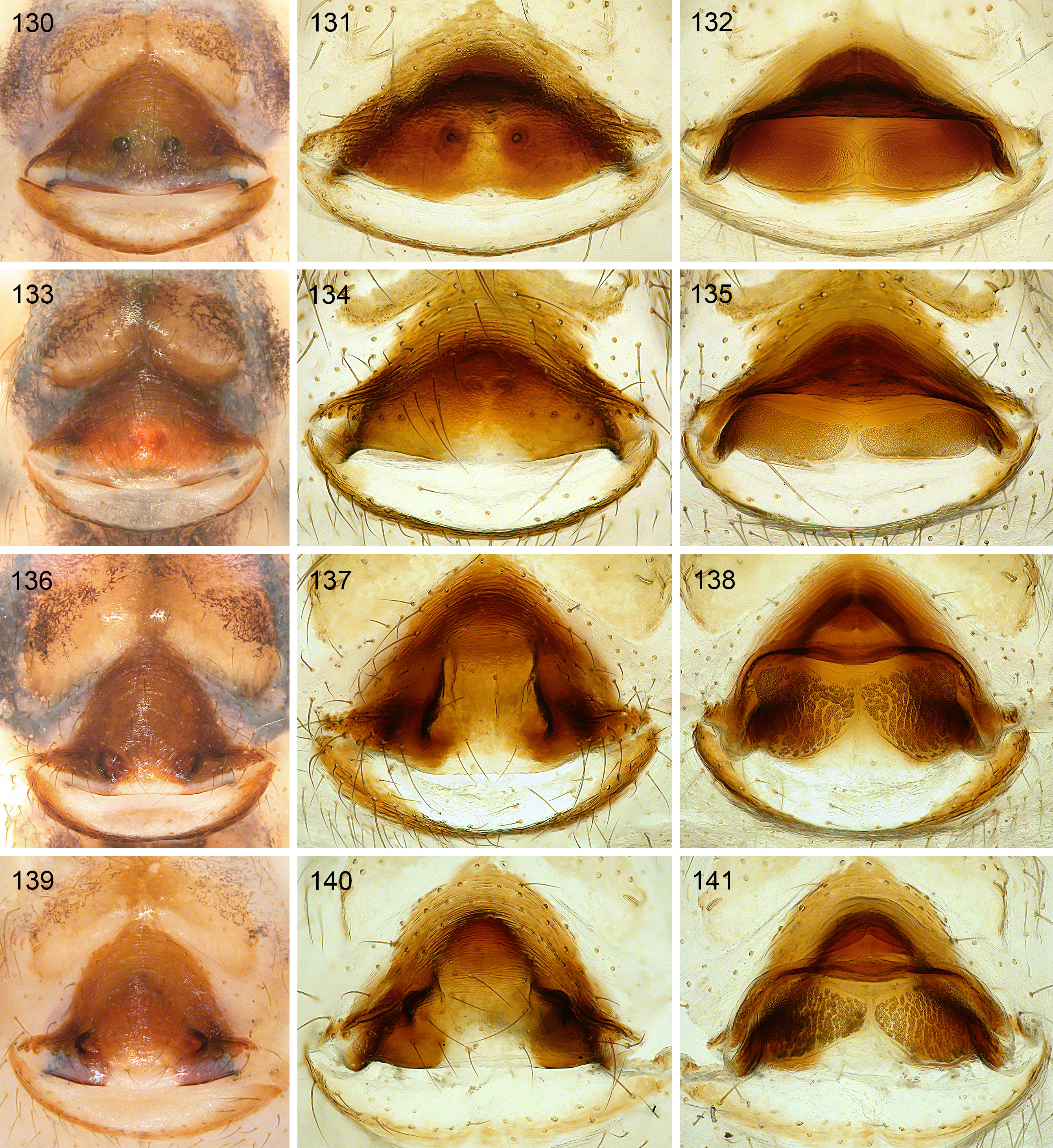
Figs 118–120 View FIGURES 118–123 , 124–129 View FIGURES 124–129 , 136–151 View FIGURES 130–141 View FIGURES 142–151
Hoplopholcus asiaeminoris Brignoli, 1978: 490 View in CoL , figs 43–49 ( ♂ ♀; only part of the specimens listed; at least fig. 46 probably represents different species; see Notes below). Topçu et al. 2005b: 289 (see Notes below). Eberle et al. 2018 (molecular data). Huber et al. 2018: fig. 6.
Diagnosis. Distinguished from known congeners by shapes of procursus and bulbal processes ( Figs 142–143, 146– 149 View FIGURES 142–151 ): distinctive weakly sclerotized obtuse process retrolaterally (arrow in Fig. 143 View FIGURES 142–151 ), ventral spine of procursus curved toward ventrally at half length, strong prolateral process, small prolateral ridge; ventral bulbal sclerite slen- der in lateral view ( Fig. 147 View FIGURES 142–151 ); tip of embolar sclerite with small cone-shaped processes (arrow in Fig. 148 View FIGURES 142–151 ). Females are difficult to distinguish externally from congeners; internal ventral arc strongly curved ( Figs 137, 140 View FIGURES 130–141 , 150 View FIGURES 142–151 ), lateral pouches of uterus externus long, with sclerotized lateral margins and visible in uncleared specimens ( Figs 136, 139 View FIGURES 130–141 ); median pouch of uterus externus large but membranous and not visible in uncleared specimens.
Type material. TURKEY, Konya: ♂ holotype , 3♂ 1♀ paratypes, MCVR, Hadim, Grotta Su Ciktigi (= Su Çıktığı Mağarası , 36.978°N, 32.401°E, 1770 m a.s.l.), 25.iv.1973 ( V. Sbordoni), examined GoogleMaps ; 6♂ paratypes, MCVR, same locality and date, leg. S. Forestiero GoogleMaps ; 4♂ 2♀ paratypes, MCVR, same locality and date, leg. P. Agnoletti; all examined GoogleMaps .
Other material examined. TURKEY, Konya: 1♂ 1♀ abdomen, ZFMK ( Ar 20917), and 2♀ in pure ethanol (one abdomen transferred to ZFMK Ar 20917), ZFMK ( Tur 48), Hadim District , Su Çıktığı Mağarası ( 36.978°N, 32.401°E, 1770 m a.s.l.), among rocks near cave, 24.vii.2016 ( H. Öztürk) GoogleMaps . 4♂ 1♀, ZFMK ( Ar 20918), and 4♀ in pure ethanol, ZFMK ( Tur 49), Hadim District, Göksu Şelalesi ( 37.029°N, 32.700°E, 790 m a.s.l.), among rocks be- low waterfalls, 25.vii.2016 ( H. Öztürk) GoogleMaps .
Karaman: 1♂ 1♀, ZFMK ( Ar 20919), and 1♂ in pure ethanol, ZFMK ( Tur 51), Ermenek District, Ermenek ( 36.642°N, 32.893°E, 1380 m a.s.l.), in small cave above Maraspoli Mağarası, 26.vii.2016 ( H. Öztürk) GoogleMaps . 2♂ 4♀ 6 juvs, ZFMK ( Ar 21304), Mut District, Sertavul Pass ( 36.897°N, 33.269°E), 1500 m a.s.l., juniper forest on steep slope, under stones, 19–20.ix.2010 ( Y.M. Marusik) GoogleMaps .
Mersin: 2♂ 2♀, ZFMK ( Ar 20920), and 2♀ in pure ethanol, ZFMK ( Tur 52), Silifke District, Cennet Çöküğü ( 36.452°N, 34.105°E, 100 m a.s.l.), among rocks at cave entrance in sinkhole, 27.vii.2016 ( H. Öztürk) GoogleMaps . 1♂ 2♀, ZFMK ( Ar 20921), and 2♀ in pure ethanol, ZFMK ( Tur 53), Silifke District, Aşağı Dünya Obruğu (= Akhayat sink- hole) ( 36.448°N, 34.010°E, 450 m a.s.l.), under rocks at base of sinkhole, 27.vii.2016 ( H. Öztürk) GoogleMaps .
Notes. Brignoli (1978) gives “Grotta Su Cikkigi” as type locality; the correct name is Grotta Su Çiktiği (this is also how it is written on three of the four labels accompanying the topotypes), or Su Çıktığı Mağarası.
In addition to the paratypes from the type locality, Brignoli (1978) listed paratypes from six further localities. I have seen all these paratypes. Most paratypes other than those from the type locality are here considered to represent a different species (see H. dim below).
It is not clear from which specimens Brignoli (1978) produced the illustrations in the original description. The strongly curved ventral spine in his fig. 46 suggests that at least this drawing was made from a paratype specimen that is now considered to represent H. dim .
I have not seen the material cited in Topçu et al. (2005b). It is said to originate from Belemedik and Sarıışık. The coordinates of Belemedik in Adana Province [ 37.36°N, 34.915°E] do not agree with the dot in the map of Topçu et al. (2005b: fig. 1); Sarıışık in Mersin Province, according to Seyyar et al. (2008), is at 37.2°N, 34.8°E. Judging from the known distributions of Hoplopholcus species in Turkey, the identifications could be correct; both localities are thus shown in the map ( Fig. 442 View FIGURE 442 ).
Redescription. Male ( paratype from type locality). MEASUREMENTS. Total length 6.5, carapace width 2.2. Distance PME-PME 150 µm; diameter PME 160 µm; distance PME-ALE 40 µm; diameter AME 100 µm; distance AME-AME 30 µm. Leg 1: 49.3 (13.7 + 0.9 + 13.6 + 18.4 + 2.7), tibia 2: 10.0, tibia 3: 7.6, tibia 4: 9.1; tibia 1 L/d: 54.
COLOR (in ethanol). Carapace ochre-yellow, ocular area slightly darker; clypeus ochre-yellow; sternum light brown with slightly darker margins and radial marks; legs ochre-yellow, with barely visible darker rings subdistally on femora and tibiae; abdomen pale gray, with pattern of black internal marks dorsally in posterior half, ventrally light brown genital area and black mark in front of spinnerets.
BODY. Habitus as in Fig. 120 View FIGURES 118–123 . Ocular area slightly elevated. Deep thoracic pit and indistinct pair of furrows diverging from pit toward posterior margin. Clypeus unmodified. Sternum wider than long (1.6/1.2), unmodified. Abdomen oval, dorso-posteriorly rounded.
CHELICERAE. As in Figs 144–145 View FIGURES 142–151 , with pair of latero-distal apophyses provided with two modified coneshaped hairs each; with fine stridulatory ridges (barely visible in dissecting microscope).
PALPS. As in Figs 124–126 View FIGURES 124–129 ; coxa with low retrolateral hump, trochanter barely protruding ventrally, femur with transversal dark line retrolaterally and distinct prolateral stridulatory pick; procursus ( Figs 142–143 View FIGURES 142–151 ) with distinct ventral ‘knee’, ventral spine long and curved toward ventrally at half length, strong pointed prolateral process, small prolateral ridge, distinctive weakly sclerotized obtuse process retrolaterally (arrow in Fig. 143 View FIGURES 142–151 ); genital bulb ( Figs 146–149 View FIGURES 142–151 ) with ventral sclerite slender in lateral view; tip of embolar sclerite with small cone-shaped processes (arrow in Fig. 148 View FIGURES 142–151 ); with distinct dorsal membranous process ( Fig. 147 View FIGURES 142–151 ).
LEGS. Femora 1 and 2 with single rows of ventral spines (femur 1: ~33–35; femur 2: ~18–20); with few weakly curved hairs on metatarsus 1; few vertical hairs; retrolateral trichobothrium of tibia 1 at 5%; prolateral trichobothrium present on all leg tibiae; tarsi without distinct pseudosegments but with many small platelets.
Male (variation). Tibia 1 in 23 other males: 11.1–19.9 (mean 13.7). Most males with two modified hairs on each cheliceral apophysis (2+2), two males symmetric 3+3, and three males asymmetric 2+3. Some specimens with dark radial marks on carapace. Some specimens with dark rings on legs distinct (also patellae dark). Males (and females) from Mersin Province with dark brown sternum. Dorsal abdominal marks variably distinct, often with distinct pair of round marks at about half length. Number of spines on femora slightly variable.
Female. In general similar to male but without spines on legs; habitus as in Figs 118–119 View FIGURES 118–123 . Most females with (few) curved hairs on metatarsi 1 and 2, rarely also tibiae 1 and 2. Tibia 1 in ten females: 7.5–13.7 (mean 10.8).
FEMALE GENITALIA. Epigynum as in Figs 127 View FIGURES 124–129 , 136, 139 View FIGURES 130–141 , internal ventral arc strongly curved and visible in uncleared specimens, pair of lateral pouches of uterus externus also usually visible in uncleared specimens; with pair of light brown weakly bulging areas in front of epigynum; posterior plate short but wide. Internal genitalia with large almost contiguous pore plates ( Figs 129 View FIGURES 124–129 , 138, 141 View FIGURES 130–141 , 151 View FIGURES 142–151 ); ventral arc strongly curved; lateral pouches of uterus externus with sclerotized lateral margins, distance between pouches variable (~ 370–440 µm), sclerotized lateral margins varying from almost parallel to strongly diverging posteriorly (compare Figs 137, 140 View FIGURES 130–141 ); median pouch of uterus externus large but membranous.
Distribution. Known from several localities in Konya, Karaman, Mersin, and Adana Provinces, Turkey ( Fig. 442 View FIGURE 442 ).
Natural history. At the type locality, no specimens (not even webs) were seen in the cave in July 2016, but the species was found under rocks and in vertical rock pits in the surrounding area. At Göksu Şelalesi the species was very abundant under large rocks below the waterfall, in large webs with a diameter of ~ 40 cm. At Cennet Çöküğü, the spiders were abundant in the twilight zone but absent from deeper parts of the cave. At disturbance, the spiders quickly retreated toward the back into holes and crevices.


| ZFMK |
Zoologisches Forschungsmuseum Alexander Koenig |
No known copyright restrictions apply. See Agosti, D., Egloff, W., 2009. Taxonomic information exchange and copyright: the Plazi approach. BMC Research Notes 2009, 2:53 for further explanation.
|
Kingdom |
|
|
Phylum |
|
|
Class |
|
|
Order |
|
|
Family |
|
|
Genus |
Hoplopholcus asiaeminoris Brignoli, 1978
| Huber, Bernhard A. 2020 |
Hoplopholcus asiaeminoris
| Topcu, A. & Demir, H. & Seyyar, O. & Turkes, T. 2005: 289 |
| Brignoli, P. M. 1978: 490 |