
Yola matsikammae, Bilton, David T., 2015
|
publication ID |
https://doi.org/ 10.11646/zootaxa.3905.3.10 |
|
publication LSID |
lsid:zoobank.org:pub:5A9DBB7C-BCAD-40B2-AFCD-0CFF2D4F643 |
|
DOI |
https://doi.org/10.5281/zenodo.6093402 |
|
persistent identifier |
https://treatment.plazi.org/id/03CBD108-FFA3-E117-96C9-02D38FD7FB33 |
|
treatment provided by |
Plazi |
|
scientific name |
Yola matsikammae |
| status |
sp. nov. |
Yola matsikammae View in CoL sp. nov.
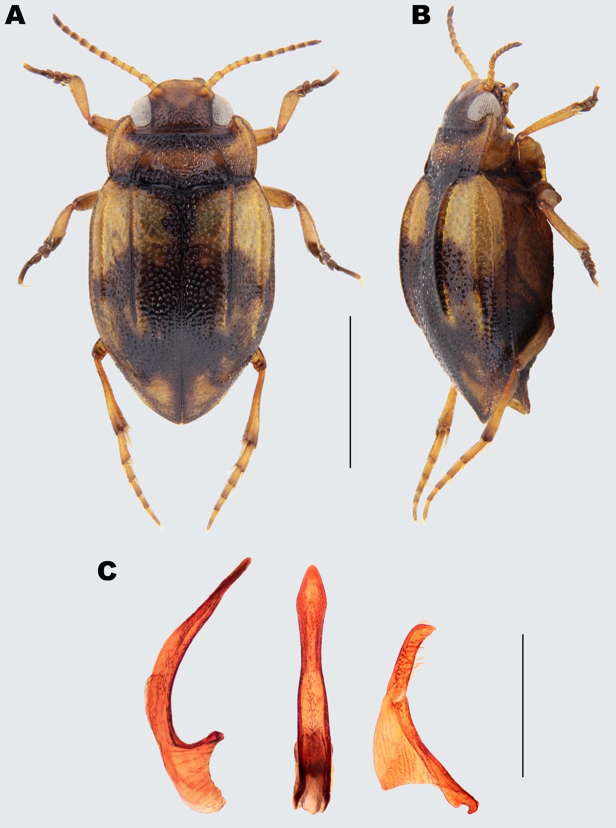
( Figs 1 View FIGURE 1 and 2 View FIGURE 2 )
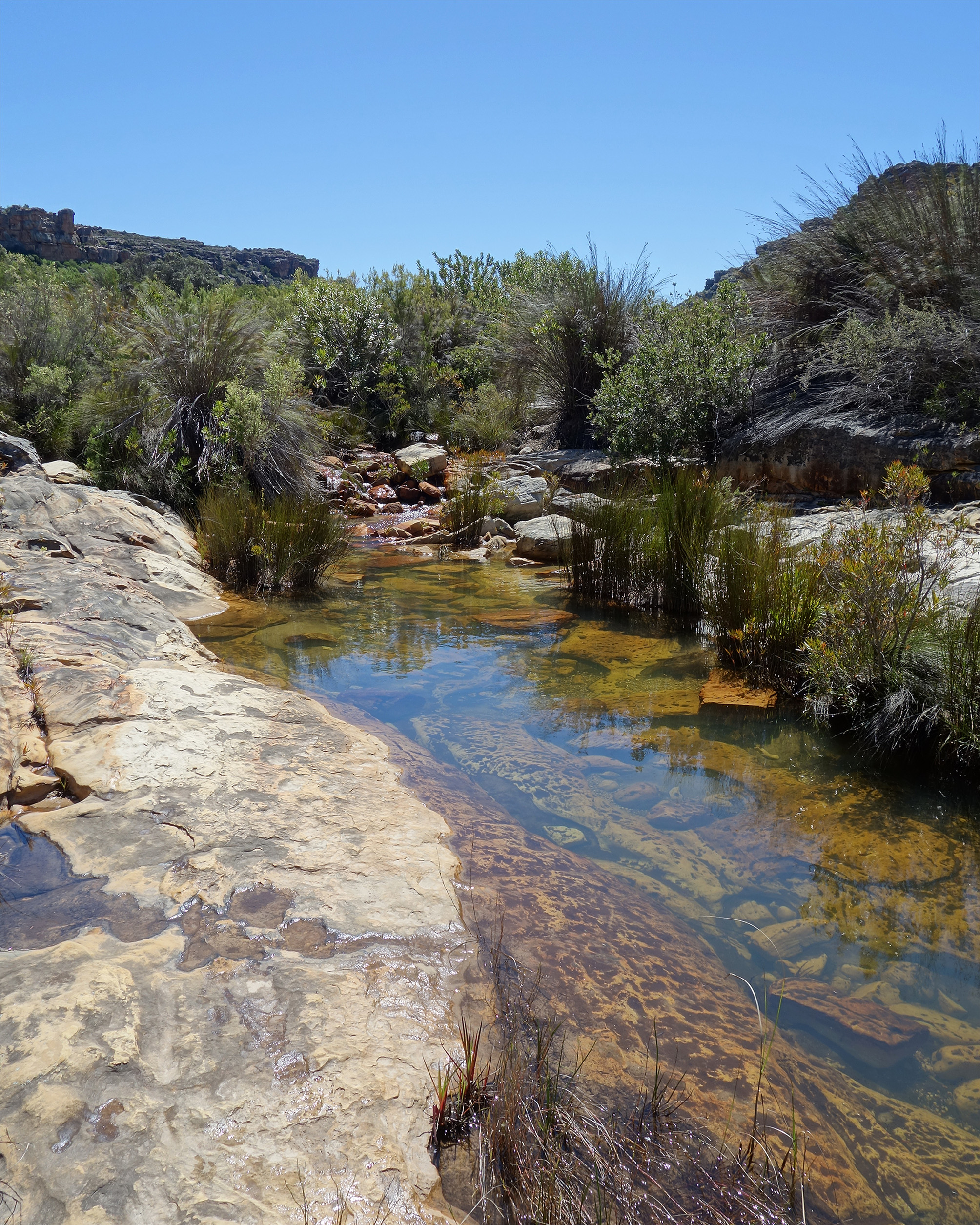
Type locality. South Africa, Western Cape, Matsikammaberg, permanent stream ca. 1 km SE of Sewefontein Farm, 31°41'48.08''S 18°49'48.43''E, 617 m. ( Fig. 2 View FIGURE 2 ).
Type material. Holotype (male): “ 22/ix/2014 South Africa WC// Matsikammaberg stream 1 km // SE of Sewefontein Farm// permanent D T Bilton leg.” (genitalia extracted and mounted in DMHF on same card) with red printed holotype label “ Holotype // Yola matsikammae sp. nov. // Bilton” ( ISAM).
Paratypes: 3♂, 9♀ same data as holotype ( CDTB, DMSA, ISAM, NMW, SANC, OUMNH). All with red printed paratype labels “ Paratype // Yola matsikammae sp. nov. // Bilton”.
Description. Size: Holotype: body length (to elytral apices) 2.3 mm; maximum width (elytra) 1.25 mm; elytral length 1.65 mm. Same values for paratypes: 2.3–2.45 mm, 1.3–1.4 mm and 1.6–1.85 mm respectively.
Colour ( Fig. 1 View FIGURE 1 ): Dorsal surface predominantly yellowish brown, straw coloured. Head and pronotum darker than ground colour of elytra. Frons darker than clypeus; infuscated along inner border of eyes. Front and hind margins of pronotum with a black band; anterior band present over central 4/5 of width, between projections of anterior angles. Posterior band more well-developed between lateral plicae, here extending for approximately 1/5 of pronotal length. Elytra with diffuse dark markings, boundaries rather ill-defined. Anterior margins with narrow dark band, laterally almost reaching shoulders. Suture dark to apex, with diffuse dark markings expanded laterally from suture; interrupted in centre and reaching lateral keel anteriorly and elytral margin posteriorly ( Fig. 1 View FIGURE 1 B). Maxillary palpi pale straw yellow, last segment infuscated. Fore and mid legs straw yellow with femuro-tibial junctions and tarsi infuscated. Hind legs same but femuro-tibial and tibio-tarsal junctions infuscated, along with apical 1/3–1/2 of tarsal segments. Antennae with segments 1–5 pale yellow, 6–10 with apices infuscated (more so in later segments) and segment 11 infuscated throughout. Venter reddish brown, abdomen dark pitchy brown, particularly towards lateral margins and apex.
Head: Broad, with large eyes. Clypeus somewhat thickened and evenly rounded, lacking anterior border, and not separated from rest of head. Head raised in front of eye, above antennal insertion, into a distinct, low tubercle. Frons slightly elevated in centre. Cervical line well marked. Head in front of cervical line with coarse, often confluent punctures, punctures particularly large mediodistally on frons; many punctures bearing small, peg-like setae. Anterior half of frons and clypeus with smaller sparse punctures. Centre of frons and clypeus shining, lacking microreticulation. Areas immediately inside eyes with shallow, isodiametric microreticulation, particularly anteriorly and around frontal tubercles, giving a duller appearance. Anterior margin of clypeus shining, but with traces of transverse microreticulation. Head behind cervical line duller, with strong transverse microreticulation.
Pronotum: Transverse rectangular. Sides rounded; broadest just before middle. Hind margins distinctly bisinuate around middle. Lateral plicae well developed, relatively deep, and open interiorly. Plicae deepest at posterior ends; sinuate and becoming shallow anteriorly, here extending to approximately 1/3 away from front margin of pronotum. Posterior 1/3 of disc between plicae depressed, especially in centre, surface here raised abruptly to level of remainder of disc. Pronotal surface shining, lacking microreticulation, but roughened due to dense, coarse punctation. Punctures largest and closest towards centre, smaller towards lateral margins. Punctures bearing flattened, white recumbent setae in centre; peg-like towards posterior and lateral margins.
Elytra: Together forming an elongate oval, with point of maximum width just behind middle. Lateral margin rounded to front angles, parallel over central 1/3, and tapered towards apex in posterior 1/2. Each elytron with three keels. Discal keel very strong, gradually elevated both anteriorly and posteriorly and of even height over central 4/ 5, occupying ca. 2/3 of elytron ( Fig. 1 View FIGURE 1 B). Median keel lower, running from shoulder to just in front of posterior end of discal keel. Lateral keel slightly lower still, same length as median keel. Elytra shining, lacking
microreticulation. Punctate, punctures close, coarse and shallow anteriorly, becoming finer and sparser posteriorly. Punctures almost confluent in anterior 1/2, between discal and medial keels. Each puncture bearing a flattened decumbent seta.
Venter: Mentum and prementum smooth and shining, lacking microreticulation. Anterior margin of prementum with short, stout bristles. Anterior and lateral margins of mentum thickened and somewhat darkened. Gula and genae shining, lacking microreticulation. Gular sutures weak; gula with scattered shallow medium punctures. Subgenal ridges well-marked, area between ridge and eye with open, isodiametric microreticulation. Pronotal hypomera smooth and shining, with scattered shallow punctures bearing long golden setae, and traces of transverse wrinkles posteriorly. Proepisternum shining, minutely wrinkled, especially posteriorly. Prosternum shining, strongly punctate, especially in centre where punctures are deeper and almost confluent in places. Punctures in centre of prosternum with long, fine golden setae. Prosternal process raised, neck of process making an angle of approx. 70° with prosternum. Process lanceolate, with broad, shallow central groove furnished with dense, coarse, punctures bearing long, decumbent setae. Neck of process with sparse small punctures bearing hairlike, golden setae. Prosternal process meeting anterior projection of metaventrite. Metaventrite and metathoracic anepisternum shining, with sparse, shallow punctures; traces of microreticulation visible towards centre of metaventrite. Punctures somewhat larger on metaventrite and deeper anteriorly, each bearing a short, flattened, decumbent seta. Metacoxa shining, with large, shallow, almost confluent punctures bearing short, flattened, decumbent setae. Elytral epipleura shining, with close, coarse, setose punctures which are almost confluent in posterior 1/2. Metacoxal lines well marked, subparallel, extending forwards as lines onto metaventrites, these converging towards its anterior margin. Metacoxal processes with shallow open central channel extending forwards onto metaventrite; foveate at junction of metacoxae and metaventrite. Processes punctate, with coarse, shallow punctures, each bearing a short, flat, recumbent seta. Apex of metacoxal processes fused to abdomen; sinuate, lobes weakly developed and not covering leg insertions. Abdominal ventrites 2–4 fused, junctions weakly visible. Abdominal ventrites shining, 2–4 densely punctate; punctures smaller and more distinct in centre, larger and more confluent laterally; bearing flattened, decumbent setae. Abdominal ventrites 5–7 with smaller, sparser punctures, each bearing a golden recumbent seta, longer than those on preceding ventrites. Abdominal ventrites 5–6 with central tuft of long, golden, hair-like setae, extending almost to abdominal apex.
Aedeagus: Median lobe characteristically shaped ( Fig. 1 View FIGURE 1 C); apex flattened and spathulate in ventral view. Parameres ( Fig. 1 View FIGURE 1 C) two-segmented, with bluntly curved, hooked tips.
Females: As males except head, pronotum and elytra entirely microreticulate between punctures, giving a duller appearance. Venter duller, with fine microreticulation throughout. Venter paler than in male, yellowish brown throughout except for neck of prosternal process and segmental junctions, which are slightly infuscated. Fore and mid tarsi slightly narrower and noticeably shorter than in males, particularly last tarsal segment.
Variability. Paratypes vary slightly in size (see above) and the development of the elytral pattern, some specimens being darker than the holotype.
Differential diagnosis and key to southern African members of the Yola bicarinata group. The new species is apparently a member of the bicarinata species group, sensu Biström (1983), having three distinct elytral keels with discal keels that descend gradually to the elytral surface ( Fig. 1 View FIGURE 1 B). Within this group Y. matsikammae sp. nov. would key to the Palaearctic Y. bicarinata (Latreille, 1804) using Biström (1983), which at 1.8–2.1 mm is smaller than the new species, as well as being more strongly rounded, and differing in genital anatomy (median lobe of aedeagus pointed and needle-like in ventral view in Y. bicarinata— see Biström, 1983). The new species can be distinguished from other southern African members of the bicarinata group as follows (for detailed descriptions and illustrations of previously described species see Biström 1983):
1 Median keel of elytra relatively low, equal in height to lateral keel; both keels indistinct. Median lobe of aedeagus with elongate, parallel-sided, needle-like apical projection in ventral view. Total length 1.8–2.0 mm. ... Yola subopaca Régimbart, 1895 View in CoL
- Median elytral keel more strongly elevated than lateral keel. Both keels distinct.................................... 2
2 Median lobe of aedeagus in lateral view with apex distinctly curved in a ventral direction. All three elytral keels distinct; discal keel higher than median keel, lateral keel lower than median. Total length 1.8–2.0 mm....... Yola dilatata Régimbart, 1906 View in CoL
- Median lobe of aedeagus straight, or curved dorsally in lateral view.............................................. 3
3 Median elytral keel only slightly more strongly elevated than lateral keel. Pronotum broadest at posterior corners, or just anterior to them. Pronotal disc flat between plicae. Median lobe of aedeagus in lateral view relatively broad almost until apex; apex at most slightly expanded and triangular in ventral view. Size smaller, total length 1.9–2.3 mm; maximum width 1.1–1.4 mm ............................................................................. Yola frontalis Régimbart, 1906 View in CoL
- Median elytral keel distinctly more strongly elevated than lateral keel. Pronotum broadest just before middle. Posterior 1/3 of pronotal disc depressed between plicae, especially in centre; surface here raised abruptly to level of remainder of disc ( Fig. 1 View FIGURE 1 A). Median lobe of aedeagus ( Fig. 1 View FIGURE 1 C) in lateral view narrowing approx. 1/6 before apex, then parallel-sided to apex; apex strongly expanded, spathulate, in ventral view. Size larger, total length 2.3–2.45 mm; maximum width 1.25–1.4 mm ....................................................................................... Yola matsikammae View in CoL sp. nov.
Superficially, in both shape and dorsal colouration, the new taxon resembles species of Sharphydrus View in CoL Omer- Cooper, 1958, especially S. brincki View in CoL , with which it was microsympatric (i.e. co-occurred in the same microhabitat). It can readily be distinguished from S. brincki View in CoL by the presence of three elytral keels rather than just the discal one. The discal keel of Y. matsikammae View in CoL sp. nov. is also much stronger, the dorsal surface shinier, and the dorsal and ventral punctures larger than in S. brincki View in CoL . In addition, the median lobe of the aedeagus lacks the tripartite apical structure characteristic of known Sharphydrus View in CoL species (see Bilton 2013). As discussed by Biström (1988) and Bilton (2013), the distinction between Yola View in CoL and Sharphydrus View in CoL is not well-defined. Bilton (2013) suggested that the tripartite structure of the median lobe of the aedeagus represented a unique synapomorphy of this group, although the inter-relationships between Sharphydrus View in CoL and Yola View in CoL , including the new species, require further exploration.
Distribution. To date known from the type locality ( Fig. 2 View FIGURE 2 ), a permanent stream surrounded by Bokkeveld Sandstone Fynbos (sensu Mucina & Rutherford 2006) on the Matsikammaberg in the northern part of the Western Cape province of South Africa. The Matsikammaberg is a striking inselberg, reaching just over 1,000 m in altitude, with 700 m high sandstone cliffs towering over the dry Knersvlakte plains of Namaqualand. The mountain forms a mesic island in an otherwise semi-arid landscape, annual rainfall reaching 550 mm in the east contrasting with as little as 50 mm per year on the plains below. The Matsikammaberg is consequently home to a diverse flora, 10% of which is regionally, and 4% locally endemic ( Helme 2004).
Etymology. Named after the Matsikammaberg, on which the type locality is situated. Matsikamma is a Khoi- San word which translates as “full of pools”, in apparent reference to the abundance of seasonal rock pools on the mountain plateau. The specific epithet is a noun in the genitive case.
Ecology. Specimens of Y. matsikammae sp. nov. were taken from the margins of deep (ca. 40–50 cm), clear pools in a permanent stream section, over sand and gravel. Here they co-occurred with a range of water beetles, including Hydaticus dregei Aubé, 1838 , Sharphydrus brincki , Canthyporus consuetus Omer-Cooper, 1965 , C. lateralis (Boheman, 1848) , C. nebulosus Omer-Cooper, 1965 , Hydropeplus montanus Omer-Cooper, 1965 , H. trimaculatus (Laporte, 1835) and Laccophilus sp. Other beetles taken in the same stream reach, but in other microhabitats, were Delevea bertrandi Reichart, 1976 , Aulonogyrus capensis (Thunberg, 1781) , A. formosus (Modeer, 1776) , A. marginatus (Aubé, 1838) , Dineutus punctatus Aubé, 1838 , Copelatus capensis Sharp, 1882 , Anacaena glabriventris Komarek, 2004 , A. reducta Komarek, 2004 , Crenitis zimmermanni Knisch, 1924 , Helochares sp., Mesoceration umbrosum Perkins, 2008, Mesoceration sp. nov. 1, Mesoceration sp. nov. 2, Prosthetops wolfbergensis Bilton, 2013 , Pneuminion endroedyi Perkins, 2004 , Pneuminion velamen Perkins, 1997 , Discozantaena genuvela Perkins & Balfour-Browne, 1994 , Elpidelmis capensis (Grouvelle, 1890) and Strina sp.
No known copyright restrictions apply. See Agosti, D., Egloff, W., 2009. Taxonomic information exchange and copyright: the Plazi approach. BMC Research Notes 2009, 2:53 for further explanation.
|
Kingdom |
|
|
Phylum |
|
|
Class |
|
|
Order |
|
|
Family |
|
|
Genus |