
Neonesidea kamiyai, Horikoshi & Nakao & Tsukagoshi, 2019
|
publication ID |
https://doi.org/ 10.11646/zootaxa.4679.3.2 |
|
publication LSID |
lsid:zoobank.org:pub:13EF227C-33FF-4E10-B863-FA2623F36198 |
|
persistent identifier |
https://treatment.plazi.org/id/03CC9E2F-FF83-DC0A-C8C9-48AEFE8ACBA0 |
|
treatment provided by |
Plazi |
|
scientific name |
Neonesidea kamiyai |
| status |
sp. nov. |
Neonesidea kamiyai View in CoL sp. nov.
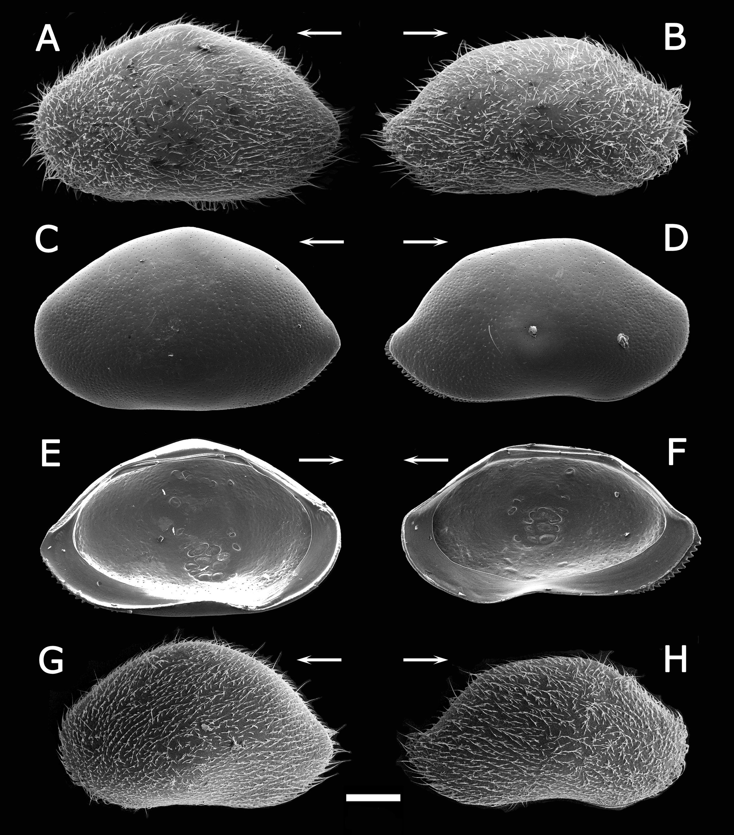
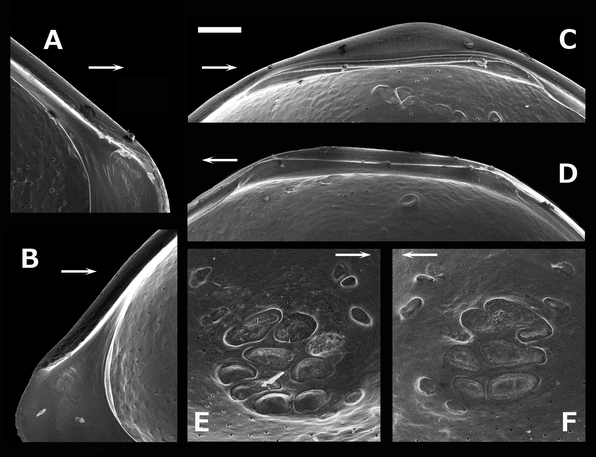
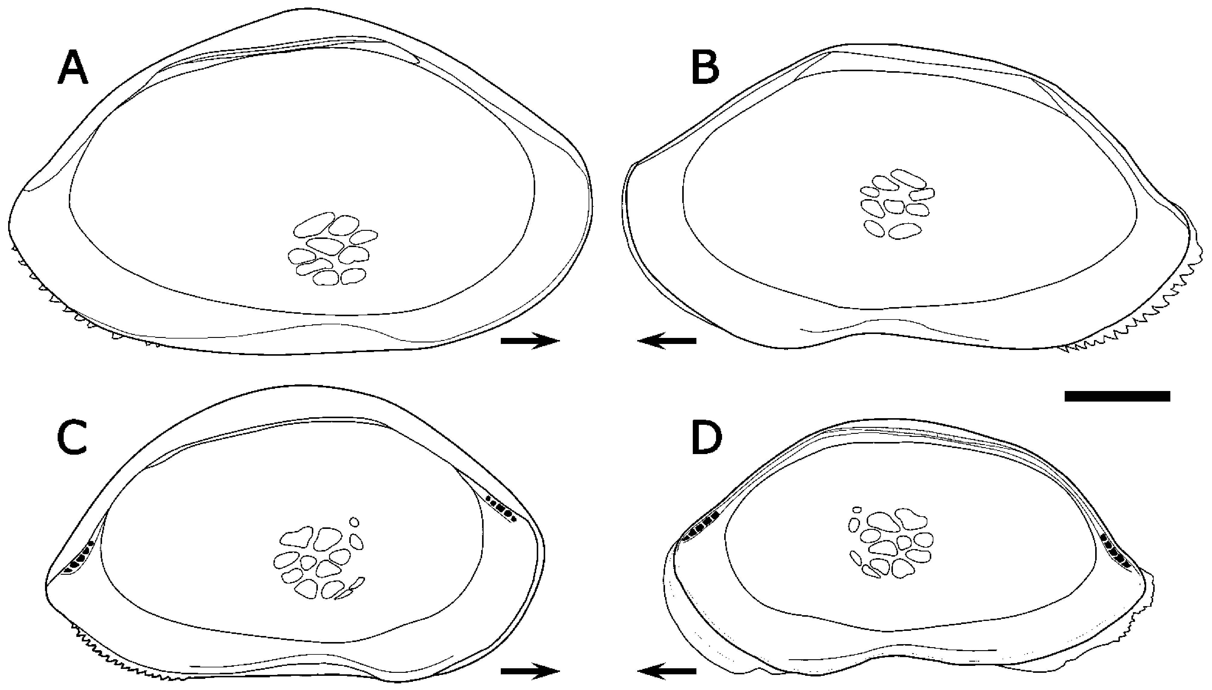
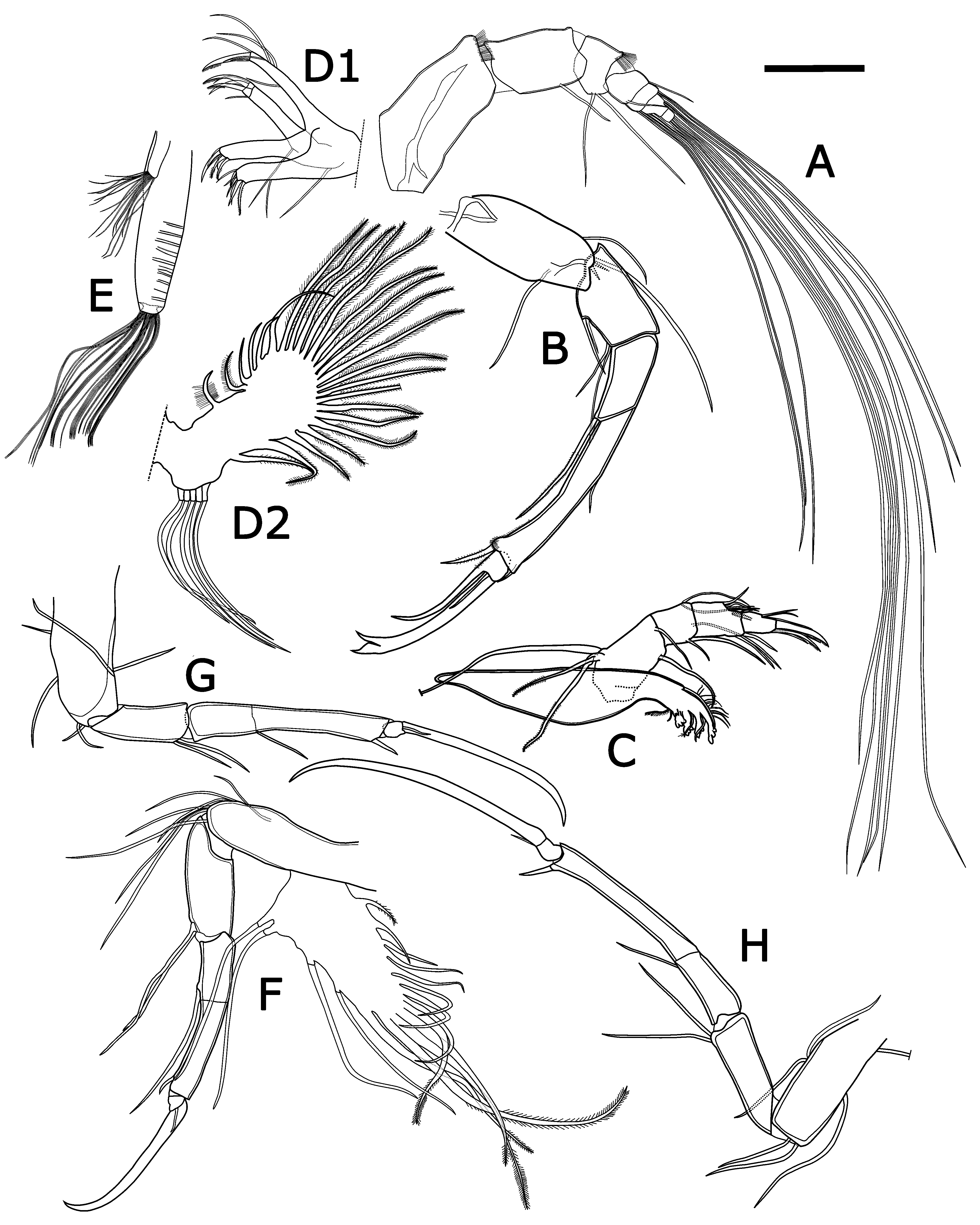
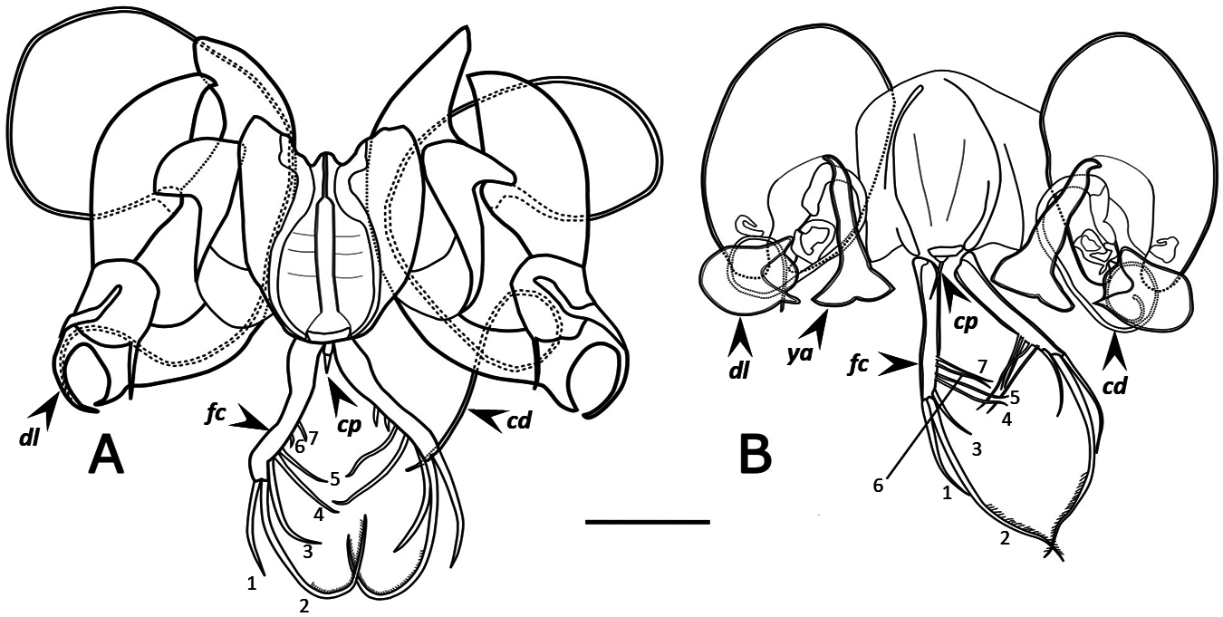
( Figs. 2 View FIGURE 2 , 3 View FIGURE 3 , 4A, B View FIGURE 4 , 5 View FIGURE 5 , 6A View FIGURE 6 )
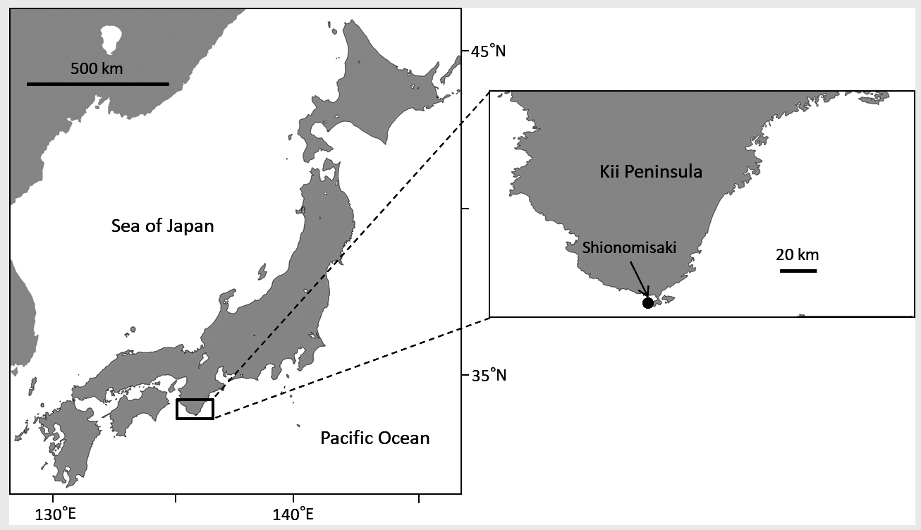
Type series. All examined specimens collected at tidal pool of rocky shore, in Shionomisaki, Wakayama Prefecture, western Honshu, Japan (33° 25′ 59″ N, 135° 45′ 45″ E; Fig. 1 View FIGURE 1 ) on 29th August 2008. Holotype: adult male ( SUM- CO-2446 ), right valve length 1.08 mm, height 0.60 mm, left valve length 1.09 mm, height 0.65mm, appendages mounted on slide and valves preserved in cardboard cell slide. Paratypes: 3 adult males ( SUM CO-2445 , 2447 and 2455) and 7 adult females (SUM-CO- 2448‒2454) .
Etymology. Named after Professor Takahiro Kamiya (Kanazawa University, Japan), in recognition of his significant contribution to our knowledge on Neonesidea species.
Diagnosis. Outline of left valve with blunt angle at center of dorsal margin. In exopodite of mandibula, dorsalmost seta very long, second one medium, third (ventral-most) one short. In male copulatory organ, anterior part of median lobe divided into two pronged, interior part longer than exterior part; posterior part of median lobe with hook-like structure; distal lobe with slender spine on tip, forming hold median lobe; basal lobe short, wide, and tip curving dorsally; copulatory duct very long and arched.
Description. Carapace ( Figs. 2 View FIGURE 2 , 3 View FIGURE 3 and 4A, B View FIGURE 4 ) streamlined sub-triangular in lateral view and surface smooth. Left valve larger than right valve and overlapping along dorsal and ventral margins with blunt angle at center of dorsal margin. Posterior margin of right and left valves finely serrated ( Fig. 2 View FIGURE 2 C‒F). Surface covered with numer- ous (more than 1000) simple type pore systems, above all several long setae, concentrating in posterior-most part. Carapace bearing dark brown colour pattern (probably in epidermis) in whole or part with large variation in living individuals. Central muscle scars consisting of 2 mandibular scars, 2 frontal scars, 3 limb scars and including 1 fulcral point in left valve ( Fig. 3E View FIGURE 3 ). Hingement simple: right valve bearing simple bar with weak dorsal groove and anterior and posterior ends bearing weak elongated teeth ( Fig. 3D View FIGURE 3 ); left valve bearing anterior and posterior elongated sockets with weak dorsal awning ( Fig. 3C View FIGURE 3 ). Very weak auxiliary dentition at upper interior side of anterior and posterior ends of both valves ( Fig. 3A, B View FIGURE 3 ).
Antennule ( Fig. 5A View FIGURE 5 ). Seven articulated podomeres, length ratio among them from proximal to distal 75: 45: 15: 15: 7: 10. Three terminal podomeres with much longer setae than total length of antennule podomeres. First podomere with assemblage of setulae at antero-distal corner. Second podomere with 1 medium seta on posterior margin. Third podomere with 2 long setae on posterior margin and assemblage of fine setae at antero-distal corner. Fourth podomere with 1 medium apical seta at both anterior and posterior corners, respectively. Fifth podomere with 2 very long setae on both, antero- and postero-distal ends, respectively. Sixth podomere with 2 and 3 very long setae on antero- and postero-distal ends, respectively. Seventh (terminal) podomere with 5 very long setae grouped together at bases on distal end.
Antenna ( Fig. 5B View FIGURE 5 ). Five articulated podomeres, length ratio among them from proximal to distal 6: 4: 3: 8: 1. First podomere (protopodite) bearing 1 long and 1 medium setae on anterior margin and postero-medium area, respectively and assemblage (reduced exopodite) of 1 long and 2 short setae in distal end (reduced exopodite). Second podomere with 1 long and 1 medium apical setae at posterior distal corner and 1 medium and 1 short apical setae at posterior proximal corner. Third podomere with 1 long setulous seta at postero-distal corner. Fourth podomere with 1 short seta on middle of anterior margin, 1 medium setulous apical seta at postero-distal corner and 1 very short apical seta at antero-distal corner, and assemblage of numerous short setulae along posteror-distal corner. Fifth (terminal) podomere with 1 large claw (terminally bifurcated in male), 1 smaller claw and 1 medium seta.
Mandibula ( Fig. 5C View FIGURE 5 ). Consisting of 5 podomeres, length ratio among them from proximal to distal 30: 10: 5: 6: 6: 4. First podomere (coxa) with masticatory part of 4 stout trilobed denticles, and several very short, thin setae on distal end. One short simple seta (endite) on anterior ledge. First podomere of palp (basis) with 1 seta on anteriodistal corner, 1 seta on middle of distal end, and branchial plate (reduced exopodite) consisting of 1 very long and 2 medium plumose setae on dorso-proximal end. Second podomere of palp (1st podomere of endopodite) with 2 setae on anterior-distal end, 1 seta on middle of distal end, and 2 long setae on middle of dorsal margin. Third podomere of palp with 4 simple setae on ledge of dorso-median area and 3 simple setae on anterio-distal end. Fourth (terminal) podomere with 2 short setae on middle of dorsal margin, 1 stout terminal claw, and 3 setae on anterio-distal end.
Maxillula ( Fig. 5D View FIGURE 5 1 View FIGURE 1 , D 2 View FIGURE 2 ). Thin branchial plate (exopodite) bearing 25 plumose setae and 5 long reflexed setae. Basal podomere bearing 1 palp (endopodite) and 3 masticatory endites. Palp with 1 and 3 medium setae on ventral and dorsal ledges, respectively, 1 stout serrated claw, and 1 medium seta on distal end. First (ventral-most) endite with 1 short seta on ventral margin of four-fifth from proximal end, 4 short, stout and 3 short, thin setae on distal end. Second endite with 6 short, stout setae on distal end. Third endite with 1 long seta on proximal end, 2 stout serrated claw, 1 short and 2 medium setae on distal end.
Brush-shaped organ (in male) ( Fig. 5E View FIGURE 5 ). Consisting of paired branches. Well-developed right branch approximately 3 times length of left one and bearing about 10 transverse lines and more than 20 long, thin setae. Left branch with less than 20 medium or short, thin setae.
Fifth limb ( Fig. 5F View FIGURE 5 ) Consisting of 5 articulated podomeres, length ratio among them from proximal to distal 13: 9: 5: 8: 1. First podomere with branchial plate (bearing 15 plumose setae and 2 reflexed setae on middle of ventral margin), 1 seta on anterior margin at two-thirds from proximal end, 2 setae on ledge on anterior margin at three-fourths from proximal end, and 2 long and 2 medium setae on distal end. Second podomere with 1 long and 1 medium apical setae at anterior distal corner. Third podomere with 1 long apical seta at anterior distal corner. Fourth podomere with 1 short seta at anterior distal corner. Fifth podomere with 1 very short seta and 1 long, stout terminal claw on distal end.
Sixth limb ( Fig. 5G View FIGURE 5 ). Consisting of 5 articulated podomeres, length ratio among them from proximal to distal 7: 5: 4: 7: 1. Third and 4th podomeres almost fused. First podomere with 1 short and 1 medium setae on anterior margin at one-third and two-thirds from proximal end, respectively, 1 short and 1 medium setae on posterior margin at three-fourths from proximal end, and 2 medium setae on antero-distal end. Second podomere with 2 medium setae on antero-distal end. Third podomere with 1 medium apical seta at antero-distal corner. Fourth podomere with 1 very short apical seta at antero-distal corner. Fifth podomere with 1 very short apical seta at antero-distal corner and 1 long, curved terminal claw on distal end.
Seventh limb ( Fig. 5H View FIGURE 5 ). Consisting of 5 articulated podomeres, length ratio among them from proximal to distal 6: 5: 3: 7: 1. First podomere with 1 long seta on anterior margin at half and three-fourths from proximal end, respectively, 2 medium setae on middle of posterior margin, 1 medium seta at anterior distal corner and on distal end, respectively. Second podomere with 2 apical setae at anterior distal corner. Third podomere with 1 medium apical seta at antero-distal corner. Fourth podomere with 1 short apical seta at antero-distal corner. Fifth podomere with 1 very short, thin apical seta at antero-distal corner, and 1 very long, curved terminal claw on distal end.
Furca ( Fig. 6B View FIGURE 6 ). Bearing 7 setae. Terminal-most 2nd seta (2) twice as long as setae of terminal-most (1) and terminal 3rd (3) ones. Proximal-most 1st and 2nd setae (6 and 7) much shorter than others.
Caudal process ( Fig. 6B View FIGURE 6 ). One simple short, stout seta.
Male copulatory organ ( Fig. 6B View FIGURE 6 ). Basal capsule elliptical and consisting of several lobes. Distal lobe forming furcal talon-like shape; outer parts arched and larger than inner part. Copulatory duct very long and arched.
Eye. Naupliar eyes present without clear cuticle lens.
Dimensions. See Table 1 View TABLE 1 .
Occurrence. Type locality and rocky shore of Tanabe Bay , Tanabe City, Wakayama Pref., Japan .
Remarks. This new species resembles Neonesidea oligodentata ( Kajiyama, 1913) , which was re-described in detail by Smith & Kamiya (2002), in most features of the carapace and appendages. The carapace lateral outline of N. kamiyai sp. nov. appears more triangular. The male copulatory organ of N. kamiyai sp. nov. is apparently different from that of N. oligodentata , described by Smith & Kamiya (2002), especially in the shape of the distal lobe and copulatory duct.
| SUM |
Stellenbosch University |
No known copyright restrictions apply. See Agosti, D., Egloff, W., 2009. Taxonomic information exchange and copyright: the Plazi approach. BMC Research Notes 2009, 2:53 for further explanation.
|
Kingdom |
|
|
Phylum |
|
|
Class |
|
|
Order |
|
|
Family |
|
|
Genus |