
Styracura schmardae ( Werner, 1904 )
|
publication ID |
https://doi.org/ 10.11646/zootaxa.4175.3.1 |
|
publication LSID |
lsid:zoobank.org:pub:2B916685-5384-4665-A759-FC8911DF3E4F |
|
DOI |
https://doi.org/10.5281/zenodo.5696862 |
|
persistent identifier |
https://treatment.plazi.org/id/03CD878D-8649-A805-50F7-66A7CB8BB47F |
|
treatment provided by |
Plazi |
|
scientific name |
Styracura schmardae ( Werner, 1904 ) |
| status |
|
Styracura schmardae ( Werner, 1904)
( Figs. 1 View FIGURE 1 , 3–6 View FIGURE 3 View FIGURE 4 View FIGURE 5 View FIGURE 6 ; Table 1 View TABLE 1 )
Trygon schmardae Werner, 1904: 298 View in CoL (original description, single specimen, not depicted; type locality: Jamaica).
Dasybatus schmardae ( Werner, 1904) .— Garman, 1913: 386 (verbatim from Werner, 1904; placed in subgenus Pastinachus View in CoL ; no specimens examined); Meek & Hildebrand, 1923: 81 (morphological description, Panama Canal, two specimens).
Dasybatus torrei Garman, 1913: 386 –388 (original description, denticles, teeth, pelvic girdle, based on single specimen from Tunas de Zaza, Cuba, not depicted; placed in subgenus Pastinachus View in CoL ).
Dasyatis schmardae ( Werner, 1904) View in CoL .— Fowler, 1931: 391 (color pattern, Trinidad); Beebe & Tee-Van, 1941: 263 (compared to H. pacifica View in CoL , described as new); Boeseman, 1948: 31 –33 (morphological description, pelvic girdle, range extension, four specimens).
Himantura schmardae ( Werner, 1904) View in CoL .— Bigelow & Schroeder, 1953: 390 –394 (morphological redescription, synonymy, distribution); Cervigón, 1992: 200 (Venezuela); Lovejoy, 1996: 220, 221, 229, 230, 246–248 (morphology, relationships, biogeography); McEachran et al., 1996: 64, 65, 71, 72, 75–81, 83 (morphology, phylogeny); McEachran & Fechhelm, 1998: 182 (Gulf of Mexico); Lovejoy et al., 1998: 421 (molecular phylogeny); Cervigón & Alcalá, 1999: 190 (Venezuela); Castro-Aguirre et al., 1999: 72 (Mexico); Camargo & Isaac, 2001: 144 (northern Brazil); McEachran & Carvalho, 2003: 568 (identification, distribution); McEachran & Aschliman, 2004: 80, 86, 88–92, 96, 98, 99, 101 (morphology, phylogeny); Carvalho et al., 2004: 10, 49, 77, 81, 82, 93, 105, 113 (morphology, relationships); Carvalho & Lovejoy, 2011: 46, 47 (molecular phylogeny); Naylor et al., 2012a: 43, 46, 48, 49 (mitochondrial DNA-based phylogeny); Naylor et al., 2012b: 79, 225 ( DNA identification); Aschliman et al., 2012a: 64, 66, 68, 70–72, 75, 77, 79, 86–88, 90, 92–94 (morphology, phylogeny); Aschliman et al., 2012b: 30, 34–36, 38, 39 (molecular phylogeny); Moral-Flores et al., 2015: 130 ( Mexico); Last et al., 2016: 352 (molecular phylogeny, classification).
Diagnosis. A species of Styracura distinguished from S. pacifica by the following combination of characters: anterior disc margin slightly more straight across (more oblique in S. pacifica ); usually greater enlarged scapular denticles (poorly developed in some specimens of S. pacifica , but always well developed in S. schmardae ); slightly larger eyes (up to one-half spiracle length in S. schmardae vs. one-third in S. pacifica ); slightly smaller preorbital length (range 15.6–20.9% DW and mean 18.3% DW in S. schmardae vs. 21.2% DW and 22.5% DW in holotype and measured specimen of S. pacifica , respectively); smaller prenarial length (range 10.8–13.5% DW and mean 12.2% DW in S. schmardae vs. 14.5% DW and 15% DW in holotype and measured specimen of S. pacifica , respectively); smaller preoral length (range 15.7–18.8% DW and mean 17.5% DW in S. schmardae vs. 20.3% DW in measured specimen of S. pacifica ); shorter distance from cloaca to caudal sting origin (range 51.8–90.9% DW and mean 69.5% DW in S. schmardae [holotype 66.5%] vs. 98.4% DW in measured specimen of S. pacifica ); and brown to grayish- or olivaceous-brown dorsal disc color (frequently purplish-gray or darker brown in S. pacifica ).
External morphology. Disc rounded to weakly rhomboidal, slightly broader than long, with average disc length 93.2% DW (range 87.2–98.6% DW). Anterior portion of disc more rounded and broader than posterior portion; posterior portion slightly more oval. Anterior margin of disc straight to weakly convex. Rostral knob small, rounded. Head relatively close to anterior disc margin. Snout relatively short; mean preorbital distance 18.3% DW (range 15.6–20.9% DW); preorbital distance about one and a half times interorbital distance. Mean prenasal distance 12.2% DW (range 10.8–13.5% DW). Mean preoral distance 17.5% DW (range 15.7–18.8% DW). Eyes small, protruding only slightly, their mean diameter 2.6% DW (range 1.8–3.8% DW). Eye diameter from four to five times smaller than interorbital distance. Spiracles very large and rectangular, obliquely positioned in relation to eyes, their mean length 7.7% DW (range 7.2–8.5% DW). Nasal curtain relatively straight, not widening significantly close to mouth. Posterior margin of nasal curtain weakly fringed, straight across. Nostrils anteroposteriorly elongate, slightly more rounded and larger anteriorly; mean distance between anterior margins of nostrils 7.4% DW (range 6.4–8.5% DW). Mouth slightly arched, with a small median concavity on lower jaw; mean mouth width 7.7% DW (range 6.9–8.5% DW). Internarial distance and mouth width about equal. Posterior to mouth integument corrugated. Five papillae on mouth floor. Teeth slightly wider than long, set in quincunx, in up to about 40 rows on upper and lower jaws; a few rows of teeth on upper jaw exposed even with mouth closed. Branchial basket quadrangular; mean distance between first pair of gill slits 20.7% DW (range 19.0–22.4% DW); mean distance between fifth pair of gill slits 15.8% DW (range 13.8–18.0% DW). Branchial basket region relatively long; mean distance between first and fifth pairs of gill slits 15.3% DW (range 14.1–16.3% DW).
Pelvic fins protrude only slightly beyond posterior margins of pectoral fins in dorsal view. Pelvic fins broadly triangular, presenting somewhat straight anterior margins and slightly convex posterior margins. Mean length of anterior pelvic margins 17.4% DW (range 15.4–20.1% DW); pelvic fins not particularly broad, mean distance between outer apices of pelvic fins (width of pelvic fins measured together) 44.4% DW (range 36.7–51% DW). Claspers very reduced in juvenile and young specimens, mean external length of clasper in examined specimens 1.7% DW (range 1.5–2.2% DW), and mean internal length 10.9% DW (range 9–12.9% DW); adult males not examined.
Tail very long, much longer than disc length; mean distance from posterior margin of cloaca to tail tip 146.7% DW (range 129.2–164.2% DW); tail length as measured from cloaca about twice snout to cloaca distance (mean 76.6% DW, range 64.3–84.9% DW). Tail wide at base, its greatest width slightly smaller than interorbital distance; mean tail width at pectoral fin insertion 10.8% DW (range 9.2–12.2% DW). Tail width tapering mildly from base to distal tip, but reduction slightly more accentuated posterior to caudal stings. Tail weakly rounded anterior to caudal stings, but nearly circular in cross section posterior to stings. Lateral tail ridges low, present from more or less level of posterior pelvic fins to well anterior to caudal sting origin. Ventral tail ridge very low, originating at about level of caudal sting origin to posterior to stings; lacking dorsal tail fold, ridge or keel. Caudal sting positioned far from tail base; mean distance between posterior margin of cloaca and caudal sting origin 69.5% DW (range 51.8–90.9% DW), very close to snout to cloaca distance. Caudal sting origin usually at more or less midlength of tail in specimens with intact tails. Caudal stings elongate, varying from 18.8–26.9% DW, nearly two times interorbital distance (10.8–15.1% DW), and much greater than preorbital snout length. Caudal stings relatively slender, their mean width 1.6% DW (range 1–2% DW). Lateral serrations between 60–70, closely positioned forming an almost continuous edge, especially closer to sting tip; serrations more developed toward sting tip but small serrations also present on sting base.
Coloration. Most specimens with a uniform brown, grayish-brown or olivaceous-brown dorsal disc, tail and pelvic fins, sometimes with outer disc contours slightly darker, and dark brown posterior two-thirds of tail. Ventral color creamy white, with lightly darker outer disc margins (sometimes with small, indistinct grayish spots and blotches), and darker posterior pelvic fins; ventral posterior tail darker brown.
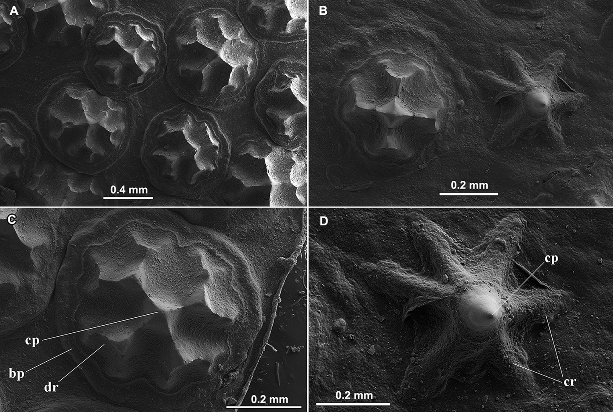
Dermal denticles. An intense shagreen of dermal denticles covers practically the entire dorsal surface of disc and tail, forming a tough, hardened exterior. Denticles slightly less numerous and less closely packed toward disc margins. Pelvic fins mostly naked. Dermal denticles of at least two main shapes: large stellate denticles with broad basal plates closely packed together on the central area of dorsal disc and all of dorsal tail, and slightly smaller asterisk-shaped denticles present on disc margins and much of tail ( Fig. 3 View FIGURE 3 ). In addition, a pair of larger scapular denticles (or spines) present above shoulders, usually one on each side, but sometimes with two pairs, one more developed than the other. Larger denticles on dorsal disc and tail have broad, subcircular to quadrangular basal plates, these frequently overlapping. Larger denticles with quadriradiate crowns, with primary ridges subdividing distally into smaller ridges, and these frequently also further subdivided. The main dichotomic crown ridges resemble a cross, and bear a central, higher, and smaller pyramidal crown plate. Greatly enlarged scapular denticles above shoulders morphologically similar to larger denticles, but with many more dichotomic ridges radiating from the crown plate, and sometimes with two central, higher, longitudinal ridges forming an inverted V-shape on crown. Smaller denticles asterisk-shaped with several high crown ridges, usually six, branching from a single elevated, acute, cylindrical crown. These denticles present a star-shaped basal plate beneath coronal ridges (in contrast to larger, stellate denticles that have a broad basal plate). On the medial dorsal region of tail, close to caudal stings, these denticles possess a more developed and acute crown with fewer ridges, resembling small thorns.
Lateral line canals. The hyomandibular canal (HMD) extends close to anterior disc margin, where numerous closely adjacent anterior subpleural tubules branch off in parallel (hyomandibular canal colored pink in Fig. 4 View FIGURE 4 ); anterior subpleural tubules present only on anterior segment of hyomandibular canal. Hyomandibular canal extends obliquely toward posterior disc; its subpleural component (spl) somewhat concave, giving off two small posterior subpleural tubules (pst), and ending in an acute triangle; jugular component (jug) more or less straight posteriorly, but broadly arched lateral to gill slits, and slightly bulging near first gill slits. Infraorbital (IOC; blue in Fig. 4 View FIGURE 4 ) canal with small, rounded infraorbital loop posteriorly, and without suborbital loop anteriorly; infraorbital canal highly sinuous, extending anteriorly to ventral midsnout length. Supraorbital canal (SPO; green) positioned medial to infraorbital canal and obliquely positioned, not sinuous; small prenasal loop present; supraorbital forming a small posterior loop just anterior to nasal curtain. Prenasal canal (anterior segment of nasal canal; NAS, yellow in Fig. 4 View FIGURE 4 ) extends in a vertical line to anterior snout tip, not connecting to its posterior oblique segment lateral to nostril. Mandibular canal (not shown in Fig. 4 View FIGURE 4 due to previous dissection) forming an inverted V-pattern just posterior to mouth (not shown in Fig. 4 View FIGURE 4 ).
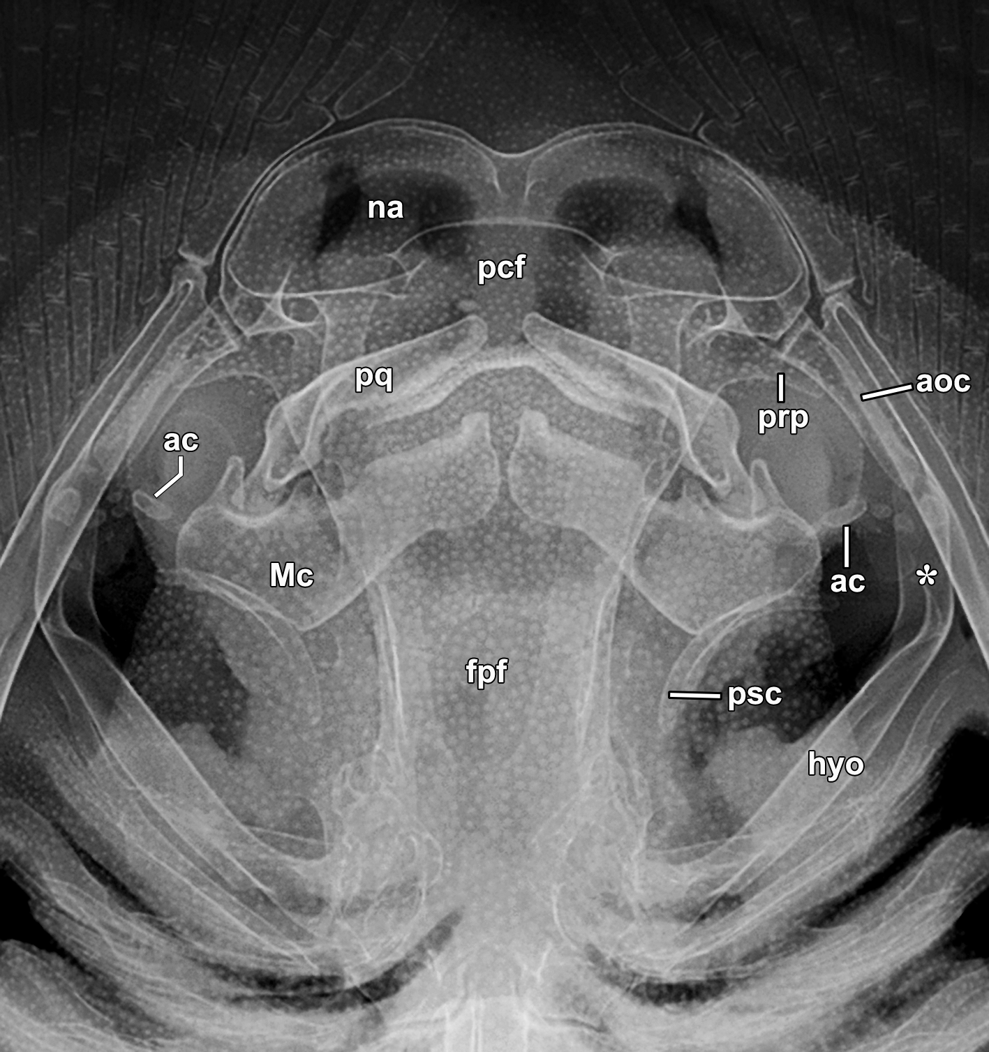
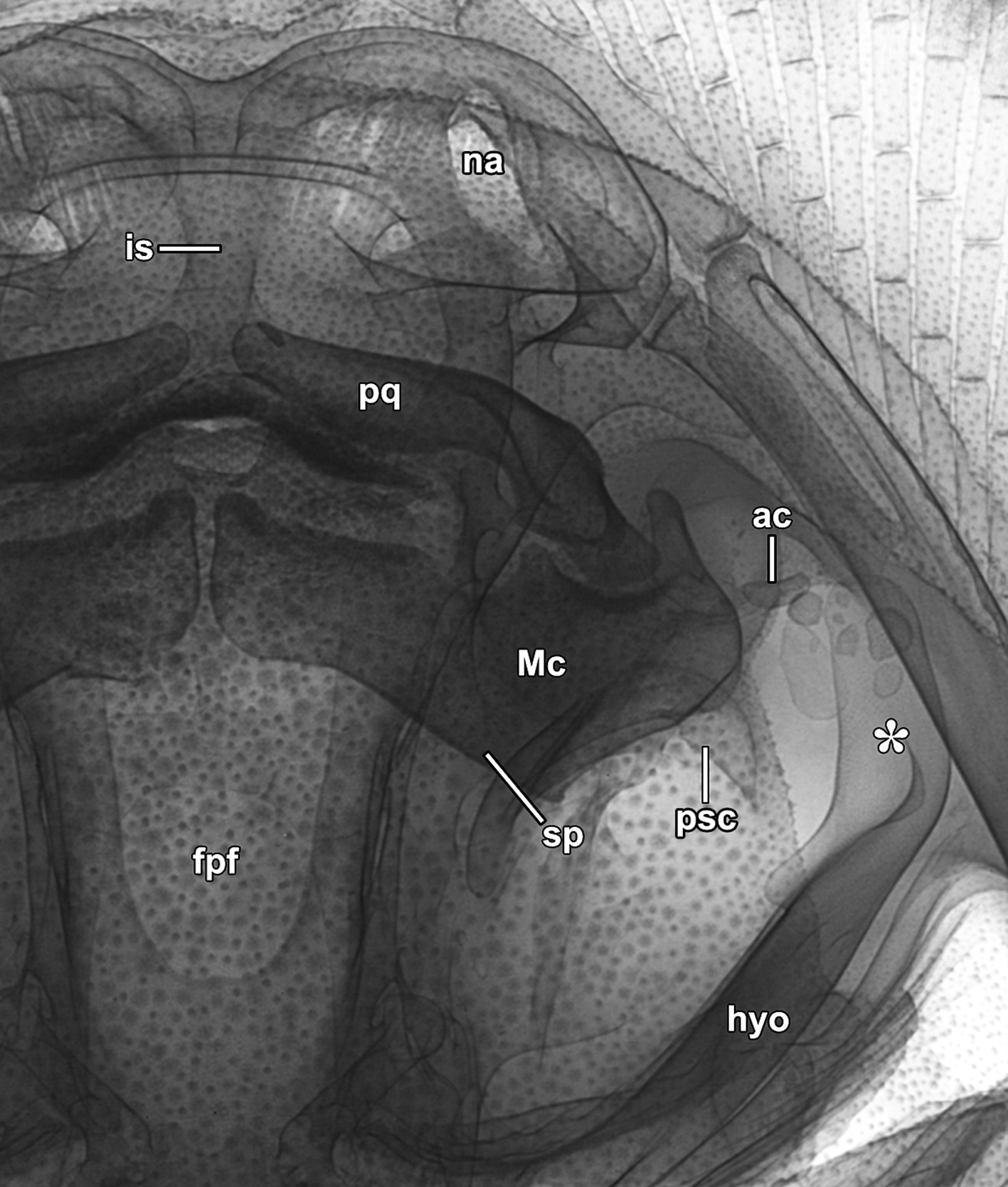
Skeletal features. Neurocranium, jaws and hyoid arch. Neurocranium longer than wide, its greatest width at level of preorbital processes; neurocranial greatest width about two-thirds its greatest length ( Figs. 5 View FIGURE 5 , 6 View FIGURE 6 ). Nasal capsules with oval and wide nasal apertures (na), with broadly rounded anterior wall, but relatively anteroposteriorly short; triangular anterior median indentation present; internasal septum (is) moderately broad. Preorbital process (prp) projecting laterally, somewhat wide, tapering only slightly. Postorbital process projecting anterolaterally, somewhat straight and rectangular, and poorly calcified, obscured in radiographs by denticles on overlying integument. Supraorbital process (sp) broadly triangular. Neurocranium most slender at its posterior fourth, at level of posterior frontoparietal fontanelle. Otic capsule very short, about as wide as orbital region. Precerebral (pcf) and frontoparietal (fpf) fontenellae about three-fourths length of neurocranium. Precerebral fontanelle relatively large, wider posteriorly, and slightly trapezoidal with rounded margins; precerebral fontanelle wider than long, just greater than half length of frontoparietal fontanelle; frontoparietal fontanelle broadly triangular, longer than wide, not medially constricted and tapering, rounded posteriorly. Antorbital cartilage (aoc) very large and laterally compressed, widest at articulation with posterolateral nasal capsule, and extending posteriorly to close to hyomandibula, at level of jaw joint. Prespiracular cartilages (psc) weakly calcified, concave and slender.
Meckel’s cartilages (Mc) greater than palatoquadrates (pq), with short and triangular dorsally projecting lateral processes posteriorly; ventral margin of lower jaws lacking distinct projection where lower jaws deflect anteriorly toward midline ( Figs. 5 View FIGURE 5 , 6 View FIGURE 6 ); ventrolateral process closer to jaw joint also absent or very reduced. Small triangular gap present between lower jaw symphyses. Jaws about equal in width to widest point of neurocranium. Palatoquadrates markedly slender, stouter at midwidth, and not as wide as lower jaws. Hyomandibulae (hyo) laterally compressed, blade-like, slender in dorsoventral view and stouter at midlength. Distal portion of hyomandibulae (where they deflect anteriorly, represented by an asterisk in Figs. 5 View FIGURE 5 , 6 View FIGURE 6 ) stout, very elongate, just under one-third hyomandibular length. Hyomandibulae tightly articulated to neurocranium, extending anterolaterally from it. Well developed and stout ligament between hyomandibulae and lower jaws, usually directed slightly posteriorly toward midline. Small calcified elements present within ligament, including a small, wider than long, obliquely positioned (also directed posteriorly toward midline in Fig. 5 View FIGURE 5 ), cylindrical angular element (ac); rounded calcified element also present adjacent to hyomandibula in some specimens, with smaller rounded cartilages in between ( Figs. 5 View FIGURE 5 , 6 View FIGURE 6 ).
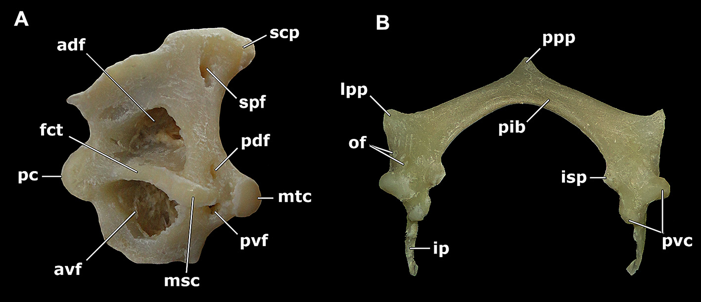
Pectoral girdle. Coracoid bar dorsoventrally flattened and laterally expanded. Scapulocoracoid in lateral view tall and triangular, with broadly inclined anterodorsal margin bearing anteriorly a triangular projection; scapulae with concave posterodorsal and posteroventral aspects ( Fig. 7 View FIGURE 7 a). Pectoral condyles situated on horizontal axis of scapular cartilage. Scapular process (scp) bearing a conspicuous fossa perforated by a small foramen (scapular process fenestra, spf) on its distal portion, broadly triangular and firmly articulated to lateral aspects of synarcual. Procondyle (pc) nearly elliptical, vertically oriented and situated at anterolateral surface of scapula. Mesocondyle (msc) slightly oval and horizontal. Metacondyle (mtc) rounded, just posterior to mesocondyle. Horizontally oriented, expanded, and slightly curved facet (fct) present in large gap between pro- and mesocondyle; facet helps support propterygium. Anterior fenestrae much greater than posterior fenestrae. Anterodorsal (adf) and anteroventral (avf) fenestrae somewhat oval and dorsal and ventral to pectoral condyles, respectively. Posterodorsal fenestra (pdf) and posteroventral fenestra (pvf) greatly reduced, about equal in size, and much smaller than anterior fenestrae.
Pelvic girdle. Puboischiadic bar (pib) relatively wide, laterally expanded and dorsoventrally flattened, with relatively oblique but straight anterior margins and posterior margin concave ( Fig. 7 View FIGURE 7 b). Five obliquely positioned obturator foramina (of) just medial to pelvic condyles (pvc) arranged in an arch along the anteroposterior axis of pelvic girdle. Prepelvic process (ppp) short and broadly triangular. Iliac processes (ip) on posterolateral corners of puboischiadic bar relatively long and wide, laterally flattened, projecting dorsally, and distally tapering. Ischial processes (isp) blunt, broadly triangular and posteromedially directed. Lateral prepelvic processes (lpp) robust, broadly triangular and anteriorly directed.
Distribution. Essentially that given in McEachran & Carvalho (2003) with the noted extension of the northern Brazilian coast (e.g. Camargo & Isaac, 2001).
Etymology. Named after zoologist and explorer Ludwig Karl Schmarda (23 August 1819 – 7 April 1908), founder of the zoological museum of the University of Graz, and later professor in the Zoological Institute of the University of Vienna (1861–1883), where he kept his collection of fishes including the specimen that became the type of Trygon schmardae . Franz Werner was on staff in the same institute for over 40 years, eventually retiring as professor in 1939 ( Salvini-Plawen & Mizzaro, 1999).
Remarks. Werner (1904: 298, 299) used the following characters to diagnose his new species: "front disc not perfectly rounded, but with small snout tip; top completely rough, especially on the pectoral fins, although sparser than the trunk; tail without enlarged thorns; scapular region with two large, round, ribbed tubercles side by side (their distance apart close to distance between nostrils); tail nearly twice as long as disc; color brown uniform, tail darker" [our translation]. Remarkably, Werner (1904: 298) alluded to the similarity between S. schmardae and " Trygon hystrix M. et H." (= Potamotrygon histrix ), and the features listed above were given to distinguish his new species from this species of Potamotrygon (Werner did not report any specimen of Potamotrygon and took his information directly from Müller & Henle, 1841); thus, a putative close relationship between S. schmardae and potamotrygonins was alluded to when S. schmardae was described more than 110 years ago. Werner's (1904) paper on the fishes of the zoological and comparative anatomy collections of the University of Vienna is very thorough, providing morphological details of dozens of species of elasmobranchs, chimaeras, basal actinopterygians, lungfishes, and even agnathans; his paper contains keys to species, and makes constant reference to the works of Cuvier, Günther, Vaillant, and especially Müller & Henle (1841). Concerning dasyatid stingrays, Werner reported on material which had been collected by P. Bleeker and L.K. Schmarda of species, if correctly identified, now classified as Dasyatinae ( Dasyatis pastinaca , Hemitrygon bennetti ), Urogymninae ( Himantura uarnak , Brevitrygon walga , and Urogymnus polylepis ), and Neotrygoninae ( Taeniura lymma ).
Garman's (1913) Dasybatus (Pastinachus) torrei clearly equals S. schmardae , a species mentioned by him but not examined. His description highlights typical characters of S. schmardae , such as the denticles (stellate, quadriradiate, though no mention of asterisk-like denticles) and their tight distribution on dorsal disc and tail, the enlarged scapular denticles, rounded disc, low ventral keel on tail, lack of dorsal keel, and olivaceous brown color; other characters mentioned are of little diagnostic value (e.g. morphology of teeth). Garman separated his new species from S. schmardae on the basis of the tail being one and a half times as long as the disc (not twice as long as he states for S. schmardae , a detail he took from Werner, 1904). There is little doubt concerning this synonymy (also Bigelow & Schroeder, 1953), especially given the relatively wide distribution of S. schmardae , a common stingray off Cuba (even featured in a 1967 Cuban postal stamp).
Both Beebe & Tee-Van (1941) and Bigelow & Schroeder (1953) provided diagnostic characters that separate S. schmardae and S. pacifica , which are similar to some characters employed in the diagnosis of S. schmardae above (eye-spiracle proportions, scapular denticles). Our comparisons in the diagnosis of S. schmardae are based on data provided in the original description of S. pacifica by Beebe & Tee-Van (1941) and on the specimen we measured in the AMNH. However, the tail is missing posterior to the caudal sting in this specimen, precluding more direct comparisons of tail length (and we are not certain how tail length was measured in Beebe & Tee-Van, 1941). The distance between the cloaca and caudal sting origin given in the diagnosis needs to be confirmed in additional specimens of S. pacifica . Beebe & Tee-Van (1941: 263) included the measurement "base of tail to first spine" (69.5% DW in holotype of S. pacifica ), but our measurement is from the cloaca (98.4% DW in AMNH 233890), farther anterior than the base of tail at pectoral fin insertions (presumably how their measurement was taken). Beebe & Tee-Van (1941) may have taken this measurement from even more posteriorly, i.e. from the junction of the pelvic fins and tail; hence their data are not directly comparable to ours. We also note that the preoral length was given as 17.7% DW in the holotype of S. pacifica ( Beebe & Tee-Van, 1941) , which places it within the range of S. schmardae (15.7–18.8% DW; Table 1 View TABLE 1 ), even though our measured specimen of S. pacifica has a greater preoral length (20.3% DW); this character needs confirmation. Further comparisons to strengthen the differences between both species are warranted as little morphological detail is available for S. pacifica (for recent depictions, see Allen & Robertson, 1994; Bussing & López S., 1994; McEachran, 1995).


| DNA |
Department of Natural Resources, Environment, The Arts and Sport |
No known copyright restrictions apply. See Agosti, D., Egloff, W., 2009. Taxonomic information exchange and copyright: the Plazi approach. BMC Research Notes 2009, 2:53 for further explanation.
|
Kingdom |
|
|
Phylum |
|
|
Class |
|
|
Order |
|
|
Family |
|
|
Genus |
Styracura schmardae ( Werner, 1904 )
| De Carvalho, Marcelo R., Loboda, Thiago S. & Da Silva, João Paulo C. B. 2016 |
Himantura schmardae (
| Naylor 2012: 43 |
| Aschliman 2012: 64 |
| Aschliman 2012: 30 |
| Carvalho 2011: 46 |
| McEachran 2004: 80 |
| Carvalho 2004: 10 |
| McEachran 2003: 568 |
| Camargo 2001: 144 |
| Cervigon 1999: 190 |
| Castro-Aguirre 1999: 72 |
| McEachran 1998: 182 |
| Lovejoy 1998: 421 |
| Lovejoy 1996: 220 |
| McEachran 1996: 64 |
| Cervigon 1992: 200 |
| Bigelow 1953: 390 |
Dasyatis schmardae (
| Boeseman 1948: 31 |
| Beebe 1941: 263 |
| Fowler 1931: 391 |
Dasybatus schmardae (
| Meek 1923: 81 |
| Garman 1913: 386 |
Dasybatus torrei
| Garman 1913: 386 |
Trygon schmardae
| Werner 1904: 298 |