
Cladorhiza corallophila, Göcke, Christian, Hestetun, Jon T., Uhlir, Carolin, Freiwald, André, Beuck, Lydia & Janussen, Dorte, 2016
|
publication ID |
https://doi.org/ 10.11646/zootaxa.4168.3.4 |
|
publication LSID |
lsid:zoobank.org:pub:3F006EA6-D22C-4CFF-A763-D60F2E5D892A |
|
DOI |
https://doi.org/10.5281/zenodo.5631045 |
|
persistent identifier |
https://treatment.plazi.org/id/03CD87C9-FFA0-AA46-DEC0-F84303FFF81F |
|
treatment provided by |
Plazi |
|
scientific name |
Cladorhiza corallophila |
| status |
sp. nov. |
Cladorhiza corallophila View in CoL sp. nov.
Diagnosis. Cladorhiza of straight upright growth without roots, with surface filaments being rounded in life, attached to skeleton of live or occasionally dead cold-water corals.
Material. 3 specimens (SMF11490 holotype, SMF11491, GenBank nr. KU508388 View Materials /391/394, SMF 11492, GenBank nr. KU508387 View Materials /390/393) on live Lophelia pertusa , sta. GeoB 14914-1-3, ROV dive 12 17°08.196'N, 16°49.466'W, 17 November 2010, 512 m water depth, Tiguent Mound Complex, two of which consist of several coalesced individuals, attached to dead framework portions of a live Lophelia pertusa colony, originally inhabited by amphipods. One separate specimen (SMF11489) and approximately five individuals on one live Lophelia pertusa colony (SaM-ID 1 430, GenBank nr. KU508386 View Materials /389/392), Timiris Mound Complex, sta. GeoB 14873-1-5, ROV dive 5, 18°57.765'N, 16°52.151'W, 10 November 2010, 485 m water depth, basal portions of sponge inside coral skeleton (coral overgrowth), co-occurring with gastropod Calliostoma grazing on Lophelia pertusa , sponges in live often inhabited by amphipods. Several dried fragmentary specimens (SMF 11410) from sta. GeoB 14902-1- 9, ROV dive 9, 17°32.848’N 16°39.703’W, 407 m water depth, Tamxat Mound Complex.
Additional material used for comparison: Cladorhiza abyssicola Sars, 1872 : BMNH [19]34.2.16.14 labelled “ Norway, From Oslo Museum, Challenger-Collection ”, BMNH 1910.1 .1.1372 labelled “ Norwegian Arctic Expedition 1876-8, Dr. A. Hansen, “ Type ”. Cladorhiza gelida Lundbeck, 1905 : ZMUC-DEM-31 (Syntype), Ingolf station 113, 69°31’N, 7°06’W, 1896, 2465 m. GoogleMaps
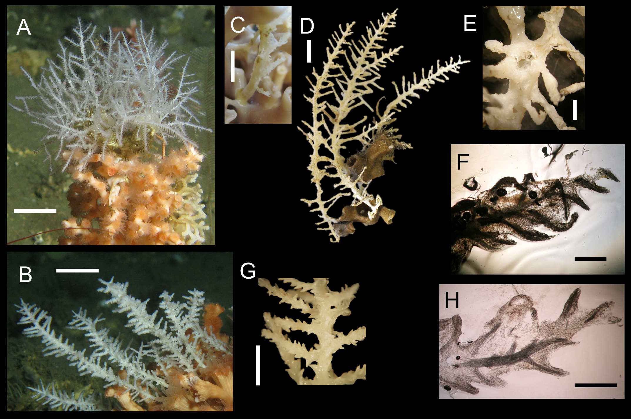
Description. The species is always found growing on cold-water corals of the species Lophelia pertusa or to a lesser degree on Madrepora oculata ( Fig. 2 View FIGURE 2 ). The sponge grows predominantly erect upright into the water column and reaches a height of at least 10 cm. It can be locally very abundant. The sponges grow upright with a continuous middle axis with a diameter of about 2 mm from which lateral processes emerge. These processes are only very rarely branching. The processes of the lower part of the sponge are longer than those of the upper part, so a continuous growth of the processes can be assumed. They can be up to about 2 cm long with a diameter about 1 mm. Often the lateral branches are arranged more or less in a plane, giving the sponge a somewhat flat or featherlike appearance. Most specimens nonetheless have their branches arranged more or less regularly around the central axis. The surface of the lateral branches is covered by small, short filaments. In this regard, the species comprises two different forms, which are especially noticeable when looking at specimens fixed in ethanol: some have the surface covered with very distinct pointed filaments about 1 mm long, while others have only very tiny surface filaments, which are almost like nodes of the sponge surface. When looking at in situ pictures of living sponges ( Fig. 2 View FIGURE 2 A, B), it becomes obvious that this character is mainly due to shrinking of the tissue in ethanol, because in life, the filaments are larger and seem to be somewhat inflated and more rounded, than in the ethanol samples. The similarity is also apparent when comparing sections of both varieties ( Fig. 2 View FIGURE 2 F, H). On closer examination the sponge surface reminds of a cork-screw ( Fig. 3 View FIGURE 3 F). Several specimens are coalesced together ( Fig. 2 View FIGURE 2 D), in these cases, the main axes of the sponges must have met and merged, sometimes forming one main axis, but more often a branching follows again. This latter case might actually derive from different individuals growing near to each other and coalescing where they met. All specimens grow on scleractinian framework-building corals, they do not have distinct basal structures, but they are connected to their substrate very strongly. When attached in between live portions of the coral skeleton, the coral tends to overgrow the attachment zones of the sponge, encalcifying subsequently its base and thus strengthening its attachment strongly. This interaction gives better support to the large growing sponge bodies in moving water and thus points to an advantage for the sponges to their life on cold-water corals. The color of the sponge alive and in ethanol is yellowish to white. Several findings of fragmented crustaceans within the sponges underline their carnivorous feeding habit ( Fig. 2 View FIGURE 2 H, 3 H, I). Additionally, ROV images show a multitude of amphipods in close vicinity to the sponge. As these are certainly not prey for the sponge, due to their large size, they might be considered as commensals, feeding on the same source as the sponges.
Skeleton. The whole sponge body, the main axis as well as the branches, is cored by a filament formed by a dense aggregation of mycalostyles, strongyles, and occasional oxeas ( Fig. 2 View FIGURE 2 F, H). Branches project laterally from the main axis, being supported by a spicular fibre with a thickness of a few megascleres. At the attachment point of the lateral processes, the coring spicules of their central fibres fan out, forming a broader attachment site. At the top of the tapering appendages the apex of some megascleres breaks through the surface of the sponge ( Fig. 3 View FIGURE 3 G). Anchorate anisochelae densely cover the whole body, especially at the ends of the branchlets. Sigmata are less common than anisochelae and often distributed along the axis, but are absent in the younger branchlets. On some SEM-pictures sigmata are found on exposed places without or under anisochelae. In Fig. 3 View FIGURE 3 H, I are entire crustacean bodies seen while being digested.
Spicules. Here, we restrict the description of spicules mainly to those of the holotype, further data can be found in tab. 1.
Megascleres
a) Mainly mycalostyles ( Fig. 3 View FIGURE 3 B) and strongyles ( Fig. 3 View FIGURE 3 A), straight or slightly curved. Sometimes hybrid and hard to define, probably blunt styles. Rarely subtylostyles. Dimensions: mycalostyles 28 0–420 (335 ± 43) L x 8 –14.4 (11.6 ± 1.5) W, N= 30; strongyles 200–410 (358 ± 52) L x 11.2–17.6 (14.2 ± 1.42) W, N= 30
b) Occasionally oxea. Dimensions: 300–440 (373.5 ± 42.7) L x 6.4–9.6 (7.8 ± 1.2) W

No known copyright restrictions apply. See Agosti, D., Egloff, W., 2009. Taxonomic information exchange and copyright: the Plazi approach. BMC Research Notes 2009, 2:53 for further explanation.
|
Kingdom |
|
|
Phylum |
|
|
Class |
|
|
Order |
|
|
Family |
|
|
Genus |