
Echinoderes unispinosus, Yamasaki & Neuhaus & George, 2018
|
publication ID |
https://doi.org/ 10.11646/zootaxa.4387.3.8 |
|
publication LSID |
lsid:zoobank.org:pub:DA75D56E-22BB-487E-8BA3-42C5C98F9E53 |
|
DOI |
https://doi.org/10.5281/zenodo.5952905 |
|
persistent identifier |
https://treatment.plazi.org/id/03CD87E0-245D-FFD2-3D86-2DF609C1FF37 |
|
treatment provided by |
Plazi |
|
scientific name |
Echinoderes unispinosus |
| status |
sp. nov. |
Echinoderes unispinosus sp. nov.
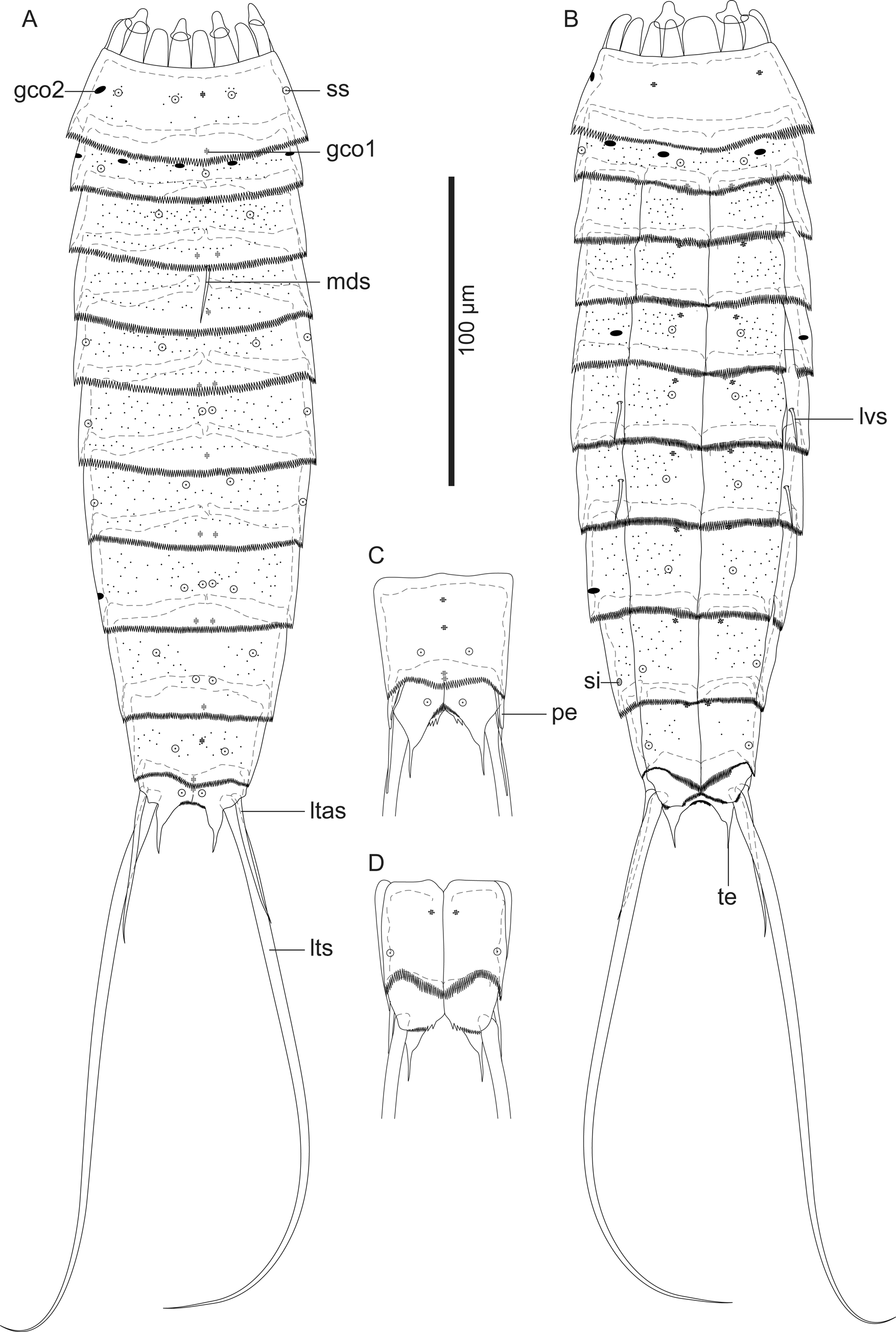
( Figs 8–10 View FIGURE 8 View FIGURE9 View FIGURE10 ; Tables 4, 5)
Diagnosis. Echinoderes with middorsal acicular spine on segment 4; lateroventral acicular spines on segments 6 and 7; type-2 glandular cell outlets present in midlateral position on segment 1, in subdorsal, laterodorsal, sublateral, and ventrolateral position on segment 2, in lateral accessory position on segment 5, and in sublateral position on segment 8; narrow pectinate fringe teeth of primary pectinate fringe similar in width on segments 1–10; tergal extension long and smoothly pointed posteriorly.
Etymology. The species name is composed of the Latin uni (one) and Latin spina (spine), referring the species with only one middorsal acicular spine.
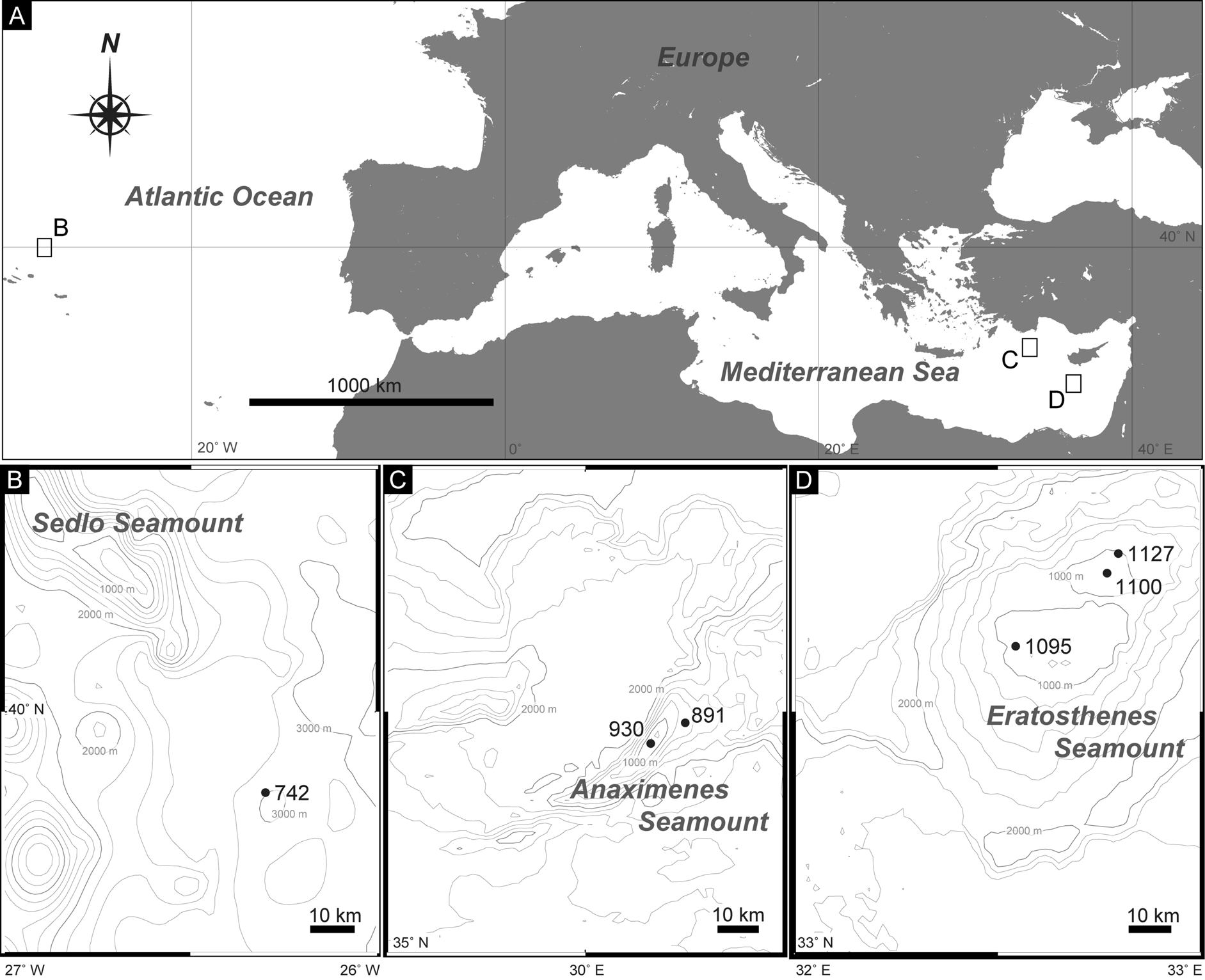
Material examined. Holotype: Adult female (ZMB 11587), collected at station 742 in the deep-sea plain near the Sedlo Seamount ( Fig. 1A, B View FIGURE1 ; Table 1), mounted as glycerol-paraffin slide on a Cobb aluminum frame.
Paratypes: Three adult females and two adult males ( ZMB 11588, 11589a–11589c), collected at the same station to the holotype. All paratypes mounted as glycerol-paraffin slides on Cobb aluminum frames.
Type locality. Deep-sea plain near the Sedlo Seamount (39°50'0.00"N, 26°17'54.00"W), 2,875 m depth ( Fig. 1A, B View FIGURE1 ; Table 1).
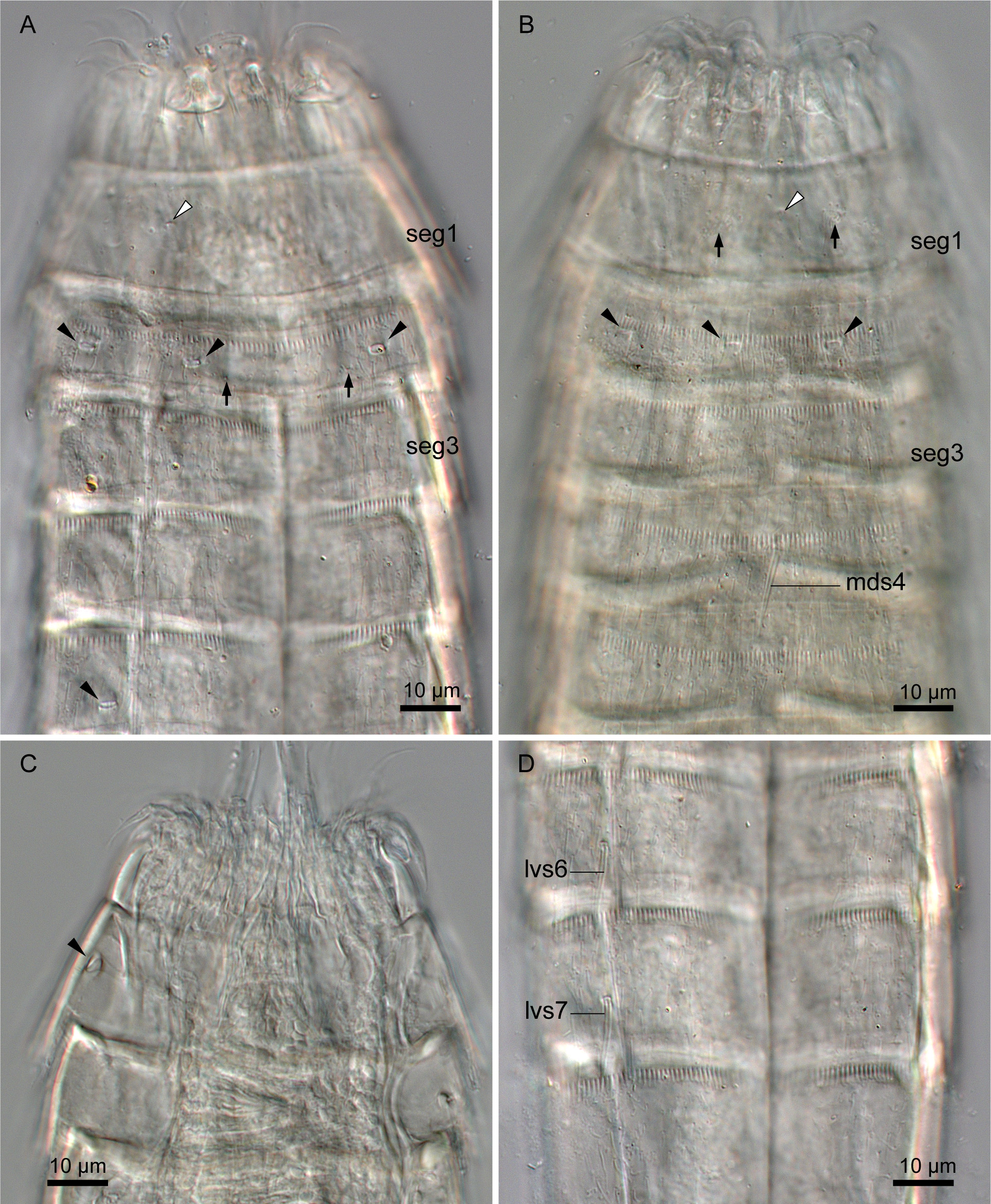
Description. Adult with head, neck, and eleven trunk segments ( Fig. 8A, B View FIGURE 8 ). See Table 4 for measurements. Table 5 indicates the positions of cuticular structures (sensory spots, glandular cell outlets, spines, and sieve plate).
Head consisting of retractable mouth cone and introvert. Mouth cone with inner oral styles and nine outer oral styles. Introvert composed of several rings of spinoscalids and one ring of trichoscalids. Exact number and arrangement of inner and outer oral styles and scalids not examined.
Neck with 16 placids ( Fig. 8A, B View FIGURE 8 ). Midventral placid broadest. Remaining placids similar in size. Two trichoscalid plates present ventrally and four dorsally.
Segment 1 consisting of complete cuticular ring. This and following nine segments with thick pachycyclus at anterior margin. Pachycyclus interrupted middorsally in segments 2–10 as well as at tergosternal junctions in segments 3–10. With few cuticular hairs irregularly arranged on the segment, especially concentrated around sensory spots ( Fig. 8A View FIGURE 8 ). All cuticular hairs on this and following nine segments thin and ca. 4–8 µm in length. Sensory spots located centrally on segment in subdorsal and laterodorsal position ( Figs 8A View FIGURE 8 , 9B View FIGURE9 ). Type-1 glandular cell outlets situated in middorsal and lateroventral position ( Figs 8A View FIGURE 8 , 9A, B View FIGURE9 ). Additional type-1 glandular cell outlets present sublaterally in four out of six examined specimens. Type-2 glandular cell outlets present in midlateral position ( Fig. 8A View FIGURE 8 , 9C View FIGURE9 ). Posterior part of this and following nine segments with primary pectinate fringe ( Figs 8A–D View FIGURE 8 , 9A, B, D View FIGURE9 ). Pectinate fringe teeth of primary pectinate fringes on this and following eight segments similar in length and width ( Fig. 9A, B, D View FIGURE9 ).
Segment 2 with complete cuticular ring as segment 1. With numerous cuticular hairs aligning on three to four lines on entire segment ( Fig. 8A, B View FIGURE 8 ). Sensory spots present in middorsal, laterodorsal, and ventromedial position ( Figs 8A, B View FIGURE 8 , 9A, B View FIGURE9 ). Type-1 glandular cell outlet in middorsal position ( Fig. 8A View FIGURE 8 ). Type-1 glandular cell outlets on ventral side not examined because free flap of preceding segment covering the relevant area. Type-2 glandular cell outlets in subdorsal, laterodorsal, sublateral, and ventrolateral position ( Figs 8A, B View FIGURE 8 , 9A, B View FIGURE9 ).
Segment 3 and following eight segments consisting of one tergal and two sternal plates. This and following seven segments entirely covered with cuticular hairs except for midventral and paraventral area ( Fig. 8A, B View FIGURE 8 ). Sensory spots present subdorsally ( Fig. 8A View FIGURE 8 ). Type-1 glandular cell outlets situated in middorsal and ventromedial position ( Fig. 8A, B View FIGURE 8 ).
Segment 4 with middorsal short acicular spine ( Figs 8A View FIGURE 8 , 9B View FIGURE9 ). No sensory spots present. Type-1 glandular cell outlets present paradorsal and ventromedial position ( Fig. 8A, B View FIGURE 8 ).
Segment 5 with sensory spots in subdorsal, midlateral, and ventromedial position ( Fig. 8A, B View FIGURE 8 ). Type-1 glandular cell outlets present in middorsal and ventromedial position ( Fig. 8A, B View FIGURE 8 ). Type-2 glandular cell outlets present in lateral accessory position ( Figs 8B View FIGURE 8 , 9A View FIGURE9 ).
Segment 6 with lateroventral short acicular spines ( Figs 8B View FIGURE 8 , 9D View FIGURE9 ). Sensory spots present in paradorsal, midlateral, and ventromedial position ( Fig. 8A, B View FIGURE 8 ). Type-1 glandular cell outlets present paradorsally and ventromedially ( Fig. 8A, B View FIGURE 8 ).
Segment 7 with lateroventral short acicular spines, as in segment 6 ( Figs 8B View FIGURE 8 , 9D View FIGURE9 ). Sensory spots present in subdorsal, midlateral, and ventromedial position ( Fig. 8A, B View FIGURE 8 ). Type-1 glandular cell outlets present in middorsal and ventromedial position ( Fig. 8A, B View FIGURE 8 ).
Segment 8 with paradorsal and ventromedial sensory spots ( Fig. 8A, B View FIGURE 8 ). Additional subdorsal sensory spots observed in two out of six examined specimens ( Fig. 8A View FIGURE 8 ). Type-1 glandular cell outlets present in paradorsal and ventromedial position ( Fig. 8A, B View FIGURE 8 ). Type-2 glandular cell outlets situated sublaterally ( Figs 8B View FIGURE 8 , 10A View FIGURE10 ).
Segment 9 with paradorsal, laterodorsal, and ventrolateral sensory spots ( Fig. 8A, B View FIGURE 8 ). Type-1 glandular cell outlets present in paradorsal and ventromedial position ( Fig. 8A, B View FIGURE 8 ). Small rounded sieve plates present in sublateral position ( Fig. 8B View FIGURE 8 ).
Segment 10 with subdorsal and ventrolateral sensory spots ( Fig. 8A–D View FIGURE 8 ). Two type-1 glandular cell outlets aligned middorsally ( Fig. 8A, C View FIGURE 8 : please note that one of the glandular cell outlets is hidden under the pectinate fringe of the previous segment in Fig. 8A View FIGURE 8 ). Additional pairs of type-1 glandular cell outlets present in ventromedial position ( Fig. 8B, D View FIGURE 8 ). Pectinate fringe teeth of primary pectinate fringe in ventromedial to midventral area longer than those on middorsal to ventrolateral area.
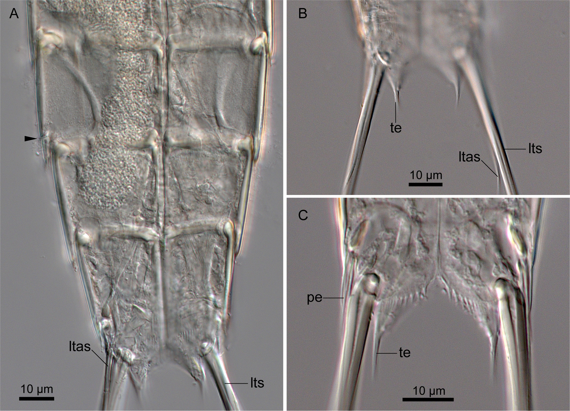
Segment 11 with lateral terminal spines ( Figs 8A–D View FIGURE 8 , 10A–C View FIGURE10 ). Lateral terminal accessory spines present only in females ( Figs 8A, B View FIGURE 8 , 10A, B View FIGURE10 ). Three pairs of penile spines present in males ( Figs 8C, D View FIGURE 8 , 10C View FIGURE10 ). Dorsal and ventral penile spines long and tube-like, whereas middle penile spines stout and triangular-shaped. Sensory spots in subdorsal position. Two type-1 glandular cell outlets present tandemly in middorsal position. Posterior parts of sternal plates curved and end with primary pectinate fringe ( Figs 8B, D View FIGURE 8 , 10B, C View FIGURE10 ). Tergal plate extending laterally and forming tergal extensions ( Figs 8A, C View FIGURE 8 , 10B, C View FIGURE10 ). Each extension pointed posteriorly with a broader base tapering towards the tip ( Fig 10C View FIGURE10 ).
Remarks. Echinoderes unispinosus sp. nov. can be easily distinguished from the other congeners by the combination of having a middorsal acicular spine only on segment 4 and lateroventral acicular spines on segments 6 and 7, and lacking any tube. No other Echinoderes species has this spine and tube pattern. Other Echinoderes with only a single middorsal acicular spine on segment 4 include 13 species: Echinoderes ajax Sørensen, 2014 ; Echinoderes annae Sørensen et al., 2016 ; Echinoderes cantabricus Pardos et al., 1998 ; Echinoderes capitatus (Zelinka, 1928) ; Echinoderes charlotteae Sørensen et al., 2016 ; Echinoderes isabelae G a Ordóñez et al., 2008; E. maxwelli ; Echinoderes ohtsukai Yamasaki & Kajihara, 2012 ; Echinoderes regina Yamasaki, 2016 ; Echinoderes reicherti Neves et al., 2016 ; E. rex ; Echinoderes serratulus Yamasaki, 2016 ; Echinoderes teretis Brown, 1999 in Adrianov & Malakhov (1999) (see Zelinka 1928; Omer-Cooper 1957; Brown 1985; Pardos et al. 1998; Adrianov & Malakhov 1999; G a Ordóñez et al. 2008; Lundbye et al. 2011; Yamasaki & Kajihara 2012; Sørensen 2014; Herranz & Leander 2016; Neves et al. 2016; Sørensen et al. 2016a, b; Yamasaki 2016). But E. unispinosus sp. nov.
is distinguishable from all these species in lacking any spine or tube laterally on segments 5 and 8. In addition, the presence of the midlateral type-2 glandular cell outlets has been known in none of the other congeners, and is a unique character for E. unispinosus sp. nov.
| ZMB |
Museum f�r Naturkunde Berlin (Zoological Collections) |
No known copyright restrictions apply. See Agosti, D., Egloff, W., 2009. Taxonomic information exchange and copyright: the Plazi approach. BMC Research Notes 2009, 2:53 for further explanation.